This website uses cookies to ensure you get the best experience on our website.


- Table of Contents
Plan chromogenic UBA6 IHC in paraffin sections with the catalog antibody at 2–5 μg/ml (datasheet A03213-2). Use the cytoplasmic spermatozoa profile and high staining in elongated or late spermatids as reference points, with caution because staining and RNA expression show very low consistency (HPA tissue IHC).

Expected localisation, validated protocols, controls and antibodies — the at-a-glance facts below, then the full design guide.
| Expected localisation | Cytoplasmic staining in spermatozoa (HPA tissue IHC) | |
| Staining pattern | Cytoplasmic spermatozoa; high in late spermatids (HPA tissue IHC) | |
| Antigen retrieval | EDTA pH 8.0 HIER, heat-mediated (datasheet A03213-2) | |
| Positive control | Testis | |
| Negative control | Adipose tissue+4 more · see all |
| Fixation | Keep paraffin-section fixation consistent. (standard IHC practice; not target-specific) | |
| Caveat | Staining has very low consistency with RNA expression (HPA tissue IHC) | |
| Regulation | Isoform 2: adult > fetal testis (UniProt) | |
| Isoform / epitope | 4 isoforms; check epitope coverage before isoform claims (UniProt) |
The catalog antibody’s IHC-P protocol (datasheet A03213-2) is accompanied by published UBA6 IHC protocols for tumour sections (PMC9478149) and human breast tissue microarrays (PMC5675648).
| Sample | Paraffin-embedded human cervical cancer tissue; fixative not specified (datasheet A03213-2) |
| Fixation | Image fixative and duration unreported (datasheet A03213-2); verify before use. |
| Sectioning | 4–5 µm sections on charged slides (standard) |
| Deparaffinisation | Xylene, graded ethanol series to water (standard) |
| Antigen retrieval | Heat retrieval: EDTA pH 8.0 (datasheet A03213-2); 20 min, 95–100 °C (standard) |
| Peroxidase block | 3% H2O2, 10 min, room temperature (standard) |
| Blocking | 10% goat serum (datasheet A03213-2) |
| Primary antibody | Rabbit anti-UBA6, 2-5 μg/ml (datasheet A03213-2) |
| Primary incubation | Overnight at 4 °C (datasheet A03213-2) |
| Detection | HRP-conjugated secondary, DAB chromogen (datasheet A03213-2) |
| Counterstain | Hematoxylin, blue, dehydrate and mount (standard) |
| Expected result | UBA6-positive staining in elongated or late spermatids of testis (HPA tissue IHC: High). HPA tissue profile: Cytoplasmic expression in spermatozoa. No signal in the no-primary control. |
In paraffin-section IHC, expect strong cytoplasmic staining in elongated or late spermatids in testis; HPA describes cytoplasmic expression in spermatozoa (HPA tissue IHC: High in elongated or late spermatids). UBA6 has no transmembrane segment (UniProt A0AVT1 topology). Treat the pattern as a guide, because HPA rates the tissue staining Approved while reporting very low consistency with RNA expression and pending external verification (HPA tissue IHC: reliability).
| Strong cytoplasmic signal follows elongated or late spermatids in testis, with identifiable cell boundaries and limited background. | This matches the clearest reported tissue positive: High staining in elongated or late spermatids and a cytoplasmic spermatozoa profile (HPA tissue IHC). Assess the stained cells against section morphology and the counterstain (general IHC practice). |
| Nuclei alone stain strongly in elongated or late spermatids, while their cytoplasm is unstained. | A nuclear-only result conflicts with the reported testis IHC profile (HPA tissue IHC: cytoplasmic expression in spermatozoa). Review morphology and controls before calling it an artifact: HPA also reports nucleoplasm and cytosol in ICC-IF, a different preparation (HPA subcellular). |
| The strongest signal is in adipocytes or adrenal glandular cells rather than late spermatids. | Those cell types were Not detected in the supplied tissue IHC observations (HPA tissue IHC: adipose tissue; adrenal gland). Consider cross-reactivity or detection-system activity, and check whether signal persists in a no-primary control (general IHC practice). |
| Brown signal spreads across tissue, empty spaces, and section edges without following cell morphology. | This is a background pattern rather than the reported cytoplasmic spermatid pattern (HPA tissue IHC). Inspect the no-primary control and review blocking, washes, chromogen development, and section handling as general chromogenic IHC checks (general IHC practice). |
| No specific signal appears in an otherwise intact testis section containing elongated or late spermatids. | That misses the reported High-positive cell population (HPA tissue IHC: testis). Before interpreting absence as biology, check that the relevant cells are present and review antibody, retrieval, detection, and counterstain conditions against the run controls (general IHC practice). |
| Which tissue and cells anchor interpretation? | Testis elongated or late spermatids provide the supplied High-positive IHC reference (HPA tissue IHC). HPA reports Not detected in several sampled cell types, including adipocytes and adrenal glandular cells; these are observed comparisons, not proof that every cell in those tissues lacks UBA6 (HPA tissue IHC). |
| How much weight should the tissue profile carry? | HPA labels its tissue IHC reliability Approved, yet reports very low consistency between staining and RNA expression, with external verification pending (HPA tissue IHC: reliability). Interpret discordant fields cautiously and retain tissue morphology and run controls in the assessment (general IHC practice). |
| Can the IF result look nuclear? | Yes. HPA ICC-IF reports enhanced nucleoplasm and cytosol localization, with images from U-251MG and HeLa BAC 5376 (HPA subcellular). That supports an IF localization discussion, but it does not replace the reported cytoplasmic spermatozoa pattern for testis IHC (HPA tissue IHC). |
| Could an isoform affect tissue comparisons? | UBA6 has four annotated isoforms; isoform 2 is predominantly expressed in testis and is higher in adult than fetal testis (UniProt A0AVT1: isoforms; tissue specificity). The supplied record does not establish which isoform produced the HPA stain, so do not assign the spermatid signal to isoform 2. |
| What do topology and antibody validation establish? | UBA6 has no signal peptide, propeptide, or transmembrane segment in the supplied record (UniProt A0AVT1: processing; topology). HPA037001 is Approved for IHC and Enhanced for ICC, but those ratings do not identify its epitope or establish retrieval or fixation sensitivity (HPA antibodies: HPA037001). |
| Situation | Likely cause | Next action |
|---|---|---|
| Testis appears negative. | The section may lack identifiable elongated or late spermatids, the reported High-positive cells (HPA tissue IHC: testis). A failed staining run is another possibility (general IHC practice). | Confirm cell-stage morphology, then check the run's positive control and the antibody and detection steps (general IHC practice). Do not score an unrepresented cell population as negative. |
| Only spermatid nuclei stain. | This differs from cytoplasmic spermatozoa staining in tissue IHC (HPA tissue IHC). Nucleoplasm localization in ICC-IF means nuclear signal alone is insufficient to identify the cause (HPA subcellular). | Recheck cell boundaries with the counterstain and compare the no-primary control (general IHC practice); report the compartment discordance rather than treating the ICC-IF result as tissue-IHC confirmation. |
| Adipocytes or adrenal glandular cells stain strongly. | Those sampled cell types were Not detected by HPA tissue IHC (HPA tissue IHC). Cross-reactivity or detection-system activity could explain an unexpected signal (general IHC practice). | Compare with the no-primary control and review staining in the reported testis-positive cells in the same run (HPA tissue IHC: testis; general IHC practice). |
| The whole section has diffuse chromogen. | Signal without cell boundaries does not match the reported cytoplasmic spermatid pattern (HPA tissue IHC). Background from blocking, washing, or detection is possible (general IHC practice). | Inspect the no-primary control; review blocking, wash steps, and chromogen development, then rescore only morphology-linked staining (general IHC practice). |
| IF shows cytosol and nucleoplasm while IHC shows cytoplasmic spermatids. | These observations come from different preparations and sampled cells: ICC-IF reports nucleoplasm and cytosol; testis tissue IHC reports cytoplasmic spermatozoa (HPA subcellular; HPA tissue IHC). | Keep the interpretation tied to each preparation's cells and compartment; use the separate IF/ICC guide for IF workflow (HPA subcellular; HPA tissue IHC). |
| A weak or variable tissue result conflicts with RNA expectations. | HPA reports low tissue RNA specificity and very low consistency between antibody staining and RNA expression (HPA tissue IHC: RNA specificity; reliability). | Describe the observed cell-level stain and controls, and flag the HPA reliability limitation; avoid inferring a validated protein-negative tissue from RNA alone (HPA tissue IHC; general IHC practice). |
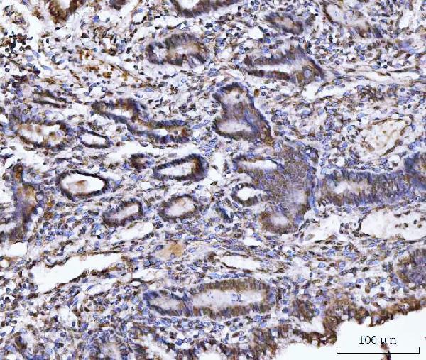
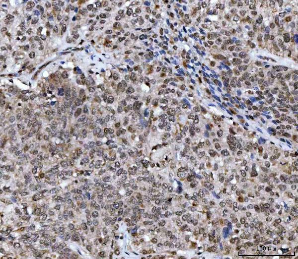


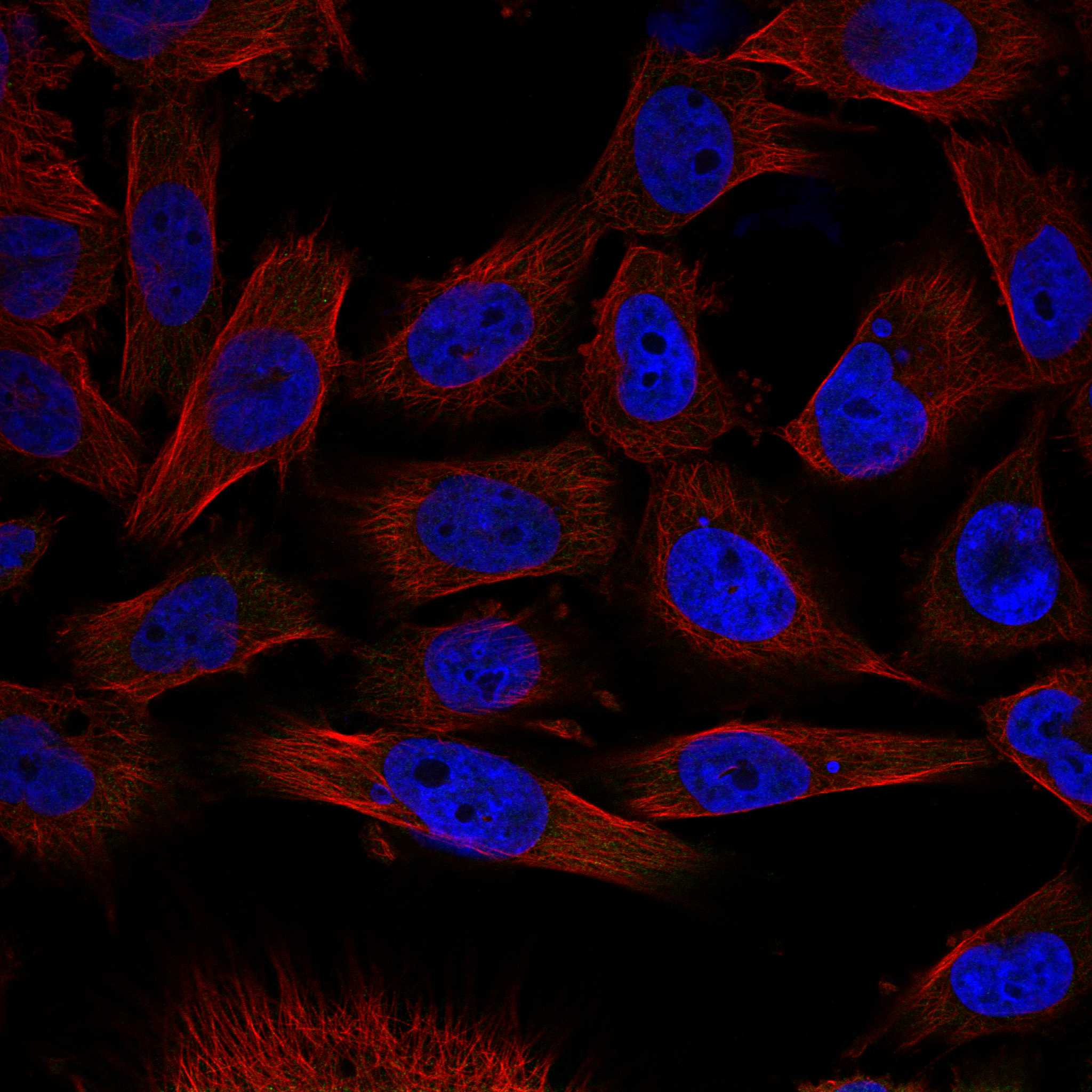
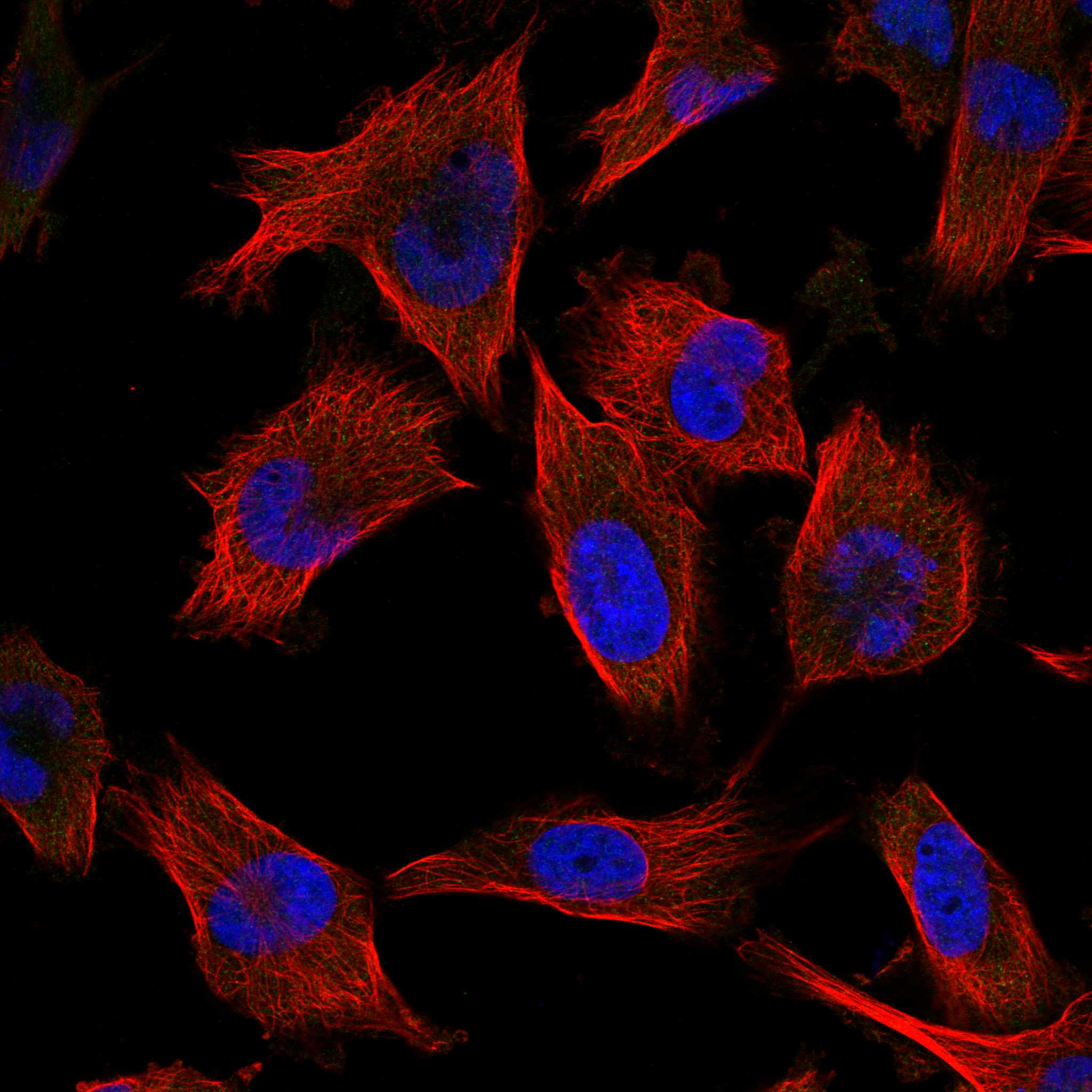
Comprehensive Human Protein Atlas IHC scoring per tissue (reliability: Approved — Very low consistency between antibody staining and RNA expression data. External characterization data supports antibody staining. Pending external verification.). Rows are taken directly from the HPA tissue chart — click any row's HPA link to view the source.
| Tissue | Cell type | Level | Evidence | Source |
|---|---|---|---|---|
| Testis | Elongated or late spermatids | High | Protein (IHC) | HPA → |
| Tissue | Cell type | Level | Evidence | Source |
|---|---|---|---|---|
| Adipose tissue | Adipocytes | Not detected | Protein (IHC) | HPA → |
| Adrenal gland | Glandular cells | Not detected | Protein (IHC) | HPA → |
| Appendix | Glandular cells | Not detected | Protein (IHC) | HPA → |
| Bone marrow | Hematopoietic cells | Not detected | Protein (IHC) | HPA → |
| Breast | Adipocytes | Not detected | Protein (IHC) | HPA → |
Troubleshoot UBA6 staining in paraffin sections by checking retrieval, tissue preservation, compartment pattern and controls before comparing signal between samples.
A03213-2 has real IHC data from human paraffin sections (IHC image captions). Its listed reactivity includes human, monkey, mouse, and rat (catalog reactivity); no IF/ICC data are supplied (catalog applications and images).
A03213-2 is listed for IHC and has images from paraffin sections of human cervical cancer, lung squamous cell carcinoma, and ovarian cancer (catalog applications; IHC image captions). It lists human, monkey, mouse, and rat reactivity, with IHC dilution specified for human tissue only (catalog reactivity; dilution data).
Which to pick: Choose A03213-2 for human paraffin-section IHC: its own captions show staining at 2 μg/ml after EDTA pH 8.0 retrieval; the fixative is unreported (IHC image captions). No supplied SKU is validated for IF/ICC, so there is no IF/ICC pick (catalog applications and images). A03213-2 lists monkey, mouse, and rat reactivity, but the supplied IHC evidence is limited to human sections; assess those species separately before tissue IHC use (catalog reactivity; IHC image captions).