This website uses cookies to ensure you get the best experience on our website.


- Table of Contents
Plan UBE2L6 chromogenic IHC on formalin-fixed paraffin sections with the IHC-validated antibody at 1:50–1:100 (datasheet: A07315-1). Assess cytoplasmic and nuclear staining in immune-cell subsets, using appendix lymphoid tissue or lymph-node germinal centers as positive references (HPA tissue IHC).

Expected localisation, validated protocols, controls and antibodies — the at-a-glance facts below, then the full design guide.
| Expected localisation | Cytoplasmic and nuclear staining in immune-cell subsets (HPA tissue IHC) | |
| Staining pattern | Immune-cell subsets show cytoplasmic and nuclear staining (HPA tissue IHC) | |
| Antigen retrieval | Citrate pH 6.0 HIER, 95–98 °C, 20 min (rule: cytoplasmic / membrane antigen) | |
| Positive control | Appendix+4 more · see all | |
| Negative control | Adipose tissue+4 more · see all |
| Fixation | Keep formalin fixation consistent across paraffin sections (datasheet: A07315-1; standard IHC practice) (selected-SKU IHC image A07315-1) | |
| Caveat | Spleen red-pulp peroxidase may raise background (HPA tissue IHC; standard IHC practice) | |
| Regulation | Regulation of IHC staining intensity is unreported (UniProt) | |
| Isoform / epitope | 2 isoforms; variant epitope coverage is unknown (UniProt) |
The catalog antibody’s IHC-P protocol (datasheet) is accompanied by 1 published UBE2L6 IHC protocol for esophageal squamous cell cancer tissue microarrays (PMC9949073).
| Sample | FFPE human hepatocarcinoma tissue (datasheet A07315-1) |
| Fixation | Image formalin-fixed; duration unreported (datasheet A07315-1); verify before use. |
| Sectioning | 4–5 µm sections on charged slides (standard) |
| Deparaffinisation | Xylene, graded ethanol series to water (standard) |
| Antigen retrieval | Heat-induced epitope retrieval in citrate buffer, pH 6.0, 20 min at 95–98 °C (standard rule: cytoplasmic / membrane antigen) |
| Peroxidase block | 3% H2O2, 10 min, room temperature (standard) |
| Blocking | 10% normal serum of the secondary host, 30 min, room temperature (standard) |
| Primary antibody | Rabbit anti-UBE2L6, 1:50-1:100 (datasheet A07315-1) |
| Primary incubation | Overnight at 4 °C (standard) |
| Detection | HRP-polymer secondary, DAB chromogen 5–10 min (standard) |
| Counterstain | Hematoxylin, blue, dehydrate and mount (standard) |
| Expected result | UBE2L6-positive staining in lymphoid tissue of appendix (HPA tissue IHC: High). HPA tissue profile: Cytoplasmic and nuclear expression, most abundant in subsets of immune cells. No signal in the no-primary control. |
In paraffin-section IHC, expect UBE2L6 staining in cytoplasm and nuclei, most abundant in subsets of immune cells (HPA tissue IHC: Enhanced reliability). High staining is reported in appendix lymphoid tissue, lymph-node and tonsil germinal-center cells, and splenic red-pulp cells (HPA tissue IHC). UBE2L6 has no transmembrane segment, so a membrane-only pattern would be unexpected (UniProt O14933 topology). HPA's Enhanced tissue reliability reflects agreement with RNA data, pending external verification (HPA tissue IHC).
| Distinct cytoplasmic and nuclear chromogen in subsets of immune cells, especially germinal-center cells. | This fits the reported tissue pattern; lymph-node and tonsil germinal-center cells stain at a High level (HPA tissue IHC). Assess compartments within identified cells rather than treating every immune cell as an equally strong reference (HPA tissue IHC: subsets of immune cells). |
| An exclusively sharp plasma-membrane rim, with little cytoplasmic or nuclear staining. | This conflicts with the reported IHC compartments and the absence of a transmembrane segment (HPA tissue IHC; UniProt O14933 topology). Suspect staining artefact or nonspecific binding; morphology alone cannot identify its cause (general IHC practice). |
| Strong staining in adipocytes or cardiomyocytes while expected immune-cell staining is absent. | Those cell types are reported as Not detected (HPA tissue IHC). Consider cross-reactivity or endogenous detection activity, then compare with controls; unexpected staining alone cannot distinguish these explanations (general IHC practice). |
| Uniform pale chromogen across cells, stroma, and empty regions of the section. | A widespread haze obscures the cell-specific pattern reported by HPA (HPA tissue IHC). In chromogenic IHC, incomplete blocking, excessive antibody or detection reagent, and inadequate washing are possible causes (general IHC practice). |
| No detectable staining in appendix lymphoid tissue or tonsil germinal-center cells. | Both are reported High and are useful positive-reference regions (HPA tissue IHC). First check that the relevant cells are present and that the run controls worked; a blank section alone does not establish absent UBE2L6 expression (general IHC practice). |
| Which tissue and cell population is scored? | HPA reports High staining in appendix lymphoid tissue, lung alveolar type I cells, lymph-node and tonsil germinal-center cells, and splenic red-pulp cells; glandular cells in adrenal gland and breast are Medium (HPA tissue IHC). Score the specified cell population, since a tissue-wide average can hide this variation (general IHC practice). |
| How is a low or absent comparator chosen? | Adipocytes and cardiomyocytes are Not detected, whereas thyroid and parathyroid glandular cells are Low (HPA tissue IHC). Use these categories as observed reference patterns, not as proof that every cell in a section must be unstained (HPA tissue IHC). |
| Which antibody supports the IHC interpretation? | CAB073407 has Enhanced IHC validation; HPA003328 has Supported IHC validation (HPA antibodies). The overall tissue profile has Enhanced reliability through agreement with RNA expression data, pending external verification (HPA tissue IHC). Record which antibody produced the result when comparing patterns. |
| Does IF/ICC show the same compartment? | HPA's ICC-IF summary places UBE2L6 in the cytosol, with approved cytosol localization, while tissue IHC describes cytoplasmic and nuclear expression (HPA subcellular; HPA tissue IHC). Interpret each assay against its own observation; the ICC-IF entry does not establish that every paraffin section must lack nuclear staining. |
| Do topology or isoforms change the expected readout? | The annotated protein lacks a signal peptide, propeptide, transmembrane segment, and glycosylation sites; UniProt lists two isoforms (UniProt O14933). These annotations provide no basis to expect a shed extracellular pattern, but they do not identify the antibody epitopes or predict isoform-specific staining (UniProt O14933). |
| Situation | Likely cause | Next action |
|---|---|---|
| A positive-reference immune region is blank. | The sampled section may lack the expected cell population, or an IHC step may have failed (general IHC practice); HPA reports High staining only for specified cells (HPA tissue IHC). | Confirm cell identity and section quality, review the run's positive and detection controls, then check the catalog antibody's IHC-P instructions for the actual staining conditions (general IHC practice). |
| Signal appears only as a membrane outline. | The pattern conflicts with cytoplasmic and nuclear tissue staining and with the lack of a transmembrane segment (HPA tissue IHC; UniProt O14933 topology). Nonspecific staining is possible (general IHC practice). | Compare serial sections and controls, inspect whether the rim tracks tissue edges, and repeat with an independently validated IHC antibody if available (general IHC practice). |
| Adipocytes or cardiomyocytes stain strongly. | Both are Not detected in the HPA tissue record (HPA tissue IHC). Cross-reactivity or endogenous detection activity could produce an unexpected chromogenic signal (general IHC practice). | Compare a no-primary control and an appropriate detection control; examine whether expected positive cells stain in the same run before assigning target expression (general IHC practice). |
| The entire section has diffuse brown background. | Excess reagent, incomplete blocking, or inadequate washing can raise nonspecific chromogen background (general IHC practice). HPA supplies no UBE2L6-specific cause for diffuse haze. | Check no-primary controls, blocking and wash steps, and antibody and detection-reagent conditions against the validated IHC-P instructions (general IHC practice). |
| Weak glandular staining seems inconsistent with a strong immune-cell control. | HPA reports Medium staining in adrenal and breast glandular cells and Low staining in several other glandular populations, alongside High staining in selected immune cells (HPA tissue IHC). | Score each identified cell population against its own HPA category, using the same run and counterstain to judge signal above background (HPA tissue IHC; general IHC practice). |
| An ICC-IF image is cytosolic, but the tissue IHC section also shows nuclear staining. | Those are the respective reported assay observations: approved cytosol localization in ICC-IF and cytoplasmic plus nuclear expression in tissue IHC (HPA subcellular; HPA tissue IHC). | Record assay and specimen type when comparing images; for this paraffin IHC readout, evaluate nuclear and cytoplasmic signal against tissue controls (HPA tissue IHC; general IHC practice). |



Comprehensive Human Protein Atlas IHC scoring per tissue (reliability: Enhanced — High consistency between antibody staining and RNA expression data. Pending external verification.). Rows are taken directly from the HPA tissue chart — click any row's HPA link to view the source.
| Tissue | Cell type | Level | Evidence | Source |
|---|---|---|---|---|
| Appendix | Lymphoid tissue | High | Protein (IHC) | HPA → |
| Lung | Alveolar cells type I | High | Protein (IHC) | HPA → |
| Lymph node | Germinal center cells | High | Protein (IHC) | HPA → |
| Spleen | Cells in red pulp | High | Protein (IHC) | HPA → |
| Tonsil | Germinal center cells | High | Protein (IHC) | HPA → |
| Tissue | Cell type | Level | Evidence | Source |
|---|---|---|---|---|
| Adipose tissue | Adipocytes | Not detected | Protein (IHC) | HPA → |
| Caudate | Glial cells | Not detected | Protein (IHC) | HPA → |
| Cerebellum | Cells in granular layer | Not detected | Protein (IHC) | HPA → |
| Cerebral cortex | Glial cells | Not detected | Protein (IHC) | HPA → |
| Heart muscle | Cardiomyocytes | Not detected | Protein (IHC) | HPA → |
Troubleshoot UBE2L6 staining in paraffin sections by checking retrieval, cell identity and compartment before comparing chromogenic signal across specimens.
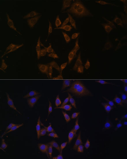
UBE2L6 antibody images cover IHC-P in formalin-fixed, paraffin-embedded human hepatocarcinoma and IF in HeLa cells (catalog image captions); IF/ICC reactivity lists human, mouse, and rat (catalog: A07315).
A07315-1 has an IHC-P image of formalin-fixed, paraffin-embedded human hepatocarcinoma stained with DAB (A07315-1 image caption). A07315 lists IF/ICC and human, mouse, and rat reactivity; its rendered IF image shows HeLa cells with DAPI nuclear staining (catalog: A07315 applications and reactivity; A07315 image caption).
Which to pick: Choose A07315-1 for human tissue IHC-P: it is listed as polyclonal, has a 1:50–1:100 IHC-P range, and its image documents formalin-fixed, paraffin-embedded human hepatocarcinoma (catalog: A07315-1 applications, reactivity, and dilution; A07315-1 image caption). Choose A07315 for IF/ICC, supported by its HeLa IF image and 1:50–1:200 IF range (catalog: A07315 applications and dilution; A07315 image caption). A07315 is the cross-species option because its listed reactivity includes human, mouse, and rat; the rendered image documents HeLa cells only (catalog: A07315 reactivity; A07315 image caption).