UBE3B / Ubiquitin-protein ligase E3B · IHC design guide

Design Immunohistochemistry for UBE3B

Plan chromogenic UBE3B IHC on paraffin sections with the catalog antibody at 1:100–1:300 (datasheet A10679). Use the HPA tissue IHC profile to assess granular cytoplasmic staining across cell types (HPA tissue IHC).

Evidence assembled Oct 2026 · For research use; verify linked source records and product datasheet before use
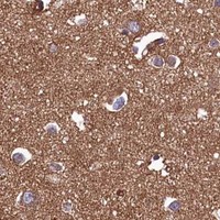
Immunohistochemistry protocol sheet for UBE3B (IHC for UBE3B): expected localisation Granular cytoplasm (HPA tissue IHC); postsynaptic density (UniProt), antibody A10679, validated IHC image, and IHC protocol steps
Printable UBE3B IHC protocol sheet — expected localisation Granular cytoplasm (HPA tissue IHC); postsynaptic density (UniProt), antibody A10679, controls and protocol steps. Open the full UBE3B IHC guide →

UBE3B Immunohistochemistry Experimental Design Guide

Expected localisation, validated protocols, controls and antibodies — the at-a-glance facts below, then the full design guide.

Must know before staining
Expected localisation Granular cytoplasm (HPA tissue IHC); postsynaptic density (UniProt)
Staining pattern Granular cytoplasmic staining across tissues (HPA tissue IHC)
Antigen retrieval Tris-EDTA pH 9.0 HIER, heat-mediated (datasheet A10679)
Positive control ⓘ Adrenal gland+4 more · see all
Negative control ⓘ None in HPA (detected in all 45 tissues); use no-primary + isotype controls
Important caveats
Reasons your staining may differ from the expected pattern.
Fixation Keep fixation consistent across paraffin sections. (standard IHC practice; not target-specific)
Caveat Smooth muscle cells and adipocytes stain weakly (HPA tissue IHC)
Regulation Widely expressed (UniProt)
Isoform / epitope 3 isoforms; check epitope coverage (UniProt)
Section 1

Recommended UBE3B IHC & IF Protocols

The catalog antibody’s IHC-P protocol is paired with two published chromogenic UBE3B IHC protocols: frozen human tissue (PMC6347389) and paraffin-embedded mouse tumors (PMC11196597).

Recommended immunohistochemistry (IHC-P) protocol parameters
SampleParaffin-embedded human tonsil tissue; fixative not specified (datasheet A10679)
FixationImage fixative and duration unreported (datasheet A10679); verify before use.
Sectioning4–5 µm sections on charged slides (standard)
DeparaffinisationXylene, graded ethanol series to water (standard)
Antigen retrievalHeat retrieval: Tris-EDTA pH 9.0 (datasheet A10679); 20 min, 95–100 °C (standard)
Peroxidase block3% H2O2, 10 min, room temperature (standard)
Blocking10% normal serum of the secondary host, 30 min, room temperature (standard)
Primary antibodyRabbit anti-UBE3B, 1:100 - 1:300 (datasheet A10679)
Primary incubationOvernight at 4 °C (standard)
DetectionHRP-polymer secondary, DAB chromogen 5–10 min (standard)
CounterstainHematoxylin, blue, dehydrate and mount (standard)
Expected resultUBE3B-positive staining in glandular cells of adrenal gland (HPA tissue IHC: High). HPA tissue profile: Granular cytoplasmic expression in all tissues. No signal in the no-primary control.
💡Decision noteFor the catalog antibody, start with heat-mediated Tris-EDTA retrieval at pH 9.0 (datasheet: A10679); citrate retrieval is reported for mouse tumor sections (PMC11196597).
Section 2

What Is the Expected UBE3B Staining Pattern?

For paraffin-section IHC, expect granular cytoplasmic UBE3B staining across tissues, with high staining in several glandular and epithelial cell populations and in bone-marrow hematopoietic cells (HPA tissue IHC). UBE3B has no transmembrane segment (UniProt Q7Z3V4 topology). The HPA tissue pattern is Approved, with medium agreement between staining and RNA data and external verification pending (HPA tissue IHC).

What am I looking at on my slide?
Granular cytoplasmic staining in adrenal or colon glandular cells, or bronchial respiratory epithelium.This fits the reported compartment and high-staining cell populations (HPA tissue IHC: granular cytoplasm; High in these cells). Assess staining within the relevant cells rather than judging the whole section by its average intensity.
Nuclear-dominant staining replaces the expected granular cytoplasmic pattern in paraffin-section IHC.Treat this as an IHC pattern mismatch and investigate staining artefact or antibody specificity (HPA tissue IHC: granular cytoplasm). Nuclear speckles are reported separately in ICC-IF, so nuclear signal alone cannot establish an artefact (HPA subcellular ICC-IF).
The strongest chromogen is in cells other than the expected high-staining population.Review cell identity, cross-reactivity and endogenous detection activity before assigning the signal to UBE3B (general IHC practice). UBE3B is broadly expressed, so staining in another cell type is not, by itself, proof of a false positive (UniProt Q7Z3V4: widely expressed).
Diffuse colour coats cells and surrounding tissue without a discernible granular cytoplasmic pattern.Score this as background until a cell-associated pattern can be resolved (general IHC practice; HPA tissue IHC: granular cytoplasm). Uneven reagent coverage, excess detection signal or inadequate blocking can obscure localisation (general IHC practice).
No signal appears in a section containing an expected high-staining cell population.First suspect a failed stain or unsuitable assay conditions, then assess tissue quality (general IHC practice). Absence conflicts with reported high staining in, for example, bone-marrow hematopoietic cells or cerebellar granular-layer cells (HPA tissue IHC).
💡Expected UBE3B appearanceCall positive IHC when the relevant cells show discernible granular cytoplasmic chromogen, especially in HPA high-staining populations; diffuse haze or dominant staining outside those cells needs review (HPA tissue IHC; general IHC practice).
How each factor affects the staining
Tissue and cell selectionHPA reports high staining in listed glandular, epithelial, hematopoietic and cerebellar granular-layer cells, but low staining in smooth-muscle cells and adipocytes (HPA tissue IHC). Use the high-staining populations to judge assay performance; low staining is not an absolute negative.
Compartment and assay contextTissue IHC shows granular cytoplasm (HPA tissue IHC), whereas ICC-IF reports approved nuclear speckles and additional mitochondrial localisation (HPA subcellular ICC-IF). Interpret each preparation against its own reported pattern; these records do not resolve the difference.
Antibody and evidence strengthHPA041012 is Approved for IHC, while the tissue profile has medium staining–RNA agreement and awaits external verification (HPA antibodies; HPA tissue IHC). Use morphology and appropriate controls when a result departs from the reported pattern (general IHC practice).
Isoforms and antigen locationUniProt lists three isoforms and no signal peptide or propeptide (UniProt Q7Z3V4: isoforms, processing). The supplied record gives no antibody epitope, so it cannot establish which isoforms the stain detects or predict an antigen-retrieval requirement.
Why is my staining missing, weak or wrong?
SituationLikely causeNext action
Little or no chromogen in an expected high-staining cell population.The stain may have failed, the section may be unsuitable, or detection may be too weak (general IHC practice); high staining is reported for the selected population (HPA tissue IHC).Check a suitable positive section and run controls; review antigen retrieval, primary-antibody dilution and detection steps under the IHC assay instructions (general IHC practice).
Diffuse colour prevents recognition of cytoplasmic granules.Nonspecific antibody binding, insufficient blocking or excessive detection signal can produce background (general IHC practice).Compare with a primary-omission control; review blocking, washes and detection exposure before scoring UBE3B (general IHC practice; HPA tissue IHC: granular cytoplasm).
Nuclear staining dominates an otherwise interpretable IHC section.It conflicts with the reported tissue-IHC pattern, although nuclear speckles are reported in ICC-IF (HPA tissue IHC; HPA subcellular ICC-IF).Check morphology and control sections, then reassess antibody specificity; do not transfer the ICC-IF localisation call directly to paraffin-section IHC (general IHC practice; HPA subcellular ICC-IF).
Strong colour appears mainly in smooth muscle or adipocytes.Those cells have low reported staining, so strong signal warrants scrutiny; neither is established as UBE3B-negative (HPA tissue IHC).Confirm the cell type and compare staining with a reported high-staining population and assay controls before calling the signal specific (HPA tissue IHC; general IHC practice).
Chromogen follows tissue deposits or appears in the primary-omission control.Endogenous detection activity, pigment or nonspecific reagent deposition can mimic cellular staining (general chromogenic IHC practice).Inspect the control and tissue morphology; address the implicated detection background before interpreting a UBE3B pattern (general chromogenic IHC practice).
Q: Should an ICC-IF image showing nuclear speckles change the IHC scoring rule?A: The reported assay contexts differ: ICC-IF shows nuclear speckles and mitochondria, while tissue IHC shows granular cytoplasm (HPA subcellular ICC-IF; HPA tissue IHC).Score paraffin-section chromogenic IHC against the tissue-IHC pattern; use the separate IF/ICC guide for fluorescence interpretation (HPA tissue IHC; HPA subcellular ICC-IF).

Sample controls for UBE3B IHC & IF

🧪Run adrenal gland first; its glandular cells should stain strongly (HPA: High in adrenal gland glandular cells). HPA detects UBE3B in all 45 scored tissues, so there is no validated negative tissue; use no-primary and isotype controls, and treat any unstained cells on the positive slide as background references rather than proven biological negatives (HPA: no negative tissue rows).
Positive control tissue: Adrenal gland (Glandular cells, HPA High)
Negative control tissue: None in HPA: UBE3B is detected in all 45 scored tissues. Use a no-primary (secondary-only) and an isotype control instead.
ICC-IF cell lines (HPA subcellular resource): HPA ICC-IF images show UBE3B in A-431, U-251MG, U2OS, with annotated localisation: Nuclear speckles (approved) (HPA subcellular).
Technical controls: Include a secondary-only slide, an isotype control matched to the primary antibody’s host species and immunoglobulin class or subclass, and a matched UBE3B knockout sample as a biological negative (standard IHC controls). For adrenal gland chromogenic IHC, quench endogenous peroxidase and compare any tissue pigment with the no-primary slide; block endogenous biotin if using avidin–biotin detection (standard IHC practice).
⚠️Feasibility: No target-specific fixation window or fixation effect is reported in the supplied evidence, and the selected A10679 paraffin-section caption does not report a fixative (A10679 caption: fixative unreported). Start retrieval optimization with Tris–EDTA at pH 9.0; the caption reports primary antibody at 1:200, 4°C overnight, for tonsil IHC, so those conditions are a starting point rather than validation for adrenal gland (A10679 caption). The evidence does not establish whether frozen sections or IF are easier; assess adrenal pigment against controls when scoring chromogenic signal (standard IHC practice).

HPA tissue IHC evidence for UBE3B

Comprehensive Human Protein Atlas IHC scoring per tissue (reliability: Approved — Medium consistency between antibody staining and RNA expression data. Pending external verification.). Rows are taken directly from the HPA tissue chart — click any row's HPA link to view the source.

Positive expression · recommended positive controls

TissueCell typeLevelEvidenceSource
Adrenal gland Glandular cells High Protein (IHC) HPA →
Appendix Glandular cells High Protein (IHC) HPA →
Bone marrow Hematopoietic cells High Protein (IHC) HPA →
Breast Glandular cells High Protein (IHC) HPA →
Bronchus Respiratory epithelial cells High Protein (IHC) HPA →

Undetected expression · recommended negative controls

TissueCell typeLevelEvidenceSource
None in HPA: UBE3B is detected in all 45 scored tissues. Use a no-primary (secondary-only) and an isotype control instead.
Section 3

Advanced UBE3B IHC Tips

Troubleshoot UBE3B staining by comparing matched paraffin sections, checking compartment patterns, and interpreting signal within the identified cell population.

Which retrieval conditions should I try first for weak UBE3B staining?
Start with heat-mediated Tris-EDTA at pH 9.0 for antigen retrieval on paraffin sections (datasheet A10679). The selected tonsil image used that buffer and a 1:200 primary incubation at 4°C overnight; its caption does not state the fixative (datasheet A10679). If staining is weak, vary heating and cooling across adjacent sections while keeping antibody and detection conditions fixed, then consider another buffer or pH as a fallback (standard IHC practice). Excessive retrieval can damage morphology or raise background, so retain the condition that preserves tissue structure and resolves the reported granular cytoplasmic pattern (standard IHC practice; HPA: granular cytoplasmic expression in all tissues).
Could fixation explain weak or uneven UBE3B staining?
UBE3B-specific fixation sensitivity is unknown because the selected paraffin-section caption does not identify its fixative (datasheet A10679). For newly collected specimens, record fixative, time before fixation, and fixation duration, then process comparison sections together (standard IHC practice). A conventional 10% neutral buffered formalin workflow can provide a documented starting point, but its suitability for this antibody must be established on the actual material (standard IHC practice). When comparing staining, hold retrieval at Tris-EDTA pH 9.0 and the primary dilution at 1:200, and assess morphology alongside signal (datasheet A10679; standard IHC practice).
How should I interpret UBE3B staining in different cell compartments?
In tissue IHC, the reported pattern is granular cytoplasmic staining across tissues, making a well-resolved cytoplasmic signal the initial comparison point (HPA: granular cytoplasmic expression in all tissues). Cellular IF also reports nuclear speckles and mitochondria, while UniProt annotates the postsynaptic density; these are observations from different contexts (HPA: approved subcellular locations; UniProt Q7Z3V4: postsynaptic density). Inspect 1 compartment at a time in intact cells and compare adjacent sections processed under identical conditions (standard IHC practice). A purely nuclear, diffuse extracellular, or tissue-edge signal warrants control review before being assigned to UBE3B (standard IHC practice; HPA: tissue IHC pattern).
Could the antibody epitope or UBE3B isoforms change the IHC pattern?
UBE3B has 3 annotated isoforms, an IQ region at residues 29–58, and a HECT domain at 702–1068 (UniProt Q7Z3V4). The supplied caption does not map the catalog antibody’s epitope, so its coverage of individual isoforms or modified sites cannot be determined here (datasheet A10679; UniProt Q7Z3V4: isoforms and modified residues). Compare the antibody’s documented immunogen region with isoform sequences before attributing differences between samples to expression (standard IHC practice). Keep retrieval, dilution, and scoring consistent, and seek independent validation when a compartment pattern conflicts with the reported tissue pattern (datasheet A10679; HPA: granular cytoplasmic tissue staining; standard IHC practice).
How can I check a UBE3B IHC finding with multiplex IF?
For a glandular-cell finding, multiplex UBE3B with an epithelial marker that identifies the cells being scored (HPA: high staining in glandular cells of several listed tissues; standard IF practice). Choose a spectrally separated, preferably far-red fluorophore for the weaker target channel and include an unstained section to assess tissue autofluorescence (standard IF practice). UBE3B has no annotated transmembrane segment, so use permeabilisation appropriate for an intracellular epitope and titrate detergent against preservation of cell structure (UniProt Q7Z3V4: topology; standard IF practice). Interpret nuclear speckles or mitochondrial signal in light of the reported cellular IF locations, without assuming that the paraffin IHC caption establishes an IF protocol (HPA: approved subcellular locations; datasheet A10679: paraffin-section IHC).
What should I change when UBE3B chromogenic staining has high background?
First compare a no-primary section with the stained section to locate background from detection chemistry or tissue rather than primary binding (standard IHC practice). Block endogenous peroxidase before a peroxidase and DAB workflow, and adjust blocking and washes to the tissue and detection system (standard IHC practice). The tonsil caption reports a 1:200 primary incubation at 4°C overnight and a 1:200 secondary incubation for 45 minutes at room temperature; use these as documented comparison conditions (datasheet A10679). If background persists, titrate antibody concentration while checking that granular cytoplasmic staining remains distinguishable from diffuse deposit (standard IHC practice; HPA: tissue IHC pattern).
How should I score UBE3B staining across paraffin sections? ⚠ ANSWER MARKED FOR VERIFICATION
Define the cell population and compartment before scoring, because the tissue IHC reference describes granular cytoplasmic staining and reports differing levels among cell types (HPA: tissue IHC profile). For identified viable cells, record the percentage positive and intensity from 0–3, then calculate an H-score from 0–300 when intensity distribution matters (standard IHC practice). Alternatively, report positive-cell density per mm² of viable tissue when cell abundance is the question (standard IHC practice). Normalise to the number of evaluable cells of the same type or to viable tissue area, and apply identical thresholds and imaging settings across sections (standard IHC practice).
What evidence distinguishes genuine UBE3B staining from artefact?
A credible tissue result should show staining associated with intact cells and a reproducible granular cytoplasmic pattern across matched sections (HPA: granular cytoplasmic expression in all tissues; standard IHC practice). Strong staining in reported glandular or hematopoietic populations is more consistent with the tissue reference than an isolated signal in adipocytes or smooth muscle cells (HPA: listed high and low cell types). Examine tissue edges and necrotic regions separately, since uneven processing and nonspecific deposit can concentrate there (standard IHC practice). Check no-primary and peroxidase-block controls when DAB appears outside the expected cells, and treat an exclusively nuclear pattern as requiring further validation (standard IHC practice; HPA: tissue IHC pattern).
Boster reagents

Best UBE3B / Ubiquitin-protein ligase E3B IHC Antibodies

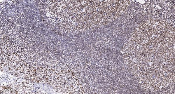
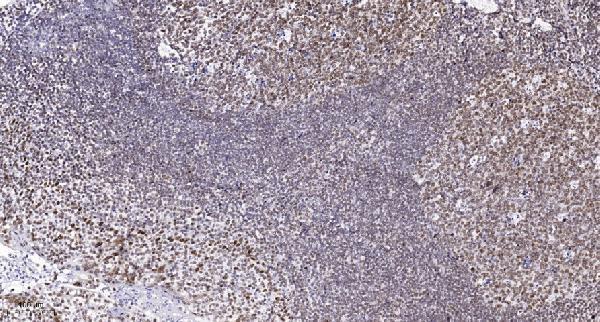
Two UBE3B antibodies have human tonsil paraffin-section IHC images (catalog: A10679 and A10679-2 image captions). Both list IF, but neither includes an IF image (catalog: applications and image captions).

Real IHC data Immunohistochemical analysis of paraffin-embedded human tonsil. 1, Antibody was diluted at 1:200(4° overnight). 2, Tris-EDTA,pH9.0 was used for antigen retrieval. 3,Secondary antibody was diluted at 1:200(room temperature, 45min).
Anti-Ubiquitin-protein ligase E3B UBE3B Antibody
Cat # A10679
Real IHC data Immunohistochemical analysis of paraffin-embedded human tonsil. 1, Antibody was diluted at 1:200 (4° overnight). 2, Tris-EDTA, pH9.0 was used for antigen retrieval. 3, Secondary antibody was diluted at 1:200 (room temperature, 45min).
Anti-Ubiquitin-protein ligase E3B UBE3B Antibody
Cat # A10679-2

A10679 lists IHC, IF and ICC for human, mouse and rat, with an IHC image from paraffin-embedded human tonsil (catalog: A10679 applications, reactivity and image caption). A10679-2 lists IHC and IF for human and mouse, with its own IHC image from paraffin-embedded human tonsil (catalog: A10679-2 applications, reactivity and image caption).

Which to pick: For human tonsil paraffin-section IHC, either SKU has its own image showing antibody at 1:200 overnight at 4°C after Tris-EDTA pH 9.0 retrieval; the fixative is unreported for both (catalog: A10679 and A10679-2 image captions). Choose A10679 for ICC because only it lists ICC; both list IF, but neither provides an IF image (catalog: applications and IF image fields). For rat samples, choose A10679 because it lists rat reactivity; it is described as polyclonal, while A10679-2 has no stated clone type (catalog: reactivity and dilution details).

Each figure is that product's own IHC / IF validation image from its datasheet.

References

  1. UniProt Consortium. UniProt entry Q7Z3V4 (UBE3B_HUMAN, Ubiquitin-protein ligase E3B).
  2. Human Protein Atlas. UBE3B tissue IHC expression (reliability: Approved).
  3. Human Protein Atlas. UBE3B subcellular location (ICC-IF): Mainly localized to the nuclear speckles. In addition localized to the mitochondria..
  4. Human Protein Atlas. UBE3B antibody validation summary (1 antibodies).
  5. Gene expression profile associated with postnatal development of pyramidal neurons in the human prefrontal cortex implicates ubiquitin ligase E3 in the pathophysiology of schizophrenia onset. Journal of psychiatric research 2018 — PMC6347389.
  6. Nrf2/UBE3B protects against acute lung injury by inhibiting ferritinophagy through the ubiquitination of NCOA4. Biology direct 2025 — PMC12265372.
  7. VHL suppresses UBE3B-mediated breast tumor growth and metastasis. Cell death & disease 2024 — PMC11196597.
  8. TRIB3 promotes MYC-associated lymphoma development through suppression of UBE3B-mediated MYC degradation. Nature communications 2020 — PMC7725785.
  9. PubMed PMID:12837265 — UniProt-cited evidence.
  10. PubMed PMID:17974005 — UniProt-cited evidence.
  11. PubMed PMID:16541075 — UniProt-cited evidence.