This website uses cookies to ensure you get the best experience on our website.


- Table of Contents
Plan chromogenic IHC for UBL3 in paraffin sections with the catalog antibody's 1:50–1:200 dilution range (datasheet). Rectal glandular cells show medium staining, but the tissue pattern awaits external verification (HPA tissue IHC).

Expected localisation, validated protocols, controls and antibodies — the at-a-glance facts below, then the full design guide.
| Expected localisation | Cytoplasmic staining (HPA tissue IHC); cell membrane (UniProt) | |
| Staining pattern | Cytoplasmic staining in several tissues and endothelial cells (HPA tissue IHC) | |
| Antigen retrieval | Citrate pH 6.0 HIER, 95–98 °C, 20 min (rule: cytoplasmic / membrane antigen) | |
| Positive control | Rectum | |
| Negative control | Adipose tissue+4 more · see all |
| Fixation | Keep fixation consistent across sections (standard IHC practice; not target-specific) | |
| Caveat | Tissue staining awaits external verification (HPA tissue IHC) | |
| Regulation | Regulation not annotated (UniProt) | |
| Isoform / epitope | No isoforms annotated; mature chain spans residues 1–114 (UniProt) |
The catalog antibody’s IHC-P protocol is accompanied by one published UBL3 immunohistochemistry protocol (PMC10295865).
| Sample | Paraffin-embedded rat testis tissue; fixative not specified (datasheet A13057) |
| Fixation | Image fixative and duration unreported (datasheet A13057); verify before use. |
| Sectioning | 4–5 µm sections on charged slides (standard) |
| Deparaffinisation | Xylene, graded ethanol series to water (standard) |
| Antigen retrieval | Heat-induced epitope retrieval in citrate buffer, pH 6.0, 20 min at 95–98 °C (standard rule: cytoplasmic / membrane antigen) |
| Peroxidase block | 3% H2O2, 10 min, room temperature (standard) |
| Blocking | 10% normal serum of the secondary host, 30 min, room temperature (standard) |
| Primary antibody | Rabbit anti-UBL3, 1:50-1:200 (datasheet A13057) |
| Primary incubation | Overnight at 4 °C (standard) |
| Detection | HRP-polymer secondary, DAB chromogen 5–10 min (standard) |
| Counterstain | Hematoxylin, blue, dehydrate and mount (standard) |
| Expected result | UBL3-positive staining in glandular cells of rectum (HPA tissue IHC: Medium). HPA tissue profile: Cytoplasmic expression in several tissues and in endothelial cells. No signal in the no-primary control. |
UBL3 is annotated at the cell membrane despite having no transmembrane segment (UniProt O95164 topology). In tissue IHC, expect mainly cytoplasmic staining across several tissues, including glandular and endothelial cells (HPA tissue IHC). Rectal glandular cells provide a documented Medium example (HPA tissue IHC). Treat this as a provisional pattern: HPA rates the tissue staining Uncertain, pending external verification (HPA tissue IHC).
| Cytoplasmic chromogen in rectal glandular cells, with distinguishable cell outlines and little surrounding deposit. | This matches the documented Medium rectal glandular staining and the broader cytoplasmic tissue profile (HPA tissue IHC). It is a useful comparison, not proof of antibody specificity: HPA rates its tissue IHC evidence Uncertain (HPA tissue IHC). |
| A strong, exclusively nuclear pattern replaces the expected cytoplasmic signal. | Exclusive nuclear staining conflicts with the tissue IHC profile (HPA tissue IHC) and the cell membrane annotation (UniProt O95164). Consider nonspecific staining or an interpretation error; inspect controls before assigning a new UBL3 location. The approved centrosome finding comes from ICC-IF, a different assay (HPA subcellular). |
| Strong staining appears in adipocytes or bronchial respiratory epithelial cells. | Those cell types were reported as Not detected in the sampled tissues (HPA tissue IHC). Check for cross-reactivity, endogenous detection activity, or misidentified cells with appropriate controls (general IHC practice). A negative HPA observation is a comparison point, not proof that every specimen must be negative. |
| Brown deposit spreads across stroma, empty spaces, or many cell types without clear cellular boundaries. | A diffuse pattern cannot be assigned confidently to the reported cytoplasmic staining in cells (HPA tissue IHC). Examine the no-primary control and reagent background; blocking, washing, and detection chemistry can influence nonspecific deposit in chromogenic IHC (general IHC practice). |
| No signal appears in rectal glandular cells that were selected as the positive comparison. | HPA reports Medium staining in this cell population (HPA tissue IHC), so first check tissue preservation, antibody and detection performance, and whether the glands are present on the section (general IHC practice). Because HPA rates IHC reliability Uncertain, absence alone does not establish UBL3 loss (HPA tissue IHC). |
| Tissue IHC evidence strength | HPA describes cytoplasmic expression in several tissues and endothelial cells but rates the tissue result Uncertain, pending external verification (HPA tissue IHC). Both listed antibodies have Uncertain IHC status (HPA antibodies: HPA053772, HPA058781). Judge weak or unexpected staining conservatively. |
| Compartment and membrane association | UniProt places UBL3 at the cell membrane and lists no transmembrane segment (UniProt O95164 topology). HPA tissue IHC describes cytoplasmic staining (HPA tissue IHC). These annotations support checking cellular localisation, but do not require a sharp membrane rim on a chromogenic section. |
| Tissue and cell context | Rectal glandular cells are Medium, while glandular cells in thyroid, adrenal, stomach, duodenum, and small intestine and lung alveolar cells are Low (HPA tissue IHC). Several other sampled cell populations are Not detected (HPA tissue IHC). Compare like cell types; whole-tissue intensity can obscure that distinction (general IHC practice). |
| Processing and epitope limits | UniProt annotates a 1–114 chain, a 115–117 propeptide, and a cysteine methyl ester at residue 114 (UniProt O95164). No antibody epitope is supplied, so these annotations cannot predict which processed form the IHC-validated antibody detects or whether retrieval changes its signal. |
| IF/ICC Q&A: should a centrosomal signal be expected here? | HPA approves centrosome localisation in ICC-IF and lists images from A-431, MCF-7, U2OS, and NIH 3T3 (HPA subcellular). That is useful context for an IF/ICC result, but it does not establish a centrosomal chromogenic pattern in paraffin tissue or resolve the Uncertain tissue IHC rating (HPA tissue IHC). |
| Situation | Likely cause | Next action |
|---|---|---|
| Rectal glandular cells are blank, including in a comparison section expected to stain. | The antibody, detection reagents, or tissue section may have failed; rectal glandular cells were Medium in HPA's sampled tissue (HPA tissue IHC; general IHC practice). | Confirm that glands are present, inspect counterstaining and run-level controls, then check antibody dilution, detection steps, and the validated assay instructions (general IHC practice). Do not infer a UBL3-specific fixation or retrieval effect from this result. |
| Most cells and extracellular spaces show similar chromogen. | Nonspecific reagent background or endogenous detection activity can obscure a cellular pattern (general IHC practice). The reported tissue pattern is cytoplasmic within cells (HPA tissue IHC). | Review a no-primary control, blocking and washes; assess endogenous activity for the chosen detection chemistry (general IHC practice). Score cellular staining only after background is separable from the tissue pattern. |
| Adipocytes or bronchial respiratory epithelial cells stain strongly. | These cell populations were Not detected in the HPA samples (HPA tissue IHC); cross-reactivity, background, or cell-identification error is possible (general IHC practice). | Verify the cell type on the counterstained section, compare a no-primary control, and seek independent antibody or orthogonal evidence before interpreting the signal as UBL3 (general IHC practice). |
| Only a crisp membrane rim is accepted as positive, so cytoplasmic cells are scored negative. | A membrane-only scoring rule overlooks HPA's cytoplasmic tissue staining (HPA tissue IHC). UniProt's membrane annotation does not prescribe the visual IHC pattern (UniProt O95164 topology). | Score identifiable cytoplasmic staining in the relevant cells against controls and document any membrane emphasis separately (HPA tissue IHC; general IHC practice). |
| A predominantly nuclear or isolated punctate tissue signal is called the expected IHC result. | Neither pattern is established as the tissue IHC profile (HPA tissue IHC). Centrosome localisation is approved in ICC-IF, not tissue IHC (HPA subcellular). | Recheck cell boundaries, counterstain, and control sections; describe the observed compartment without assigning it to UBL3 until independently supported (general IHC practice; HPA tissue IHC). |
| Low-intensity tissue staining varies between sections or readers. | Several listed tissue cell types are Low and tissue IHC reliability is Uncertain (HPA tissue IHC), making a faint result difficult to distinguish from background. | Compare the same cell population across sections, apply a consistent scoring threshold and include an interpretable positive comparison and background control (general IHC practice). Report borderline staining as equivocal rather than forcing a positive call. |
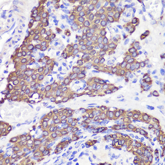
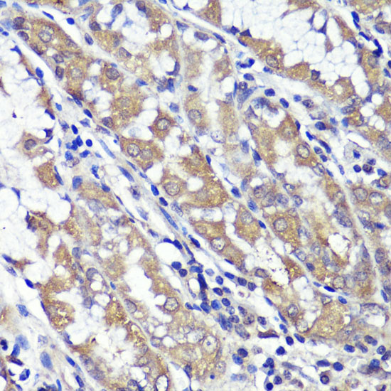


Comprehensive Human Protein Atlas IHC scoring per tissue (reliability: Uncertain — Pending external verification. Antibody staining in cells/structures not annotated, view images.). Rows are taken directly from the HPA tissue chart — click any row's HPA link to view the source.
| Tissue | Cell type | Level | Evidence | Source |
|---|---|---|---|---|
| Rectum | Glandular cells | Medium | Protein (IHC) | HPA → |
| Tissue | Cell type | Level | Evidence | Source |
|---|---|---|---|---|
| Adipose tissue | Adipocytes | Not detected | Protein (IHC) | HPA → |
| Bone marrow | Hematopoietic cells | Not detected | Protein (IHC) | HPA → |
| Breast | Adipocytes | Not detected | Protein (IHC) | HPA → |
| Bronchus | Respiratory epithelial cells | Not detected | Protein (IHC) | HPA → |
| Caudate | Glial cells | Not detected | Protein (IHC) | HPA → |
Troubleshoot UBL3 staining in paraffin sections by checking retrieval, compartment, controls and cell-specific scoring before interpreting chromogenic signal.
A13057 has IHC images of paraffin-embedded rat testis, human stomach, and mouse brain (catalog IHC image captions). No IF image is supplied (catalog: IF images).
A13057 is listed for IHC and reacts with human, mouse, and rat samples (catalog: applications and reactivity). Its IHC images show paraffin-embedded rat testis, human stomach, and mouse brain (catalog IHC image captions).
Which to pick: For tissue IHC, choose A13057; its own captions document paraffin sections stained at 1:100 after microwave retrieval in 10 mM PBS at pH 7.2, but do not report the fixative (catalog IHC image captions). No SKU can be recommended as IF/ICC validated because A13057 has no listed IF/ICC application or IF image (catalog: applications and IF images). For cross-species IHC, A13057 lists human, mouse, and rat reactivity; its clone identity is unreported (catalog: reactivity and clone).