This website uses cookies to ensure you get the best experience on our website.


- Table of Contents
Plan UBTD1 chromogenic IHC using mainly cytoplasmic tissue staining as a starting expectation (HPA tissue IHC). The catalog antibody's IHC dilution is 1:100–1:300 (datasheet); interpret staining with controls because HPA rates the tissue evidence uncertain (HPA tissue IHC).

Expected localisation, validated protocols, controls and antibodies — the at-a-glance facts below, then the full design guide.
| Expected localisation | Mainly cytoplasmic in several tissues (HPA tissue IHC) | |
| Staining pattern | Mainly cytoplasmic in adipocytes and squamous epithelia (HPA tissue IHC) | |
| Antigen retrieval | Citrate pH 6.0 HIER, 95–98 °C, 20 min (rule: cytoplasmic / membrane antigen) | |
| Positive control | Adipose tissue+4 more · see all | |
| Negative control | Appendix+4 more · see all |
| Fixation | Matched tissue-IHC evidence does not establish the fixation claim. Validate the specimen-specific method before use. (selected-SKU IHC image A16697-1) | |
| Caveat | Antibody staining and RNA expression show low consistency (HPA tissue IHC) | |
| Regulation | TP53 downstream target (UniProt) | |
| Isoform / epitope | No isoforms or processing annotated; epitope effects unknown (UniProt) |
Compare the catalog antibody's IHC-P protocol with the published UBTD1 colorectal tissue protocol (PMC11297151).
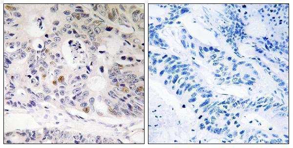
| Sample | Paraffin-embedded human colon carcinoma tissue; fixative not specified (datasheet A16697-1) |
| Fixation | Image fixative and duration unreported (datasheet A16697-1); verify before use. |
| Sectioning | 4–5 µm sections on charged slides (standard) |
| Deparaffinisation | Xylene, graded ethanol series to water (standard) |
| Antigen retrieval | Heat-induced epitope retrieval in citrate buffer, pH 6.0, 20 min at 95–98 °C (standard rule: cytoplasmic / membrane antigen) |
| Peroxidase block | 3% H2O2, 10 min, room temperature (standard) |
| Blocking | 10% normal serum of the secondary host, 30 min, room temperature (standard) |
| Primary antibody | Rabbit anti-UBTD1, 1:100 - 1:300 (datasheet A16697-1) |
| Primary incubation | Overnight at 4 °C (standard) |
| Detection | HRP-polymer secondary, DAB chromogen 5–10 min (standard) |
| Counterstain | Hematoxylin, blue, dehydrate and mount (standard) |
| Expected result | UBTD1-positive staining in adipocytes of adipose tissue (HPA tissue IHC: High). HPA tissue profile: Mainly cytoplasmic expression in several different tissue types. No signal in the no-primary control. |
In paraffin sections, expect mainly cytoplasmic UBTD1 staining, with High staining reported in adipocytes, cardiomyocytes, and selected epithelial and stromal cells (HPA tissue IHC). HPA rates the tissue IHC pattern Uncertain because staining has low consistency with RNA expression (HPA tissue IHC). UniProt does not annotate a subcellular location or a transmembrane segment, so it cannot independently confirm the observed compartment (UniProt Q9HAC8).
| Cytoplasmic staining in adipocytes from adipose tissue or breast, cardiomyocytes, or lung alveolar cells. | These are reported High cell populations and fit the mainly cytoplasmic tissue profile (HPA tissue IHC). Score the named cells rather than treating every cell in the section as an expected positive. The IHC evidence remains Uncertain (HPA tissue IHC). |
| Strong nuclear-only staining, or an exclusively sharp membrane outline, in otherwise expected-positive cells. | This does not match the mainly cytoplasmic tissue pattern (HPA tissue IHC). Review morphology, counterstain, and control sections before assigning UBTD1 positivity. HPA reports additional plasma-membrane localization in ICC-IF, so a membrane component alone cannot establish an artefact (HPA subcellular ICC-IF). |
| Prominent staining in appendix glandular cells or bone-marrow hematopoietic cells. | These cell populations were Not detected by HPA tissue IHC. Check whether the signal follows cell boundaries and persists after control checks; cross-reactivity or endogenous chromogen activity are possible general IHC explanations. Because HPA rates the IHC pattern Uncertain, this mismatch alone does not prove either cause (HPA tissue IHC). |
| Uniform pale color across stroma, empty spaces, and several unrelated cell types. | That distribution is harder to interpret as cell-specific UBTD1 staining than cytoplasmic signal in the named positive cells (HPA tissue IHC). In general chromogenic IHC practice, residual detection reagent, incomplete blocking, or excess primary antibody can produce diffuse background; compare a no-primary control. |
| No staining in adipocytes from adipose tissue despite an otherwise readable section. | Adipocytes are reported High in this tissue (HPA tissue IHC). A negative result merits a technical check, but does not by itself establish absent UBTD1: the tissue IHC reliability is Uncertain (HPA tissue IHC). Compare morphology and controls before changing the biological interpretation. |
| Tissue and cell selection | HPA reports High staining in esophageal and oral squamous epithelium, nasopharyngeal respiratory epithelium, ovarian stroma, and several other listed cells, while selected glandular and hematopoietic cells are Not detected (HPA tissue IHC). Compare the specified cell type within each tissue. |
| Strength of the tissue evidence | The tissue pattern is rated Uncertain because antibody staining and RNA expression show low consistency; RNA tissue specificity is low (HPA tissue IHC). Treat a single positive or negative section as a provisional observation, particularly when the cell type differs from the HPA listing. |
| Topology and processing | UniProt lists no transmembrane segment, signal peptide, propeptide, or glycosylation site and describes one chain spanning residues 1–227 (UniProt Q9HAC8). These annotations provide no basis for predicting a secreted or shed staining pattern; they do not identify the antibody epitope. |
| Antibody-specific support | For HPA034825, HPA lists IHC as Uncertain and ICC as Approved (HPA antibodies). An ICC localization result helps frame a compartment question but does not upgrade validation of chromogenic paraffin-section staining. |
| Situation | Likely cause | Next action |
|---|---|---|
| Expected-positive adipocytes show no chromogenic signal. | The section may have a detection or staining-workflow problem; HPA also rates tissue IHC Uncertain (HPA tissue IHC). | In general IHC practice, verify section integrity, retrieval and detection controls, and primary-antibody application. Recheck adipocytes in the section before concluding that the target is absent. |
| Signal fills the section rather than outlining identifiable cells. | Diffuse background can arise from nonspecific antibody binding or incomplete washing in general IHC practice. | Inspect a no-primary control and tissue morphology. Review blocking, wash steps, antibody concentration, and development time; retain only cell-resolved staining when comparing with HPA's cytoplasmic profile (HPA tissue IHC). |
| Color appears in an HPA Not detected cell population. | Cross-reactivity or endogenous detection activity is possible in general chromogenic IHC practice; HPA's negative listing is itself part of an Uncertain IHC profile (HPA tissue IHC). | Compare a no-primary control and check the detection system's endogenous-activity controls. Describe the observed cell type and compartment, then assess it alongside an HPA High cell population. |
| Most color is nuclear although the expected cells are present. | A nuclear-dominant pattern conflicts with HPA's mainly cytoplasmic tissue profile; UniProt supplies no location annotation to resolve it (HPA tissue IHC; UniProt Q9HAC8). | Recheck the counterstain and no-primary control, then compare the same cell type across sections. Record persistent nuclear staining as a discrepancy rather than scoring it as the expected pattern. |
| A weak signal is seen only in glandular cells. | Some glandular populations are Low, while appendix, epididymis, and parathyroid glandular cells are Not detected; the tissue and cell identity matter (HPA tissue IHC). | Identify the exact glandular population and compare it with the corresponding HPA entry. Use an expected-positive cell population as a staining reference before interpreting weak color. |
| Can ICC-IF images settle an ambiguous IHC compartment pattern? | HPA's ICC-IF shows mainly vesicles, with additional plasma membrane and cytosol, while tissue IHC is described as mainly cytoplasmic (HPA subcellular ICC-IF; HPA tissue IHC). | Use the ICC-IF finding as context only. Judge the paraffin section by its cell-resolved tissue pattern and controls; HPA lists HPA034825 as ICC Approved but IHC Uncertain (HPA antibodies). |


Comprehensive Human Protein Atlas IHC scoring per tissue (reliability: Uncertain — Low consistency between antibody staining and RNA expression data.). Rows are taken directly from the HPA tissue chart — click any row's HPA link to view the source.
| Tissue | Cell type | Level | Evidence | Source |
|---|---|---|---|---|
| Adipose tissue | Adipocytes | High | Protein (IHC) | HPA → |
| Breast | Adipocytes | High | Protein (IHC) | HPA → |
| Esophagus | Squamous epithelial cells | High | Protein (IHC) | HPA → |
| Heart muscle | Cardiomyocytes | High | Protein (IHC) | HPA → |
| Lung | Alveolar cells | High | Protein (IHC) | HPA → |
| Tissue | Cell type | Level | Evidence | Source |
|---|---|---|---|---|
| Appendix | Glandular cells | Not detected | Protein (IHC) | HPA → |
| Bone marrow | Hematopoietic cells | Not detected | Protein (IHC) | HPA → |
| Caudate | Glial cells | Not detected | Protein (IHC) | HPA → |
| Cerebellum | Cells in granular layer | Not detected | Protein (IHC) | HPA → |
| Epididymis | Glandular cells | Not detected | Protein (IHC) | HPA → |
Troubleshoot UBTD1 staining in paraffin sections using the catalog antibody’s tissue image, reported expression patterns, and matched controls.
A16697-1 has a paraffin-section IHC image from human colon carcinoma (catalog image caption); IF/ICC is listed without an IF image (catalog applications; catalog IF image alts). Human, mouse and rat reactivity is listed (catalog reactivity).
A16697-1 has an IHC image of paraffin-embedded human colon carcinoma tissue with a peptide-blocked comparison (catalog image caption). The same SKU lists IHC, IF and ICC applications and human, mouse and rat reactivity; no IF image is provided (catalog applications; catalog reactivity; catalog IF image alts).
Which to pick: For tissue IHC, choose A16697-1 at the listed 1:100–1:300 range: its image documents paraffin-embedded human colon carcinoma, but the fixative is unreported (catalog IHC dilution; catalog image caption). For IF/ICC, A16697-1 lists both applications and is polyclonal, though no IF image is provided (catalog applications; catalog dilution record; catalog IF image alts). For mouse or rat samples, A16697-1 lists reactivity with both species, while its IHC image documents human tissue only (catalog reactivity; catalog image caption).