This website uses cookies to ensure you get the best experience on our website.


- Table of Contents
Plan chromogenic UCN staining in paraffin sections using the catalog antibodies’ IHC dilutions of 1:50–1:200 (datasheet: A01807) or 1:100–1:300 (datasheet: A01807-1). Interpret cytoplasmic and plasma staining (HPA tissue IHC) in light of UCN secretion (UniProt).

Expected localisation, validated protocols, controls and antibodies — the at-a-glance facts below, then the full design guide.
| Expected localisation | Cytoplasm and plasma signal (HPA tissue IHC) | |
| Staining pattern | Broad cytoplasmic staining with plasma staining (HPA tissue IHC) | |
| Antigen retrieval | Citrate pH 6.0 HIER, 95–98 °C, 20 min (rule: cytoplasmic / membrane antigen) | |
| Positive control | Adipose tissue+4 more · see all | |
| Negative control | None in HPA (detected in all 45 tissues); use no-primary + isotype controls |
| Fixation | Keep fixation consistent across sections (standard IHC practice; not target-specific) | |
| Caveat | Secretion can separate protein and RNA tissue patterns (HPA tissue IHC) | |
| Regulation | Released in response to stress (UniProt) | |
| Isoform / epitope | No isoforms; signal peptide and propeptide are processed (UniProt) |
Compare the catalog antibody’s paraffin-section IHC protocol with four published UCN chromogenic IHC workflows (datasheet; PMC10761541; PMC2826606; PMC2982926; PMC3168230).
| Sample | Paraffin-embedded human colorectal carcinoma tissue; fixative not specified (datasheet A01807) |
| Fixation | Image fixative and duration unreported (datasheet A01807); verify before use. |
| Sectioning | 4–5 µm sections on charged slides (standard) |
| Deparaffinisation | Xylene, graded ethanol series to water (standard) |
| Antigen retrieval | Heat-induced epitope retrieval in citrate buffer, pH 6.0, 20 min at 95–98 °C (standard rule: cytoplasmic / membrane antigen) |
| Peroxidase block | 3% H2O2, 10 min, room temperature (standard) |
| Blocking | 10% normal serum of the secondary host, 30 min, room temperature (standard) |
| Primary antibody | Rabbit anti-UCN, 1:50-1:200 (datasheet A01807) |
| Primary incubation | Overnight at 4 °C (standard) |
| Detection | HRP-polymer secondary, DAB chromogen 5–10 min (standard) |
| Counterstain | Hematoxylin, blue, dehydrate and mount (standard) |
| Expected result | UCN-positive staining in adipocytes of adipose tissue (HPA tissue IHC: Medium). HPA tissue profile: Ubiquitous cytoplasmic expression with additional plasma staining. No signal in the no-primary control. |
UCN is a secreted protein with no transmembrane segment (UniProt P55089 topology). In paraffin sections, expect predominantly cytoplasmic staining with additional plasma staining (HPA tissue IHC: Approved). Medium staining is reported in adrenal glandular cells, adipocytes and caudate neuronal cells (HPA tissue IHC: Medium). Skin keratinocytes and follicular sheaths are also reported sites (UniProt P55089 tissue specificity). Because UCN is secreted, the site of protein staining need not match the site of RNA expression (HPA tissue IHC: reliability description).
| Cytoplasmic chromogen in adrenal glandular cells, with possible plasma staining (HPA tissue IHC: Medium; profile). | This fits the reported UCN pattern (HPA tissue IHC: Approved). Judge signal by its location and the stained cells together: plasma staining can accompany the cellular pattern, while UCN secretion means protein and RNA maps may differ (UniProt P55089 subcellular location; HPA tissue IHC: reliability description). |
| Strong, predominantly nuclear staining, with little cytoplasmic signal. | Treat this as a compartment mismatch: the reported tissue pattern is cytoplasmic, and ICC-IF places UCN mainly in vesicles with additional plasma membrane localisation (HPA tissue IHC: profile; HPA subcellular: approved). Review controls and staining distribution before scoring it as UCN. |
| Staining is concentrated in an unexpected cell population while expected cells are unstained. | Consider antibody cross-reactivity or endogenous chromogenic activity (general IHC practice). Compare the same slide with its expected cell populations: adrenal glandular cells, adipocytes and caudate neuronal cells have Medium staining in HPA; rectal glandular and lymph-node germinal-center cells are reported Low, not negative (HPA tissue IHC). |
| Broad, diffuse colour obscures cell boundaries or appears similarly across compartments. | This is difficult to score as a cellular UCN pattern (HPA tissue IHC: cytoplasmic profile). Secreted UCN can also produce plasma staining, so extracellular colour alone does not prove artefact (UniProt P55089 subcellular location; HPA tissue IHC: profile). Use controls to assess nonspecific background (general IHC practice). |
| No visible signal in an HPA-reported Medium tissue, such as adrenal glandular cells (HPA tissue IHC). | The result does not establish absent UCN. Check whether the control section and detection worked, then assess antibody concentration and antigen retrieval as general IHC variables (general IHC practice). The supplied sources give no target-specific fixation sensitivity or retrieval condition for UCN. |
| Secretion and processing (UniProt P55089) | UCN has a signal peptide at residues 1–25, a propeptide at 26–82 and an amidated residue at 122 (UniProt P55089 processing). Antibody epitope location is unspecified here, so these annotations cannot predict which processed forms the antibody detects or prescribe a retrieval condition. |
| Tissue and cell context (HPA tissue IHC; UniProt P55089 tissue specificity) | HPA reports Medium staining in several tissues and Low staining in rectal glandular and lymph-node germinal-center cells; it lists no negative tissue (HPA tissue IHC). UniProt additionally reports skin structures and colonic lamina propria plasma cells (UniProt P55089 tissue specificity). Do not confuse those cells with HPA's plasma staining. |
| Evidence and antibody choice (HPA tissue IHC; HPA antibodies) | The tissue profile has Approved reliability, with a caution that RNA and protein locations may differ for secreted proteins (HPA tissue IHC). CAB010911 is Approved for IHC; the supplied antibody list does not report an Enhanced IHC result (HPA antibodies). These ratings support interpretation, not a guaranteed result in every section. |
| Antigen retrieval (general IHC practice) | Retrieval conditions can be assessed with a positive control when chromogenic IHC signal is weak (general IHC practice). Neither supplied source identifies a UCN-specific retrieval condition or shows that fixation changes UCN epitope access, sensitivity or tissue positivity; avoid assigning a target-specific cause to a weak result. |
| IF/ICC Q: Where should UCN signal appear? | A: Mainly in vesicles, with additional plasma membrane localisation (HPA subcellular: approved). HPA lists ICC-IF images from SK-MEL-30, THP-1 and U2OS, and HPA069971 as Approved for ICC (HPA subcellular; HPA antibodies). These ICC-IF observations are a localisation reference, not a paraffin-section protocol. |
| Situation | Likely cause | Next action |
|---|---|---|
| Adrenal glandular cells show no stain despite a working counterstain (HPA tissue IHC: Medium). | A counterstain alone does not verify antibody detection; low assay sensitivity or an unsuitable staining condition remains possible (general IHC practice). | Run a reported Medium tissue as a positive control and check the antibody and detection steps (HPA tissue IHC: Medium; general IHC practice). Optimise retrieval only as a general IHC variable; UCN-specific retrieval and fixation effects are unreported here. |
| Diffuse colour persists in a section processed without primary antibody. | The primary antibody cannot account for colour in that control; detection-system background or endogenous activity is possible (general IHC practice). | Check blocking and detection controls; if using peroxidase chromogen, assess endogenous peroxidase blocking (general IHC practice). Reassess cellular UCN staining against the reported cytoplasmic pattern (HPA tissue IHC: profile). |
| Many unrelated cells stain equally, obscuring the expected distribution. | Nonspecific antibody binding or excessive chromogen development may reduce cell-level discrimination (general IHC practice). HPA's broad cytoplasmic profile means breadth alone is insufficient to diagnose artefact (HPA tissue IHC: profile). | Compare positive and no-primary controls, review antibody concentration and detection time, and score by cell type and compartment (general IHC practice; HPA tissue IHC: profile). |
| Nuclear colour dominates while cytoplasmic staining is absent. | This conflicts with HPA's cytoplasmic tissue profile and vesicular ICC-IF localisation (HPA tissue IHC: profile; HPA subcellular: approved). | Check no-primary and positive controls, then review chromogen and counterstain separation before interpreting nuclear colour as target signal (general IHC practice). |
| A Low tissue stains more strongly than an HPA-reported Medium tissue. | Section handling, detection conditions or biological variation may affect comparisons (general IHC practice); HPA's Low and Medium labels describe its reported staining, not fixed thresholds for every run (HPA tissue IHC). | Compare matched sections within the same run and confirm the stained cell types before changing the UCN interpretation (general IHC practice; HPA tissue IHC). |
| Protein staining appears away from the cell population suggested by RNA data. | UCN is secreted, and HPA explicitly expects RNA and protein tissue locations to differ (UniProt P55089 subcellular location; HPA tissue IHC: reliability description). | Assess cytoplasmic cell staining and possible plasma staining against the HPA protein profile; use staining controls to investigate a discordant result rather than treating RNA location alone as the IHC reference (HPA tissue IHC: profile; general IHC practice). |


Comprehensive Human Protein Atlas IHC scoring per tissue (reliability: Approved — Secreted protein, tissue location of RNA and protein is expected to differ.). Rows are taken directly from the HPA tissue chart — click any row's HPA link to view the source.
| Tissue | Cell type | Level | Evidence | Source |
|---|---|---|---|---|
| Adipose tissue | Adipocytes | Medium | Protein (IHC) | HPA → |
| Adrenal gland | Glandular cells | Medium | Protein (IHC) | HPA → |
| Appendix | Glandular cells | Medium | Protein (IHC) | HPA → |
| Bone marrow | Hematopoietic cells | Medium | Protein (IHC) | HPA → |
| Breast | Adipocytes | Medium | Protein (IHC) | HPA → |
| Tissue | Cell type | Level | Evidence | Source |
|---|---|---|---|---|
| None in HPA: UCN is detected in all 45 scored tissues. Use a no-primary (secondary-only) and an isotype control instead. | ||||
Troubleshoot chromogenic UCN staining in paraffin sections by checking retrieval, controls, cellular location, and scoring before interpreting tissue differences.
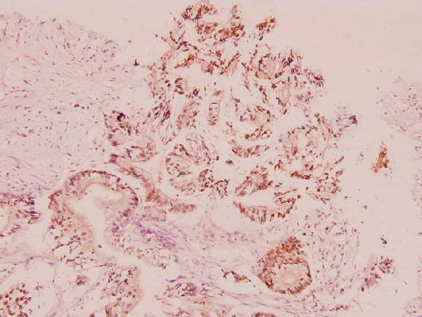
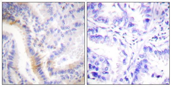
Two anti-UCN antibodies have real IHC figures from paraffin-embedded human carcinoma tissue; both list Human, Mouse and Rat reactivity, while one also lists IF/ICC applications (catalog; IHC image captions).
A01807 lists IHC and shows staining in paraffin-embedded human colorectal carcinoma tissue at 1:100 (catalog; A01807 IHC image caption). A01807-1 lists IHC, IF and ICC and shows staining in paraffin-embedded human lung carcinoma tissue with a peptide-blocked comparison (catalog; A01807-1 IHC image caption).
Which to pick: For tissue IHC, choose either SKU according to the relevant illustrated sample: A01807 for paraffin-embedded colorectal carcinoma tissue or A01807-1 for paraffin-embedded lung carcinoma tissue (respective IHC image captions). For IF/ICC, choose A01807-1 because those applications are listed only for it; no IF figure is supplied (catalog). Both list Human, Mouse and Rat reactivity, but their figures show human tissue only; neither IHC caption reports the fixative (catalog; respective IHC image captions).