This website uses cookies to ensure you get the best experience on our website.


- Table of Contents
Plan chromogenic paraffin IHC for USP24 around the cytoplasmic staining reported in most cell types (HPA tissue IHC). Compare high staining in hepatocytes with undetected staining in adipocytes (HPA tissue IHC), and titrate the IHC-validated antibody across 1:100–1:300 (datasheet).

Expected localisation, validated protocols, controls and antibodies — the at-a-glance facts below, then the full design guide.
| Expected localisation | Cytoplasmic staining in most cell types (HPA tissue IHC) | |
| Staining pattern | Cytoplasmic in most cell types; texture unspecified (HPA tissue IHC) | |
| Antigen retrieval | Citrate pH 6.0 HIER, 95–98 °C, 20 min (rule: cytoplasmic / membrane antigen) | |
| Positive control | Appendix+4 more · see all | |
| Negative control | Adipose tissue+4 more · see all |
| Fixation | Keep formalin fixation consistent across paraffin sections. (standard IHC practice; not target-specific) | |
| Caveat | Adipocytes can be unstained despite the broad profile (HPA tissue IHC) | |
| Regulation | Expression regulation unreported (UniProt) | |
| Isoform / epitope | No annotated isoforms, signal peptide or processed chain (UniProt) |
The catalog antibody’s IHC-P protocol is accompanied by three published USP24 IHC protocols for paraffin sections (PMC10394155; PMC13433775; PMC11461744).
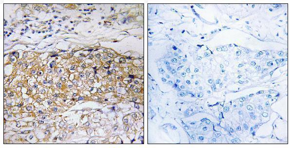
| Sample | Paraffin-embedded human breast carcinoma tissue; fixative not specified (datasheet A08241) |
| Fixation | Image fixative and duration unreported (datasheet A08241); verify before use. |
| Sectioning | 4–5 µm sections on charged slides (standard) |
| Deparaffinisation | Xylene, graded ethanol series to water (standard) |
| Antigen retrieval | Heat-induced epitope retrieval in citrate buffer, pH 6.0, 20 min at 95–98 °C (standard rule: cytoplasmic / membrane antigen) |
| Peroxidase block | 3% H2O2, 10 min, room temperature (standard) |
| Blocking | 10% normal serum of the secondary host, 30 min, room temperature (standard) |
| Primary antibody | Rabbit anti-USP24, 1:100 - 1:300 (datasheet A08241) |
| Primary incubation | Overnight at 4 °C (standard) |
| Detection | HRP-polymer secondary, DAB chromogen 5–10 min (standard) |
| Counterstain | Hematoxylin, blue, dehydrate and mount (standard) |
| Expected result | USP24-positive staining in glandular cells of appendix (HPA tissue IHC: High). HPA tissue profile: Cytoplasmic expression in most cell types. No signal in the no-primary control. |
USP24 is reported as cytoplasmic in most cell types by tissue IHC, with high staining in several glandular populations, hepatocytes and trophoblastic cells (HPA tissue IHC). HPA rates this tissue profile Approved, with medium agreement between staining and RNA data and external verification pending (HPA tissue IHC). UniProt lists no transmembrane segment or annotated subcellular location, so the IHC pattern should be judged against the observed HPA tissue images (UniProt Q9UPU5 topology; HPA tissue IHC).
| Clear cytoplasmic staining in colon glandular cells or liver hepatocytes, with readable cell boundaries and little surrounding deposit. | This matches cell populations scored High by HPA tissue IHC. Compare staining within the relevant tissue architecture; HPA's Approved rating still carries medium RNA agreement and pending external verification (HPA tissue IHC). |
| Predominantly sharp membranous or extracellular staining, while expected cellular cytoplasm remains pale. | This differs from the reported cytoplasmic tissue IHC profile (HPA tissue IHC). Treat it as a possible staining artefact and review controls and morphology. A nuclear component alone needs separate assessment because HPA ICC-IF supports nucleoplasmic localization (HPA subcellular ICC-IF). |
| Strong staining in adipocytes, skeletal myocytes or soft-tissue fibroblasts, especially beside unstained expected-positive cells. | Those populations are listed as Not detected in HPA tissue IHC. Unexpected signal raises possible cross-reactivity or endogenous detection activity (general IHC practice); it does not prove either cause, and HPA negatives are reference observations, not universal exclusions. |
| A uniform brown haze covers cells and stroma, or tissue edges are darker than intact central regions. | Diffuse deposit obscures the cell-specific pattern needed for interpretation (general IHC practice). Review background and detection controls before assigning USP24 positivity; a broad haze cannot establish the cytoplasmic pattern reported by HPA tissue IHC. |
| No convincing cellular staining appears in colon glandular cells, liver hepatocytes or another HPA high-expression population. | An absent result in a reference-positive population warrants a run-level check before a biological negative call (HPA tissue IHC; general IHC practice). HPA reports high staining in these cells, but its medium-consistency rating limits certainty for any single specimen (HPA tissue IHC). |
| Tissue and cell population | HPA tissue IHC scores appendix, colon, duodenum and gallbladder glandular cells, liver hepatocytes, pancreatic exocrine cells, nasopharyngeal respiratory epithelial cells and placental trophoblastic cells High. Its Not detected calls apply to specified cells, including adipocytes and skeletal myocytes, rather than every cell in those tissues (HPA tissue IHC). |
| Assay-specific compartment evidence | Tissue IHC describes predominantly cytoplasmic expression, whereas ICC-IF supports nucleoplasm as the main location and cytosol as an additional location (HPA tissue IHC; HPA subcellular ICC-IF). Interpret each pattern in its assay context; the ICC-IF finding does not by itself validate a nuclear chromogenic IHC result. |
| Antibody evidence | HPA lists HPA026723 and HPA028428 as Approved for IHC and ICC; HPA067124 is Approved for ICC, with no IHC status supplied (HPA antibodies). The overall tissue IHC profile is Approved but has medium staining–RNA consistency and awaits external verification (HPA tissue IHC). |
| Topology and processing | UniProt reports no transmembrane segment, signal peptide or propeptide, and annotates one chain spanning residues 1–2620 (UniProt Q9UPU5). These annotations give no basis to expect a membrane-restricted or secreted USP24 pattern; they do not establish fixation sensitivity or an antibody epitope. |
| IF/ICC Q&A: What pattern should I expect? | HPA ICC-IF supports mainly nucleoplasmic USP24 with additional cytosolic localization and lists images from A-431, U-251MG, U2OS and SK-MEL-30 (HPA subcellular ICC-IF). That is an assay-specific observation; this IHC section supplies no IF/ICC protocol. |
| Situation | Likely cause | Next action |
|---|---|---|
| A reference-positive colon or liver section is blank. | The expected cells stain High in HPA tissue IHC, so a blank run may reflect a workflow or detection problem; the payload does not identify a USP24-specific failure mechanism (HPA tissue IHC; general IHC practice). | Confirm that the reference cells are present, then check primary-antibody application, detection reagents and the run's positive control (general IHC practice). Repeat before scoring the specimen negative. |
| Cytoplasmic signal is faint in a HPA high-expression population. | Weak intensity may arise from the staining workflow or specimen variation; HPA's medium staining–RNA consistency does not guarantee strong staining in every section (HPA tissue IHC; general IHC practice). | Compare with a concurrently processed reference-positive section. Review the documented IHC dilution and retrieval conditions for the antibody being used; no target-specific settings are supplied here (general IHC practice). |
| Brown precipitate or diffuse haze makes cell boundaries unreadable. | Background from detection reagents or nonspecific staining can obscure a cellular chromogenic signal (general IHC practice). HPA's cytoplasmic profile cannot be confirmed from diffuse deposit (HPA tissue IHC). | Inspect a no-primary control and check blocking, washes and detection conditions. Score USP24 only where cellular morphology remains interpretable (general IHC practice). |
| Unexpected staining concentrates in skeletal myocytes, adipocytes or soft-tissue fibroblasts. | HPA lists these specified populations as Not detected; cross-reactivity or endogenous detection activity is possible, but the staining pattern alone cannot distinguish them (HPA tissue IHC; general IHC practice). | Compare the unexpected cells with adjacent morphology and a no-primary control; review endogenous enzyme blocking if an enzyme-based detector is used (general IHC practice). |
| Signal outlines cell membranes or appears outside cells instead of in cytoplasm. | This distribution conflicts with HPA's tissue IHC profile; UniProt reports no transmembrane segment, but neither source identifies the cause of the deposit (HPA tissue IHC; UniProt Q9UPU5 topology). | Check whether the pattern persists in a no-primary control and whether extracellular staining follows damaged tissue or precipitate. Reassess against reference-positive cellular staining (general IHC practice; HPA tissue IHC). |
| A nuclear IHC signal seems to conflict with the cytoplasmic IHC reference. | HPA reports cytoplasmic tissue IHC and supported nucleoplasmic ICC-IF localization. These assay observations differ, so nuclear IHC signal cannot be accepted or rejected from compartment alone (HPA tissue IHC; HPA subcellular ICC-IF). | Examine cytoplasmic staining in reference-positive cells and compare control sections. Record nuclear signal separately, with its assay and antibody, pending independent validation (HPA tissue IHC; general IHC practice). |


Comprehensive Human Protein Atlas IHC scoring per tissue (reliability: Approved — Medium consistency between antibody staining and RNA expression data. Pending external verification.). Rows are taken directly from the HPA tissue chart — click any row's HPA link to view the source.
| Tissue | Cell type | Level | Evidence | Source |
|---|---|---|---|---|
| Appendix | Glandular cells | High | Protein (IHC) | HPA → |
| Colon | Glandular cells | High | Protein (IHC) | HPA → |
| Duodenum | Glandular cells | High | Protein (IHC) | HPA → |
| Gallbladder | Glandular cells | High | Protein (IHC) | HPA → |
| Liver | Hepatocytes | High | Protein (IHC) | HPA → |
| Tissue | Cell type | Level | Evidence | Source |
|---|---|---|---|---|
| Adipose tissue | Adipocytes | Not detected | Protein (IHC) | HPA → |
| Bone marrow | Hematopoietic cells | Not detected | Protein (IHC) | HPA → |
| Hippocampus | Glial cells | Not detected | Protein (IHC) | HPA → |
| Ovary | Ovarian stroma cells | Not detected | Protein (IHC) | HPA → |
| Skeletal muscle | Myocytes | Not detected | Protein (IHC) | HPA → |
Troubleshoot USP24 staining in paraffin sections by checking retrieval, cell compartment, controls and scoring before interpreting chromogenic signal.
A08241 has IHC image evidence in paraffin-embedded human breast carcinoma (IHC image caption); IF/ICC applications and human and mouse reactivity are catalog-listed (catalog: applications and reactivity).
A08241 will render with an IHC image of paraffin-embedded human breast carcinoma and a peptide-blocked comparison (IHC image caption). IF and ICC are listed applications, but the payload provides no IF image (catalog: applications and IF image alts).
Which to pick: Choose A08241 for paraffin-section tissue IHC: its own image caption shows human breast carcinoma, while the fixative is unreported (IHC image caption). The same rabbit polyclonal antibody is listed for IF/ICC and for human and mouse reactivity, but its image evidence is limited to human tissue IHC (catalog: host, clone, applications and reactivity; IHC image caption; IF image alts).