This website uses cookies to ensure you get the best experience on our website.


- Table of Contents
Plan USP5 chromogenic IHC in paraffin sections using the catalog antibody’s tissue protocol and a testis control (datasheet A04550-1; HPA tissue IHC). Compare cytoplasmic and nuclear staining with the reported tissue pattern, while accounting for its uncertain reliability (HPA tissue IHC).

Expected localisation, validated protocols, controls and antibodies — the at-a-glance facts below, then the full design guide.
| Expected localisation | Cytoplasmic and nuclear staining in several tissues (HPA tissue IHC) | |
| Staining pattern | Cytoplasmic and nuclear; high in seminiferous duct cells (HPA tissue IHC) | |
| Antigen retrieval | EDTA pH 8.0 HIER, heat-mediated (datasheet A04550-1) | |
| Positive control | Cerebellum+4 more · see all | |
| Negative control | Salivary gland+2 more · see all |
| Fixation | Matched tissue-IHC evidence does not establish the fixation claim. Validate the specimen-specific method before use. (selected-SKU IHC image A04550-1) | |
| Caveat | Low staining–RNA concordance; validate the tissue pattern (HPA tissue IHC) | |
| Regulation | No abundance regulator annotated (UniProt) | |
| Isoform / epitope | 2 isoforms (Long, Short); epitope coverage is unknown (UniProt) |
The catalog antibody protocol uses EDTA retrieval (datasheet A04550-1). The published USP5 IHC protocols below describe colorectal cancer (PMC6592179), human tumor tissue (PMC6991651), and NSCLC (PMC9826599).
| Sample | Paraffin-embedded human breast cancer tissue; fixative not specified (datasheet A04550-1) |
| Fixation | Image fixative and duration unreported (datasheet A04550-1); verify before use. |
| Sectioning | 4–5 µm sections on charged slides (standard) |
| Deparaffinisation | Xylene, graded ethanol series to water (standard) |
| Antigen retrieval | Heat retrieval: EDTA pH 8.0 (datasheet A04550-1); 20 min, 95–100 °C (standard) |
| Peroxidase block | 3% H2O2, 10 min, room temperature (standard) |
| Blocking | 10% goat serum (datasheet A04550-1) |
| Primary antibody | Rabbit anti-USP5, 2-5 μg/ml (datasheet A04550-1) |
| Primary incubation | Overnight at 4 °C (datasheet A04550-1) |
| Detection | HRP-conjugated secondary, DAB chromogen (datasheet A04550-1) |
| Counterstain | Hematoxylin, blue, dehydrate and mount (standard) |
| Expected result | USP5-positive staining in purkinje cells of cerebellum (HPA tissue IHC: High). HPA tissue profile: Cytoplasmic and nuclear expression in several tissues, including testis, cerebral cortex and skin. No signal in the no-primary control. |
USP5 should show cytoplasmic and nuclear staining in several tissues, with strong staining reported in cerebellar Purkinje cells and cells in testicular seminiferous ducts (HPA: tissue IHC). A membrane-bound pattern is not expected (UniProt P45974: cytoplasm, nucleus; no transmembrane segment). Interpret tissue results cautiously: HPA rates its IHC evidence Uncertain because antibody staining has low consistency with RNA expression (HPA: IHC reliability).
| Cytoplasmic and nuclear staining in Purkinje cells or cells in seminiferous ducts. | This matches HPA's High staining calls in those cell populations and its reported cytoplasmic and nuclear tissue pattern (HPA: tissue IHC). Compare compartments within identifiable cells; the HPA reliability rating remains Uncertain, so a matching image alone does not establish antibody specificity (HPA: IHC reliability). |
| Signal restricted to cell membranes or extracellular spaces, without convincing cytoplasmic or nuclear staining. | That distribution conflicts with the reported locations and lack of a transmembrane segment (UniProt P45974: subcellular location, topology; HPA: tissue IHC). Treat it as a possible staining artefact and review morphology, controls and detection conditions before assigning it to USP5 (general IHC practice). |
| Strong staining in salivary glandular cells, smooth muscle cells or soft tissue fibroblasts. | HPA reports USP5 as Not detected in those specific cell populations (HPA: tissue IHC). Unexpected signal warrants a check for cross-reactivity or endogenous detection activity (general IHC practice). These HPA observations are comparison points, not proof that every cell in those tissues must be negative (HPA: tissue IHC, Uncertain reliability). |
| Diffuse color over the section, including spaces outside identifiable cells. | A widespread deposit does not resolve the expected intracellular pattern (UniProt P45974: cytoplasm, nucleus; HPA: tissue IHC). Consider nonspecific detection, incomplete blocking or inadequate washing, and compare a no-primary control processed with the same detection reagents (general IHC practice). |
| No convincing signal in Purkinje cells or cells in seminiferous ducts. | Those are HPA's reported High staining examples, making them useful checks for a weak run (HPA: tissue IHC). Review tissue preservation, retrieval, antibody application and detection controls before interpreting the absence as biology (general IHC practice). HPA's Uncertain IHC rating limits how definitive any single comparison can be (HPA: IHC reliability). |
| IHC evidence strength | HPA labels tissue IHC Uncertain because antibody staining and RNA expression have low consistency; external verification is pending (HPA: IHC reliability). Use its cell-level pattern as an expectation, then weigh morphology and controls before treating intensity differences as biological differences (general IHC practice). |
| Intracellular distribution | UniProt places USP5 in the cytoplasm, nucleus and stress granules, with no transmembrane segment (UniProt P45974: subcellular location, topology). HPA tissue IHC describes cytoplasmic and nuclear staining; its routine tissue profile does not establish a stress-granule pattern in a given section (HPA: tissue IHC). |
| Cell population and tissue context | HPA calls staining High in Purkinje cells and cells in seminiferous ducts, Medium in several listed glandular, hematopoietic, respiratory epithelial and neuronal populations, and Not detected in selected cell populations (HPA: tissue IHC). Score the identified population rather than assigning one intensity to an entire organ (general IHC practice). |
| Isoform coverage | UniProt lists 2 isoforms, Long and Short (UniProt P45974: isoforms). The supplied evidence gives no antibody epitope or isoform-specific IHC validation, so a staining difference cannot be attributed to one isoform from this record alone (UniProt P45974: isoforms; HPA: antibody validation). |
| IF/ICC Q&A: where should signal appear? | Nucleoplasm and cytosol are HPA's approved ICC-IF locations (HPA: subcellular). That supports an intracellular localisation check, while HPA rates the listed antibody's IHC evidence Uncertain and its ICC evidence Approved (HPA: antibody validation). ICC-IF image cell lines do not establish an IHC tissue intensity (HPA: subcellular; HPA: tissue IHC). |
| Situation | Likely cause | Next action |
|---|---|---|
| The expected positive cell population is blank. | A missed IHC step, weak detection or unsuitable processing can suppress visible staining (general IHC practice); HPA reports High staining in Purkinje cells and cells in seminiferous ducts (HPA: tissue IHC). | Confirm the correct cell population on the section, then check the run's positive control, retrieval record, antibody application and detection sequence (general IHC practice). Avoid declaring that USP5 is absent from a specimen on the basis of one failed run (HPA: IHC reliability, Uncertain). |
| Nuclei alone or cytoplasm alone stain. | HPA describes both compartments in tissue IHC, although the apparent balance can vary with cell morphology and detection conditions (HPA: tissue IHC; general IHC practice). | Check that the apparent compartment is inside intact, identifiable cells and compare with a positive tissue area on the same run (general IHC practice). Record the observed distribution; do not force a dual-compartment score when one compartment is unclear (HPA: tissue IHC). |
| Membrane rims or extracellular deposits dominate. | The pattern conflicts with USP5's cytoplasmic and nuclear annotation and its lack of a transmembrane segment (UniProt P45974: subcellular location, topology). | Inspect section edges and deposits, compare the no-primary control, and review blocking, washing and detection reagents (general IHC practice). Reassess intracellular staining before calling the rim USP5-positive (UniProt P45974: subcellular location). |
| Cells reported as not detected show strong color. | HPA reports no detection in salivary glandular cells, smooth muscle cells and soft tissue fibroblasts; cross-reactivity or endogenous detection activity is possible (HPA: tissue IHC; general IHC practice). | Verify cell identity and compare no-primary and detection controls; investigate persistent cell-associated signal with an independent specificity check if available (general IHC practice). Treat the HPA comparison cautiously because its IHC reliability is Uncertain (HPA: IHC reliability). |
| Diffuse background obscures cell boundaries. | Nonspecific reagent binding, endogenous detection activity or inadequate washing can produce broad chromogenic background (general IHC practice). | Review blocking and washes, and compare a no-primary control under the same chromogenic detection conditions (general IHC practice). Score only signal that can be assigned to identifiable cytoplasm or nuclei (HPA: tissue IHC; general IHC practice). |
| IF/ICC localisation and tissue IHC impressions disagree. | HPA approves nucleoplasm and cytosol as ICC-IF locations, but rates tissue IHC Uncertain; the assays and specimens provide different kinds of evidence (HPA: subcellular; HPA: antibody validation). | Check each result against its own controls and cell morphology (general IHC/IF practice). Use the IF/ICC result to assess localisation, while retaining HPA's tissue-specific IHC intensity calls and their reliability caveat for the paraffin section (HPA: subcellular; HPA: tissue IHC). |
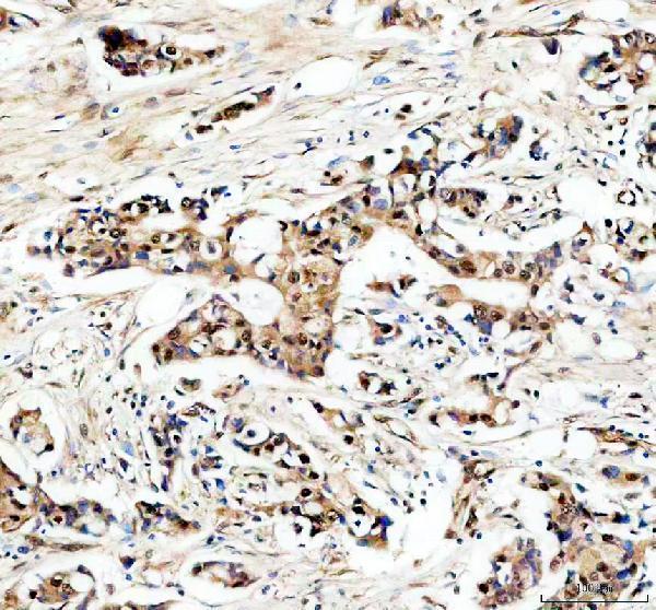
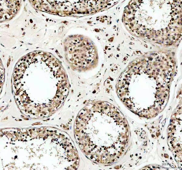
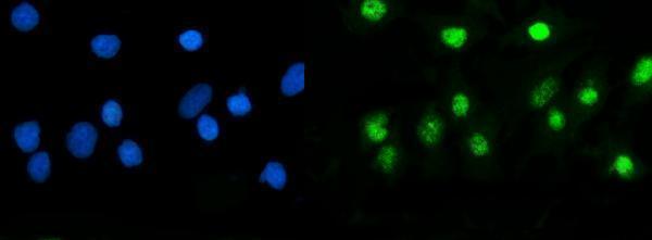
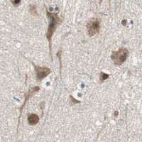

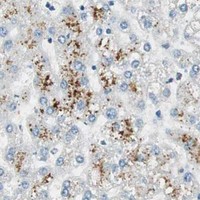


Comprehensive Human Protein Atlas IHC scoring per tissue (reliability: Uncertain — Low consistency between antibody staining and RNA expression data. Pending external verification.). Rows are taken directly from the HPA tissue chart — click any row's HPA link to view the source.
| Tissue | Cell type | Level | Evidence | Source |
|---|---|---|---|---|
| Cerebellum | Purkinje cells | High | Protein (IHC) | HPA → |
| Testis | Cells in seminiferous ducts | High | Protein (IHC) | HPA → |
| Adrenal gland | Glandular cells | Medium | Protein (IHC) | HPA → |
| Bone marrow | Hematopoietic cells | Medium | Protein (IHC) | HPA → |
| Breast | Glandular cells | Medium | Protein (IHC) | HPA → |
These questions address chromogenic USP5 staining in paraffin sections, using the A04550-1 tissue example and the stated limits of the localisation evidence.
The catalog includes USP5 antibodies for human paraffin-section IHC and A549-cell IF/ICC, with images for A04550-1 (A04550-1 image captions). Both list human, mouse, and rat reactivity (catalog).
A04550-1 has IHC images from human breast cancer and testis paraffin sections and an IF image from A549 cells (A04550-1 image captions). M04550-3 lists IF/ICC and human, mouse, and rat reactivity, but has no supplied IHC or IF image (catalog: M04550-3).
Which to pick: Choose A04550-1 for tissue IHC: its images document human paraffin sections with EDTA retrieval at pH 8.0 and primary antibody at 2 μg/ml; the fixative is unreported (A04550-1 IHC image captions). For IF/ICC, A04550-1 has an A549-cell IF image at 5 μg/ml, while rabbit monoclonal M04550-3 lists IF/ICC at 1:50–1:200 without a supplied image (A04550-1 IF image caption; catalog: M04550-3). Both list human, mouse, and rat reactivity, but the supplied tissue IHC images establish conditions only for human samples with A04550-1 (catalog; A04550-1 IHC image captions).