This website uses cookies to ensure you get the best experience on our website.


- Table of Contents
Plan chromogenic VIPR2 IHC on paraffin sections using membranous staining as the expected tissue pattern (HPA tissue IHC). Compare high-staining bronchial basal cells with glandular cells of the adrenal gland, where staining was not detected (HPA tissue IHC).

Expected localisation, validated protocols, controls and antibodies — the at-a-glance facts below, then the full design guide.
| Expected localisation | Membranous tissue staining (HPA tissue IHC) | |
| Staining pattern | Membranous staining in pneumocytes, smooth muscle and endothelium (HPA tissue IHC) | |
| Antigen retrieval | EDTA pH 8.0 HIER, heat-mediated (datasheet A03768-1) | |
| Positive control | Adipose tissue+4 more · see all | |
| Negative control | Adrenal gland+4 more · see all |
| Fixation | Keep fixation consistent across sections (standard IHC practice; not target-specific) | |
| Caveat | Staining and RNA show medium consistency; verify specificity (HPA tissue IHC) | |
| Regulation | CD4+ T cells express VIPR2; CD8+ T cells do not (UniProt) | |
| Isoform / epitope | 2 isoforms; check extracellular vs cytoplasmic epitope (UniProt) |
The catalog antibody provides the paraffin-section IHC protocol (datasheet A03768-1). The published option describes chromogenic IHC on free-floating brain sections (PMC4757782).
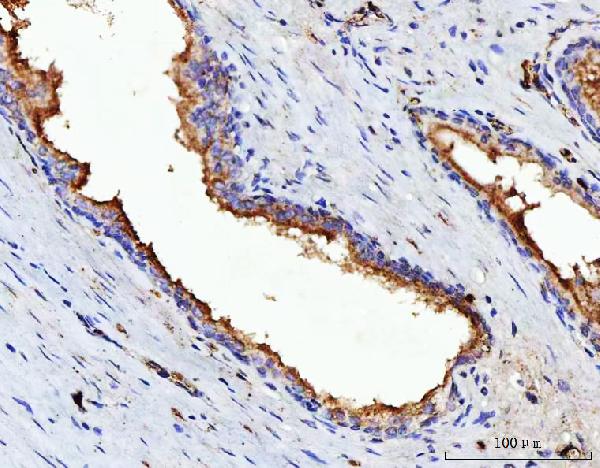
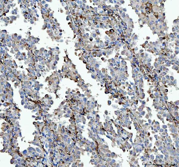
| Sample | Paraffin-embedded human prostate cancer tissue; fixative not specified (datasheet A03768-1) |
| Fixation | Image fixative and duration unreported (datasheet A03768-1); verify before use. |
| Sectioning | 4–5 µm sections on charged slides (standard) |
| Deparaffinisation | Xylene, graded ethanol series to water (standard) |
| Antigen retrieval | Heat retrieval: EDTA pH 8.0 (datasheet A03768-1); 20 min, 95–100 °C (standard) |
| Peroxidase block | 3% H2O2, 10 min, room temperature (standard) |
| Blocking | 10% goat serum (datasheet A03768-1) |
| Primary antibody | Rabbit anti-VIPR2, 2-5 μg/ml (datasheet A03768-1) |
| Primary incubation | Overnight at 4 °C (datasheet A03768-1) |
| Detection | HRP-conjugated secondary, DAB chromogen (datasheet A03768-1) |
| Counterstain | Hematoxylin, blue, dehydrate and mount (standard) |
| Expected result | VIPR2-positive staining in adipocytes of adipose tissue (HPA tissue IHC: High). HPA tissue profile: Membranous expression in most tissues, including pneumocytes, smooth muscle cells and endothelial cells. No signal in the no-primary control. |
VIPR2 is a cell-membrane receptor with 7 transmembrane segments (UniProt P41587 topology). In paraffin-section IHC, expect predominantly membranous staining in selected cells, including pneumocytes, smooth muscle cells, endothelial cells, adipocytes, bronchial basal cells and glomerular cells (HPA tissue IHC). HPA rates its tissue IHC evidence Approved, with medium consistency between antibody staining and RNA expression; interpret individual positive cells and compartments with that limitation in mind (HPA tissue IHC).
| Clear membrane-associated staining in adipocytes or bronchial basal cells, with limited surrounding background. | This fits the expected compartment (UniProt P41587 topology; HPA tissue IHC). HPA reports High staining in adipocytes in adipose tissue and breast, and in bronchial basal cells (HPA tissue IHC). Compare the relevant cell population, rather than calling an entire mixed tissue uniformly positive. |
| Strong nuclear staining, or uniform cytoplasmic color without a discernible membrane pattern. | That distribution does not match the annotated cell-membrane location or the predominantly membranous tissue pattern (UniProt P41587; HPA tissue IHC). Treat it as suspect staining; review morphology, the negative control and detection conditions before assigning it to VIPR2 (general IHC practice). |
| Prominent staining in adrenal glandular cells or bone-marrow hematopoietic cells. | HPA reports VIPR2 staining as Not detected in those specified cell populations (HPA tissue IHC). An unexpected signal may reflect cross-reactivity or endogenous detection activity; check a no-primary control and whether the signal follows the same compartmental pattern as the positive tissue (general IHC practice). |
| Diffuse color covers many structures, obscures cell boundaries or appears on the no-primary control. | This is background rather than an interpretable membrane pattern (general IHC practice; HPA tissue IHC profile). Examine blocking, washing, detection reagent and chromogen development; use the no-primary control to locate signal arising after the primary-antibody step (general IHC practice). |
| No convincing membrane signal in an expected positive cell population. | First confirm that the section contains the intended cells: HPA reports High staining in bronchial basal cells, glomerular cells and placental decidual cells (HPA tissue IHC). If present, review the IHC-validated antibody, retrieval and detection workflow (general IHC practice). HPA approval does not guarantee staining in every specimen (HPA tissue IHC reliability). |
| Membrane topology and epitope access (UniProt P41587 topology). | VIPR2 has extracellular and cytoplasmic regions around 7 transmembrane segments (UniProt P41587 topology). The antibody epitope is not supplied here, so topology alone cannot specify an antigen-retrieval condition or predict staining strength in paraffin sections. |
| Signal peptide and mature chain (UniProt P41587 processing). | The annotated signal peptide spans residues 1–23; the receptor chain spans 24–438 (UniProt P41587 processing). Without the antibody epitope, this record cannot establish whether processing affects recognition in IHC. |
| Glycosylation and isoforms (UniProt P41587). | UniProt lists 3 glycosylation sites at residues 58, 88 and 92, and 2 isoforms (UniProt P41587). These annotations do not identify which form the catalog antibody detects or explain a particular IHC intensity. |
| Tissue and cell-population choice (HPA tissue IHC). | HPA reports High staining in glomerular cells and placental decidual cells, Medium in endometrial stromal cells, and Not detected in adrenal glandular cells (HPA tissue IHC). Score the annotated cell type; a whole-section average can conceal the relevant pattern. |
| IF/ICC Q: Should focal-adhesion staining define a positive paraffin IHC result? | A: No. HPA reports focal adhesion sites in ICC-IF, while its tissue IHC profile is membranous (HPA subcellular ICC-IF; HPA tissue IHC). The listed antibody's ICC status is Uncertain (HPA antibodies); assess IF/ICC on its own guide page. |
| Situation | Likely cause | Next action |
|---|---|---|
| Expected positive tissue has no visible staining. | The sampled field may lack the relevant cells, or the IHC workflow may have failed (general IHC practice). HPA's High label applies to specified cells, such as bronchial basal cells, rather than every cell in bronchus (HPA tissue IHC). | Locate the annotated cells on the counterstained section, then check the IHC-validated antibody, antigen-retrieval run, primary incubation and detection controls (general IHC practice). Avoid inferring target-specific fixation sensitivity from this result. |
| Signal is widespread in a tissue expected to be weak or negative. | HPA reports Not detected in adrenal glandular cells and Low in bladder urothelial cells (HPA tissue IHC). Broad signal there raises concern for nonspecific primary binding or endogenous detection activity (general IHC practice). | Compare cell identity and compartment with a high-staining reference section; inspect no-primary and detection controls, blocking and chromogen development (general IHC practice). Treat HPA's 'Not detected' category as an observation, not proof of biological absence. |
| Only a nuclear or uniformly cytoplasmic pattern appears. | The pattern conflicts with VIPR2's cell-membrane annotation and HPA's membranous tissue profile (UniProt P41587; HPA tissue IHC). Background or antibody cross-reactivity is possible (general IHC practice). | Recheck the cell boundaries, compare a known HPA-high cell population, and review no-primary controls and detection conditions before scoring the section positive (HPA tissue IHC; general IHC practice). |
| Staining obscures the membranes that need scoring. | Excess chromogen development, insufficient washing or detection background can reduce contrast (general IHC practice). This prevents assessment of the expected membranous pattern (HPA tissue IHC). | Review development time and wash steps, then inspect the no-primary control. Score only cells whose boundaries and compartment remain distinguishable (general IHC practice). |
| A medium or low HPA category is being called a failed experiment. | HPA reports Medium staining in endometrial stromal and esophageal squamous cells, and Low staining in oral squamous and bladder urothelial cells (HPA tissue IHC). These categories differ from its high-staining examples. | Compare like cell types and use a separate HPA-high reference population to check assay performance; retain the reported intensity category when interpreting the test tissue (HPA tissue IHC; general IHC practice). |
| Two sections or antibodies give different-looking patterns. | HPA rates the tissue IHC evidence Approved but describes only medium consistency with RNA expression, and the listed antibody is Approved for IHC (HPA tissue IHC; HPA antibodies). Those ratings do not resolve every discordant cell-level result. | Compare the same annotated cell populations, compartment, controls and staining conditions across sections; report unresolved disagreement instead of treating one unexpected pattern as confirmed VIPR2 (HPA tissue IHC; general IHC practice). |


Comprehensive Human Protein Atlas IHC scoring per tissue (reliability: Approved — Medium consistency between antibody staining and RNA expression data. Antibody staining in cells/structures not annotated, view images.). Rows are taken directly from the HPA tissue chart — click any row's HPA link to view the source.
| Tissue | Cell type | Level | Evidence | Source |
|---|---|---|---|---|
| Adipose tissue | Adipocytes | High | Protein (IHC) | HPA → |
| Breast | Adipocytes | High | Protein (IHC) | HPA → |
| Bronchus | Basal cells | High | Protein (IHC) | HPA → |
| Kidney | Cells in glomeruli | High | Protein (IHC) | HPA → |
| Placenta | Decidual cells | High | Protein (IHC) | HPA → |
| Tissue | Cell type | Level | Evidence | Source |
|---|---|---|---|---|
| Adrenal gland | Glandular cells | Not detected | Protein (IHC) | HPA → |
| Appendix | Glandular cells | Not detected | Protein (IHC) | HPA → |
| Bone marrow | Hematopoietic cells | Not detected | Protein (IHC) | HPA → |
| Caudate | Glial cells | Not detected | Protein (IHC) | HPA → |
| Cerebellum | Cells in granular layer | Not detected | Protein (IHC) | HPA → |
Troubleshoot VIPR2 staining in paraffin sections by checking retrieval, cell identity and membrane localisation against the catalog antibody’s tissue image and reference patterns (datasheet A03768-1; HPA tissue IHC; UniProt P41587).
A03768-1 has real IHC data from paraffin sections of human prostate cancer, lung cancer and tonsil (IHC captions). No IF/ICC data are supplied (catalog).
A03768-1 is listed for IHC and has images from paraffin sections of human prostate cancer, lung cancer and tonsil (catalog; IHC captions). It is listed as reactive with human and monkey, but the supplied IHC images show human tissue only (catalog; IHC captions).
Which to pick: Choose the polyclonal A03768-1 for human paraffin-section IHC; its captions document EDTA retrieval at pH 8.0 and 2 μg/ml primary antibody, while the catalog gives an IHC range of 2–5 μg/ml (catalog; IHC captions). Its paraffin-section captions do not report the fixative (IHC captions). No IF/ICC option is validated here; A03768-1 lists monkey reactivity, but the supplied IHC images are from human tissue only (catalog; IHC captions).