This website uses cookies to ensure you get the best experience on our website.


- Table of Contents
Use this guide to plan paraffin-section WDHD1 IHC around its broad nuclear staining (HPA tissue IHC). Lymph node germinal center cells show high staining, and the catalog antibody has an IHC dilution range of 1:100–1:300 (HPA tissue IHC; datasheet A06240).

Expected localisation, validated protocols, controls and antibodies — the at-a-glance facts below, then the full design guide.
| Expected localisation | Broad nuclear staining (HPA tissue IHC) | |
| Staining pattern | Nuclear staining across many tissue cell types (HPA tissue IHC) | |
| Antigen retrieval | Tris-EDTA pH 9.0 HIER, heat-mediated (datasheet A06240) | |
| Positive control | Appendix+4 more · see all | |
| Negative control | Adipose tissue+4 more · see all |
| Fixation | Keep fixation consistent across sections (standard IHC practice; not target-specific) | |
| Caveat | Staining and RNA expression show medium consistency (HPA tissue IHC) | |
| Regulation | Expression regulation not annotated (UniProt) | |
| Isoform / epitope | 2 isoforms; epitope coverage is unspecified (UniProt) |
The catalog antibody protocol uses Tris-EDTA pH 9.0 heat retrieval (datasheet A06240). The published IHC protocols below describe nasopharyngeal and breast tumour tissue microarrays (PMC9811654; PMC7680459).
| Sample | Paraffin-embedded human tonsil tissue; fixative not specified (datasheet A06240) |
| Fixation | Image fixative and duration unreported (datasheet A06240); verify before use. |
| Sectioning | 4–5 µm sections on charged slides (standard) |
| Deparaffinisation | Xylene, graded ethanol series to water (standard) |
| Antigen retrieval | Heat retrieval: Tris-EDTA pH 9.0 (datasheet A06240); 20 min, 95–100 °C (standard) |
| Peroxidase block | 3% H2O2, 10 min, room temperature (standard) |
| Blocking | 10% normal serum of the secondary host, 30 min, room temperature (standard) |
| Primary antibody | Rabbit anti-WDHD1, 1:100-1:300 (datasheet A06240) |
| Primary incubation | Overnight at 4 °C (standard) |
| Detection | HRP-polymer secondary, DAB chromogen 5–10 min (standard) |
| Counterstain | Hematoxylin, blue, dehydrate and mount (standard) |
| Expected result | WDHD1-positive staining in glandular cells of appendix (HPA tissue IHC: High). HPA tissue profile: Ubiquitous nuclear expression. No signal in the no-primary control. |
WDHD1 is a nuclear protein localized to the nucleoplasm, with no transmembrane segment (UniProt O75717). In paraffin section IHC, expect nuclear staining across multiple tissues, including high staining in appendix and rectal glandular cells, endometrial stromal cells, and lymph node germinal center cells (HPA tissue IHC). HPA describes the overall pattern as ubiquitous nuclear expression, while reporting some cell types as undetected; its tissue IHC reliability is Enhanced, with medium consistency between staining and RNA data (HPA tissue IHC).
| Distinct nuclear staining in appendix or rectal glandular cells, endometrial stromal cells, or lymph node germinal center cells. | This matches cell types scored High in HPA tissue IHC and the nucleoplasmic location assigned by UniProt O75717. Compare nuclei with neighboring cells on the same section; an HPA High designation does not mean every cell in that tissue must stain equally. |
| Signal is predominantly cytoplasmic, membranous, or extracellular, with little nuclear signal. | That distribution conflicts with WDHD1 nucleoplasmic localization and its lack of a transmembrane segment (UniProt O75717; HPA subcellular ICC-IF). Treat it as a questionable IHC result and reassess staining specificity and detection before assigning it to WDHD1. |
| Strong staining appears in adipocytes, cardiomyocytes, or glial cells in caudate or hippocampus. | HPA scores those specific cell populations Not detected in the listed tissues (HPA tissue IHC). Check cell identity and nuclear localization; persistent signal could reflect antibody cross-reactivity or endogenous detection activity (general IHC practice). These scores do not make the entire tissues absolute negative controls. |
| Broad color covers nuclei and surrounding tissue, obscuring cell boundaries. | A diffuse deposit cannot establish the nucleoplasmic pattern reported for WDHD1 (UniProt O75717; HPA subcellular ICC-IF). Excess antibody, incomplete blocking, or detection background are possible general IHC causes; inspect a no-primary control before scoring cells. |
| No nuclear signal appears in glandular cells of an appendix or rectum section selected as a positive comparator. | Both cell populations are scored High by HPA tissue IHC, so a blank result warrants a workflow check. Confirm tissue preservation and that a general nuclear counterstain shows identifiable cells, then review the validated IHC antibody and detection steps (general IHC practice). |
| Compartment and tissue context | UniProt places WDHD1 in the nucleoplasm, and HPA reports a ubiquitous nuclear profile (UniProt O75717; HPA tissue IHC). HPA also scores particular adipocytes, cardiomyocytes, and glial populations Not detected, so interpret staining by identified cell type rather than assuming uniform positivity throughout a section. |
| IHC antibody validation | HPA lists HPA001122 as IHC Enhanced; HPA000632 has ICC Enhanced but no listed IHC validation (HPA antibodies). The tissue profile has Enhanced reliability with medium staining–RNA consistency (HPA tissue IHC). These are validation summaries, not guarantees that every section or detection setup will reproduce the pattern. |
| Isoforms, modifications, and epitope limits | UniProt lists 2 isoforms, 15 modified residues, and one annotated chain spanning residues 1–1129 (UniProt O75717). Without an epitope location in the supplied record, their effects on this antibody’s IHC staining cannot be predicted; do not infer isoform-specific staining or a processing-dependent pattern. |
| Antigen retrieval | Target-specific fixation effects are not established by the supplied assay evidence. Verify with a matched IHC source before attributing a result to fixation. |
| IF/ICC cross-check — where should fluorescence appear? | In the nucleoplasm: HPA reports an enhanced nucleoplasmic ICC-IF location and lists images from A-431, U-251MG, and U2OS cells (HPA subcellular ICC-IF). That supports a localization cross-check; it does not supply an IF protocol here or transfer ICC validation to the IHC antibody. |
| Situation | Likely cause | Next action |
|---|---|---|
| A known HPA High cell population is blank. | A missed detection step, unsuitable antibody condition, or absent identifiable cells may explain a blank section (general IHC practice). HPA High is an observed tissue score, not a guaranteed result in every preparation (HPA tissue IHC). | Check the IHC-validated antibody identity, section integrity, counterstain, and detection controls; compare with an appendix or rectum section containing the specified glandular cells (HPA antibodies; HPA tissue IHC). |
| The strongest signal lies outside nuclei. | The distribution conflicts with the reported nucleoplasmic location (UniProt O75717; HPA subcellular ICC-IF). Background deposition or cross-reactivity is possible (general IHC practice). | Check nuclear counterstain alignment and a no-primary control, then reassess whether any specific nuclear staining remains (general IHC practice). |
| High color appears in a cell population HPA scores Not detected. | Cell identification may be uncertain, or signal may reflect cross-reactivity or endogenous detection activity (general IHC practice). HPA reports specific negative cell populations, including adipocytes and cardiomyocytes (HPA tissue IHC). | Identify the stained cells morphologically, verify their compartment, and compare no-primary and detection controls before calling them WDHD1 positive (general IHC practice). |
| The entire section has diffuse chromogen. | Diffuse deposition can mask the expected nuclear pattern (UniProt O75717). Excess antibody or insufficient blocking or washing are general IHC possibilities, not WDHD1-specific findings (general IHC practice). | Use a no-primary control; review blocking, washing, antibody concentration, and detection exposure while retaining a known HPA High cell population for comparison (general IHC practice; HPA tissue IHC). |
| Signal differs between lymphoid tissue and other tissues. | HPA reports tissue-enhanced RNA in lymphoid tissue, but its protein IHC profile is described as ubiquitous nuclear with variable cell-level scores (HPA tissue IHC). RNA enrichment alone cannot predict a section’s chromogen intensity. | Score nuclear staining in the named cell populations and record cell identity and intensity separately; compare with the HPA tissue IHC scores rather than ranking whole sections by RNA specificity. |
| ICC-IF appears nucleoplasmic but paraffin IHC is equivocal. | HPA’s enhanced ICC-IF localization and IHC antibody validation apply to different listed antibodies: HPA000632 and HPA001122, respectively (HPA antibodies; HPA subcellular ICC-IF). | Use the ICC-IF finding as a compartment check, then judge paraffin IHC against the IHC antibody’s validation and HPA cell-specific tissue observations (HPA antibodies; HPA tissue IHC). |
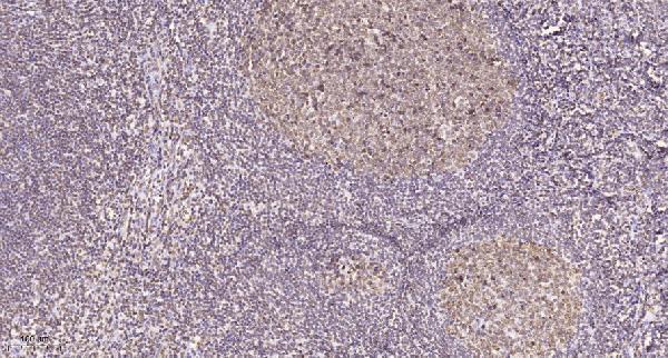
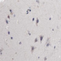

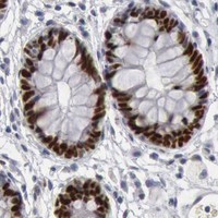
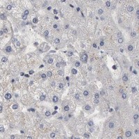


Comprehensive Human Protein Atlas IHC scoring per tissue (reliability: Enhanced — Medium consistency between antibody staining and RNA expression data.). Rows are taken directly from the HPA tissue chart — click any row's HPA link to view the source.
| Tissue | Cell type | Level | Evidence | Source |
|---|---|---|---|---|
| Appendix | Glandular cells | High | Protein (IHC) | HPA → |
| Cervix | Glandular cells | High | Protein (IHC) | HPA → |
| Duodenum | Glandular cells | High | Protein (IHC) | HPA → |
| Endometrium | Cells in endometrial stroma | High | Protein (IHC) | HPA → |
| Lymph node | Germinal center cells | High | Protein (IHC) | HPA → |
| Tissue | Cell type | Level | Evidence | Source |
|---|---|---|---|---|
| Adipose tissue | Adipocytes | Not detected | Protein (IHC) | HPA → |
| Adrenal gland | Glandular cells | Not detected | Protein (IHC) | HPA → |
| Caudate | Glial cells | Not detected | Protein (IHC) | HPA → |
| Cerebellum | Cells in granular layer | Not detected | Protein (IHC) | HPA → |
| Cerebral cortex | Endothelial cells | Not detected | Protein (IHC) | HPA → |
Troubleshoot WDHD1 staining in paraffin sections by checking retrieval, nuclear localisation, controls and scoring before interpreting differences between specimens.
A06240 has a human tonsil paraffin-section IHC image (catalog image caption); IF is listed without an image (catalog applications). Human and mouse reactivity is listed (catalog reactivity).
A06240 has a human tonsil paraffin-section IHC image and lists IF, with no IF image supplied (catalog image caption; catalog applications; catalog IF images). A06240-1 lists human reactivity and IHC, with no IHC image supplied (catalog reactivity; catalog applications; catalog IHC images).
Which to pick: For tissue IHC, choose A06240 when a documented paraffin-section example matters: its human tonsil image used Tris-EDTA retrieval at pH 9.0 and a 1:200 primary dilution overnight at 4°C; the fixative is unreported (A06240 image caption). For IF, A06240 lists that application at 1:50, but has no IF image; ICC validation is unreported (A06240 catalog applications; catalog IF dilution; catalog IF images). For human and mouse work, A06240 lists both species, while A06240-1 lists human only; both list a rabbit host, and neither reports clonality (catalog reactivity; catalog host; catalog clone).