WDR1 / WD repeat-containing protein 1 · IHC design guide

Design Immunohistochemistry for WDR1

Plan WDR1 chromogenic IHC in paraffin sections using the catalog antibody’s documented conditions (datasheet M04814-1). Compare cytoplasmic and membranous staining across cell types using HPA tissue controls (HPA tissue IHC).

Evidence assembled Oct 2026 · For research use; verify linked source records and product datasheet before use
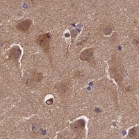
Immunohistochemistry protocol sheet for WDR1 (IHC for WDR1): expected localisation Cytoplasmic and membranous tissue staining (HPA tissue IHC), antibody M04814-1, validated IHC image, and IHC protocol steps
Printable WDR1 IHC protocol sheet — expected localisation Cytoplasmic and membranous tissue staining (HPA tissue IHC), antibody M04814-1, controls and protocol steps. Open the full WDR1 IHC guide →

WDR1 Immunohistochemistry Experimental Design Guide

Expected localisation, validated protocols, controls and antibodies — the at-a-glance facts below, then the full design guide.

Must know before staining
Expected localisation Cytoplasmic and membranous tissue staining (HPA tissue IHC)
Staining pattern Widespread cytoplasmic and membranous staining (HPA tissue IHC)
Antigen retrieval EDTA pH 8.0 HIER, heat-mediated (datasheet M04814-1)
Positive control ⓘ Appendix+4 more · see all
Negative control ⓘ Liver+2 more · see all
Important caveats
Reasons your staining may differ from the expected pattern.
Fixation Keep fixation consistent across sections (standard IHC practice; not target-specific)
Caveat Cell-type mix can shift apparent staining intensity (HPA tissue IHC)
Regulation Specific expression regulators unreported (UniProt)
Isoform / epitope 2 isoforms; check epitope coverage; no extracellular domain (UniProt)
Section 1

Recommended WDR1 IHC & IF Protocols

The catalog antibody protocol uses EDTA pH 8.0 heat retrieval (datasheet M04814-1). One published WDR1 IHC protocol provides staining conditions for FFPE sections (PMC7492282).

Recommended immunohistochemistry (IHC-P) protocol parameters
SampleParaffin-embedded human liver cancer tissue; fixative not specified (datasheet M04814-1)
FixationImage fixative and duration unreported (datasheet M04814-1); verify before use.
Sectioning4–5 µm sections on charged slides (standard)
DeparaffinisationXylene, graded ethanol series to water (standard)
Antigen retrievalHeat retrieval: EDTA pH 8.0 (datasheet M04814-1); 20 min, 95–100 °C (standard)
Peroxidase block3% H2O2, 10 min, room temperature (standard)
Blocking10% goat serum (datasheet M04814-1)
Primary antibodyMouse monoclonal (clone 5C11C8) anti-WDR1, 2-5 μg/ml (datasheet M04814-1)
Primary incubationOvernight at 4 °C (datasheet M04814-1)
DetectionHRP-conjugated secondary, DAB chromogen (datasheet M04814-1)
CounterstainHematoxylin, blue, dehydrate and mount (standard)
Expected resultWDR1-positive staining in glandular cells of appendix (HPA tissue IHC: High). HPA tissue profile: Ubiquitous cytoplasmic and membranous expression. No signal in the no-primary control.
💡Decision noteStart with EDTA pH 8.0 heat retrieval for the catalog antibody (datasheet M04814-1); the published protocol does not specify retrieval (PMC7492282).
Section 2

What Is the Expected WDR1 Staining Pattern?

WDR1 should show mainly cytoplasmic staining, with membranous or junctional staining in some cells (HPA: ubiquitous cytoplasmic and membranous expression; UniProt O75083: cytoplasm, cytoskeleton and cell junction; no transmembrane segment). High staining is reported in colon and appendix glandular cells, among others (HPA: High). Tissue IHC reliability is Approved, with medium agreement between staining and RNA data and external verification pending (HPA: reliability description).

What am I looking at on my slide?
High cytoplasmic staining in colon or appendix glandular cells, possibly with a membranous outline.This fits the reported tissue and compartment pattern (HPA: High in colon and appendix glandular cells; ubiquitous cytoplasmic and membranous expression). Judge the cells within intact tissue architecture: nearby weak cells need not stain equally because the HPA levels are assigned by cell type (HPA: tissue IHC).
Staining is confined to nuclei, with little cytoplasmic signal in otherwise positive cells.A nucleus-only pattern conflicts with the supplied locations (UniProt O75083: cytoplasm, cytoskeleton and cell junction; HPA: cytoplasmic and membranous tissue expression). Treat it as a suspect result and check antibody specificity, staining conditions and the positive control before interpreting it as WDR1.
Strong staining appears in liver cholangiocytes or ovarian stroma cells while the expected positive cells are weak.Those cells are reported as Not detected (HPA: liver cholangiocytes; ovarian stroma cells). This reversal raises concern for cross-reactivity or detection background, including endogenous activity in chromogenic IHC (general IHC practice). It does not establish that WDR1 is absent from every cell in either tissue.
Color is spread across the section, including spaces between cells, without a discernible cellular pattern.This is diffuse background rather than the reported cellular pattern (HPA: cytoplasmic and membranous tissue expression). Review the no-primary control and staining conditions; tissue-wide color alone cannot assign WDR1 to a cell type (general IHC practice).
Colon glandular cells show no signal, although the counterstain preserves recognizable glands.Colon glandular cells are a reported High-staining population (HPA: colon glandular cells). A blank result therefore calls for a run check, including the antibody, antigen retrieval and detection controls (general IHC practice), before concluding that the specimen lacks WDR1. One section cannot override the atlas profile.
💡Expected WDR1 appearanceA convincing positive is High cytoplasmic staining in colon or appendix glandular cells, with possible membranous staining (HPA: High in those cells; cytoplasmic and membranous profile); nucleus-only signal or widespread color without cellular boundaries is suspect against that profile (HPA: tissue IHC).
How each factor affects the staining
Tissue and cell choiceUse a reported High population, such as colon or appendix glandular cells, to judge a positive result (HPA: High). Compare named cells rather than whole organs: liver cholangiocytes are Not detected, while other liver cells are not classified by that entry (HPA: liver cholangiocytes).
Subcellular interpretationCytoplasm and cell junctions are annotated locations (UniProt O75083). WDR1 has no transmembrane segment (UniProt O75083: topology), so membranous staining can be assessed as a location pattern without assuming WDR1 spans the membrane.
Antibody evidenceThe tissue profile is Approved but has medium staining-to-RNA agreement and awaits external verification (HPA: reliability description). HPA036434, HPA070293 and CAB020804 each have Approved IHC status (HPA: antibodies); these entries alone do not establish Enhanced IHC validation.
Isoforms and processingTwo isoforms are listed, and the annotated chain spans residues 1–606, with no signal peptide or propeptide (UniProt O75083: isoforms and processing). No epitope position is supplied, so this record cannot predict which isoforms a particular antibody detects or assign a staining difference to processing.
IF/ICC Q&A: Where should signal appear?Cytosol and cell junctions are supported ICC-IF locations; plasma membrane is uncertain (HPA: subcellular). This answers localisation only. It does not supply an IF/ICC protocol or show that an IHC-P staining result transfers to IF/ICC (HPA: antibody application status).
Why is my staining missing, weak or wrong?
SituationLikely causeNext action
Known-positive glandular cells are blank.A reported High population failed to stain (HPA: colon or appendix glandular cells); the run or specimen may need review (general IHC practice).Confirm the tissue and cell identity, then review the catalog antibody’s IHC-P conditions, antigen retrieval and detection controls (general IHC practice). Do not infer target-specific fixation sensitivity; none is reported in the supplied sources.
Only nuclei stain.The compartment differs from the supplied cytoplasmic, cytoskeletal and junctional locations (UniProt O75083; HPA: tissue profile).Check a reported High control for cytoplasmic staining and assess the no-primary control; interpret persistent nucleus-only color cautiously (HPA: High in colon glandular cells; general IHC practice).
A Not detected cell population stains strongly.Signal in cholangiocytes or ovarian stroma cells conflicts with their HPA entries (HPA: Not detected) and may reflect nonspecific binding or detection activity (general IHC practice).Compare the exact cell population with a High control and a no-primary control; review blocking and detection steps if background persists (HPA: tissue IHC; general IHC practice).
Diffuse color obscures cell boundaries.Background can prevent assessment of the expected cellular distribution (HPA: cytoplasmic and membranous profile; general IHC practice).Inspect a no-primary control, check wash and blocking steps, and adjust development using the validated IHC-P procedure (general IHC practice). Score WDR1 only where cells and compartments remain distinguishable.
Membrane staining dominates and cytoplasm is hard to see.Membranous tissue staining is reported, but plasma membrane localisation in ICC-IF is uncertain (HPA: tissue profile; subcellular).Check for cytoplasmic signal in a reported High cell population and compare cellular morphology and controls before calling a membrane-only pattern specific (HPA: colon or appendix glandular cells; general IHC practice).
An IF/ICC image differs from the paraffin-section result.The readouts use different applications; HPA lists supported ICC for HPA070293, while its tissue profile describes IHC (HPA: antibodies; tissue IHC).Interpret the images within their applications: compare IF/ICC with supported cytosol and junction locations, and IHC with the tissue cell pattern (HPA: subcellular; tissue IHC). Use the separate IF/ICC guide for that workflow.

Sample controls for WDR1 IHC & IF

🧪Run appendix first: its glandular cells should stain (HPA: High in appendix glandular cells). Run liver cholangiocytes as the negative tissue (HPA: Not detected in liver cholangiocytes); on the appendix slide, treat non-glandular cells as internal negatives only where they show no specific signal, since no internal-negative cell type is identified in the supplied HPA rows.
Positive control tissue: Appendix (Glandular cells, HPA High)
Negative control tissue: Liver (HPA Not detected)
ICC-IF cell lines (HPA subcellular resource): HPA ICC-IF images show WDR1 in BJ [Human fibroblast], U-251MG, U2OS, with annotated localisation: Plasma membrane (uncertain), Cell Junctions (supported), Cytosol (supported) (HPA subcellular).
Technical controls: Include a no-primary (secondary-only) slide, a mouse IgG isotype control matched to the primary antibody where its isotype is known, and WDR1-knockout tissue or cells as a biological negative (selected IHC caption: mouse anti-WDR1; standard IHC practice). Check the appendix no-primary slide for endogenous peroxidase activity before interpreting DAB signal (selected IHC caption: HRP/DAB detection; standard IHC practice).
⚠️Feasibility: A target-specific fixation window or fixation effect is unreported, and the selected M04814-1 tissue-IHC caption does not state a fixative (selected IHC caption). The reported paraffin-section method uses heat retrieval in EDTA at pH 8.0; retrieval dependence has not been established, and the caption does not establish fixation (selected IHC caption). The supplied evidence does not show that frozen sections or IF are easier, or establish an appendix-specific artefact; assess glandular DAB background against the no-primary slide (selected IHC caption: HRP/DAB; standard IHC practice). The selected M04814-1 tissue-IHC caption documents paraffin sections, but does not specify the fixative (selected-SKU IHC image M04814-1).

HPA tissue IHC evidence for WDR1

Comprehensive Human Protein Atlas IHC scoring per tissue (reliability: Approved — Medium consistency between antibody staining and RNA expression data. Pending external verification.). Rows are taken directly from the HPA tissue chart — click any row's HPA link to view the source.

Positive expression · recommended positive controls

TissueCell typeLevelEvidenceSource
Appendix Glandular cells High Protein (IHC) HPA →
Breast Glandular cells High Protein (IHC) HPA →
Caudate Neuronal cells High Protein (IHC) HPA →
Cerebellum Cells in granular layer High Protein (IHC) HPA →
Cerebral cortex Glial cells High Protein (IHC) HPA →

Undetected expression · recommended negative controls

TissueCell typeLevelEvidenceSource
Liver Cholangiocytes Not detected Protein (IHC) HPA →
Ovary Ovarian stroma cells Not detected Protein (IHC) HPA →
Seminal vesicle Glandular cells Not detected Protein (IHC) HPA →
Section 3

Advanced WDR1 IHC Tips

Troubleshoot WDR1 staining in paraffin sections by checking retrieval, tissue preservation, compartment, controls and scoring before interpreting chromogenic signal.

How should I retrieve WDR1 in paraffin sections when staining is weak?
Start with heat-mediated antigen retrieval in EDTA at pH 8.0 for paraffin sections (datasheet M04814-1). The selected tissue image used this retrieval before incubation with 2 μg/mL antibody overnight at 4°C (datasheet M04814-1). If signal is weak, check that sections remain attached, retrieval solution covers them, and the same heating and cooling procedure is applied across the run (standard IHC practice). Compare any adjusted retrieval condition on adjacent sections with an established positive control, because stronger staining alone can reflect increased background (standard IHC practice). Record the condition used for every comparison (standard IHC practice).
Could fixation explain inconsistent WDR1 staining between paraffin blocks?
Target-specific fixation sensitivity is unknown: the selected paraffin-section caption does not state a fixative (datasheet M04814-1). Record each block’s fixative and processing history, then stain matched sections together under the same retrieval and detection conditions (standard IHC practice). Differences in preservation can alter morphology and antigen accessibility, so assess those features before assigning a weak result to WDR1 abundance (standard IHC practice). Use a positive-control section in each run and compare like cell populations where possible (standard IHC practice). Neither the reported tissue pattern nor WDR1’s cytoplasmic location establishes a WDR1-specific fixation effect (HPA: tissue IHC; UniProt O75083: subcellular location).
Which WDR1 staining compartments should I accept in chromogenic IHC?
Expect predominantly cytoplasmic staining, with possible junction-associated or peripheral signal (UniProt O75083: subcellular location; HPA: tissue IHC profile). WDR1 is also annotated at the cytoskeleton and podosome, while cell-junction and cytosolic locations have supporting subcellular evidence (UniProt O75083: subcellular location; HPA: subcellular). Assess those patterns within intact cells rather than treating any brown deposit at a tissue boundary as specific (standard IHC practice). Strong nuclear-only staining needs independent confirmation because the supplied localisation evidence does not support a nuclear assignment (UniProt O75083: subcellular location; HPA: subcellular). Include a matched negative control when judging faint peripheral staining (standard IHC practice).
Can this antibody distinguish WDR1 isoforms or modified epitopes in tissue?
The record lists 2 WDR1 isoforms, but the selected IHC caption does not identify the antibody epitope (UniProt O75083: isoforms; datasheet M04814-1). Do not interpret a staining difference as isoform-specific without an epitope map or independently validated isoform-specific reagent (standard IHC practice). WDR1 has annotated acetylated residues and a phosphotyrosine at residue 238, yet the caption provides no modification-specific validation (UniProt O75083: modified residues; datasheet M04814-1). Compare staining with an independent antibody or an appropriate loss-of-target control when an epitope-dependent conclusion matters (standard IHC practice). Report this assay as total WDR1 immunoreactivity unless specificity is established (standard IHC practice).
How should I check WDR1 localisation by IF alongside this IHC assay?
For the separate IF/ICC assay, multiplex WDR1 with a marker for the cell population under study, such as a glandular-cell marker when examining glandular staining (HPA: high in appendix, breast and colon glandular cells; standard IF practice). Choose spectrally separated fluorophores and consider a far-red WDR1 channel if tissue autofluorescence interferes with shorter wavelengths (standard IF practice). WDR1 is cytosolic and junction-associated, with no transmembrane segment; permeabilise for a cytosolic epitope, while confirming the antibody’s actual epitope accessibility empirically (UniProt O75083: location and topology; standard IF practice). Include single-label and no-primary controls to assess bleed-through and background (standard IF practice).
How can I reduce diffuse brown background without losing WDR1 signal?
First check the no-primary control and inspect whether the deposit follows cells or pools over damaged tissue (standard IHC practice). The selected paraffin-section example used 10% goat serum blocking, an overnight primary incubation at 4°C, and DAB development (datasheet M04814-1). Keep blocking, washing and development consistent while titrating primary-antibody concentration around the documented 2 μg/mL condition on adjacent sections (datasheet M04814-1; standard IHC practice). Block endogenous peroxidase before HRP detection and limit DAB development when background rises in the controls (standard IHC practice). Judge improvement against preserved cytoplasmic staining and tissue morphology (HPA: tissue IHC profile; standard IHC practice).
What should I score when comparing WDR1 staining across sections? ⚠ ANSWER MARKED FOR VERIFICATION
Define the cell population and compartment before scoring, because WDR1 staining can be cytoplasmic and peripheral (HPA: tissue IHC profile; UniProt O75083: subcellular location). Within matched regions, report an H-score or the percentage of positive cells using a fixed intensity threshold (standard IHC practice). If the biological question concerns infiltrating cells, positive-cell density per mm² is an alternative, provided the analysed area is defined consistently (standard IHC practice). Normalise counts to the relevant cell population or viable tissue area, and keep retrieval, imaging and DAB development constant across comparisons (standard IHC practice). Exclude folds, edges and necrotic regions by prespecified rules (standard IHC practice).
When is a WDR1-positive IHC result credible rather than artefactual?
A credible result shows cell-associated cytoplasmic or junctional staining in preserved tissue and exceeds the matched negative-control signal (UniProt O75083: subcellular location; HPA: tissue IHC profile; standard IHC practice). Compare cell identity with context: HPA reports high staining in several glandular populations but no detection in liver cholangiocytes (HPA: appendix, breast, colon and liver tissue IHC). The selected image demonstrates staining in a paraffin section of human liver cancer tissue; it does not establish positivity in every liver cell type (datasheet M04814-1). Treat edge staining, necrotic deposits and residual endogenous peroxidase signal as suspect until controls and morphology support specificity (standard IHC practice).
Boster reagents

Best WDR1 / WD repeat-containing protein 1 IHC Antibodies

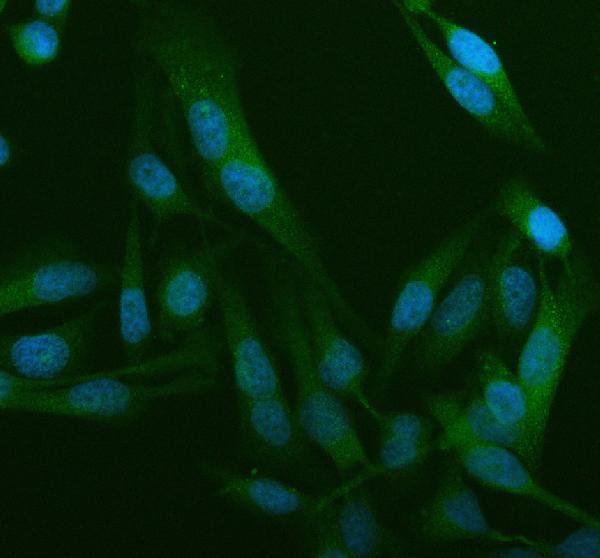
Anti-WDR1 antibodies have IHC images from human liver and breast cancer paraffin sections and an IF image from HeLa cells (M04814-1 and PB9962 image captions).

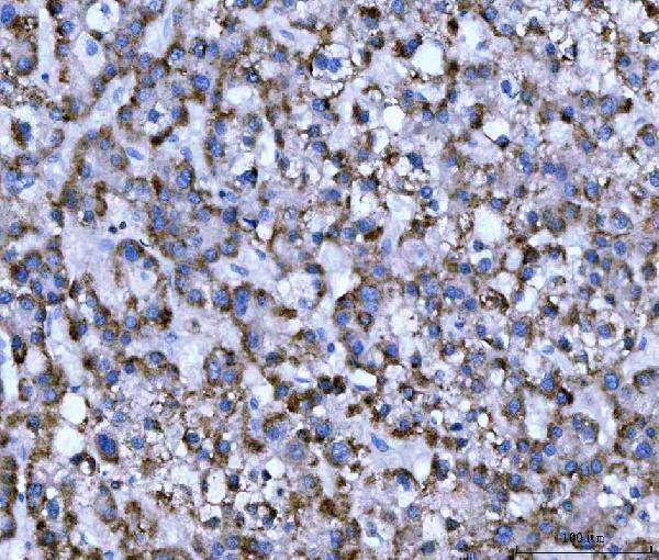
Real IHC data IHC analysis of WDR1 using anti-WDR1 antibody (M04814-1). WDR1 was detected in a paraffin-embedded section of human liver cancer tissue. Heat mediated antigen retrieval was performed in EDTA buffer (pH 8.0, epitope retrieval solution). The tissue section was blocked with 10% goat serum. The tissue section was then incubated with 2 μg/ml mouse anti-WDR1 Antibody (M04814-1) overnight at 4°C. Peroxidase Conjugated Goat Anti-mouse IgG was used as secondary antibody and incubated for 30 minutes at 37°C. The tissue section was developed using HRP Conjugated Mouse IgG Super Vision Assay Kit (Catalog # SV0001) with DAB as the chromogen.
Anti-WDR1 Antibody ® (monoclonal, 5C11C8)
Cat # M04814-1
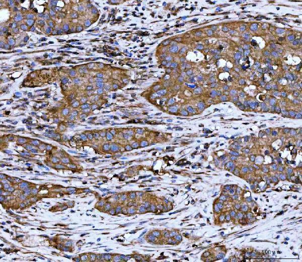
Real IHC data IHC analysis of WDR1 using anti-WDR1 antibody (PB9962). WDR1 was detected in a paraffin-embedded section of human breast cancer tissue. Heat mediated antigen retrieval was performed in EDTA buffer (pH 8.0, epitope retrieval solution). The tissue section was blocked with 10% goat serum. The tissue section was then incubated with 2 μg/ml rabbit anti-WDR1 Antibody (PB9962) overnight at 4°C. Peroxidase Conjugated Goat Anti-rabbit IgG was used as secondary antibody and incubated for 30 minutes at 37°C. The tissue section was developed using HRP Conjugated Rabbit IgG Super Vision Assay Kit (Catalog # SV0002) with DAB as the chromogen.
Anti-WDR1 Antibody ®
Cat # PB9962

M04814-1 has an IHC image from a human liver cancer paraffin section (M04814-1 IHC image caption). PB9962 has an IHC image from a human breast cancer paraffin section and an IF image from HeLa cells (PB9962 image captions).

Which to pick: For tissue IHC, choose M04814-1 for a mouse monoclonal or PB9962 for a rabbit antibody; each has its own human paraffin-section IHC image (catalog hosts; M04814-1 and PB9962 IHC image captions). For IF/ICC, choose PB9962, which lists both applications and has a HeLa-cell IF image (PB9962 catalog and IF image caption). Both list Human, Mouse and Rat reactivity, while their pictured IHC experiments use human tissue; the fixative is unreported in both paraffin-section captions (catalog reactivity; M04814-1 and PB9962 IHC image captions).

Each figure is that product's own IHC / IF validation image from its datasheet.

References

  1. UniProt Consortium. UniProt entry O75083 (WDR1_HUMAN, WD repeat-containing protein 1).
  2. Human Protein Atlas. WDR1 tissue IHC expression (reliability: Approved).
  3. Human Protein Atlas. WDR1 subcellular location (ICC-IF): Localized to the plasma membrane, cell junctions and cytosol..
  4. Human Protein Atlas. WDR1 antibody validation summary (3 antibodies).
  5. WD repeat-containing protein 1 maintains β-Catenin activity to promote pancreatic cancer aggressiveness. British journal of cancer 2020 — PMC7492282.
  6. Systematic pan-cancer analysis reveals context-dependent prognostic and immunological roles of WDR1 with divergent effects in renal and gastric cancers. Frontiers in immunology 2026 — PMC13366190.
  7. WD Repeat Domain 1 Deficiency Inhibits Neointima Formation in Mice Carotid Artery by Modulation of Smooth Muscle Cell Migration and Proliferation. Molecules and cells 2020 — PMC7468582.
  8. PubMed PMID:10036186 — UniProt-cited evidence.
  9. PubMed PMID:14702039 — UniProt-cited evidence.
  10. PubMed PMID:15815621 — UniProt-cited evidence.