WDR74 / WD repeat-containing protein 74 · IHC design guide

Design Immunohistochemistry for WDR74

Use skeletal or smooth muscle as medium-staining references for WDR74 IHC-P (HPA tissue IHC). This guide pairs the catalog antibody’s 5 μg/mL starting concentration (datasheet) with a nuclear/nucleolar expectation (UniProt) and flags the uncertain tissue-IHC reliability (HPA tissue IHC).

Evidence assembled Oct 2026 · For research use; verify linked source records and product datasheet before use
Immunohistochemistry protocol sheet for WDR74 (IHC for WDR74): expected localisation Expected nuclear/nucleolar; tissue compartment unverified (UniProt), antibody A14044, validated IHC image, and IHC protocol steps
Printable WDR74 IHC protocol sheet — expected localisation Expected nuclear/nucleolar; tissue compartment unverified (UniProt), antibody A14044, controls and protocol steps. Open the full WDR74 IHC guide →

WDR74 Immunohistochemistry Experimental Design Guide

Expected localisation, validated protocols, controls and antibodies — the at-a-glance facts below, then the full design guide.

Must know before staining
Expected localisation Expected nuclear/nucleolar; tissue compartment unverified (UniProt)
Staining pattern Medium muscle-cell; low myoepithelial staining (HPA tissue IHC)
Antigen retrieval Tris-EDTA pH 9.0 HIER, 95–98 °C, 20 min (rule: nuclear antigen)
Positive control ⓘ Skeletal muscle+1 more · see all
Negative control ⓘ Adipose tissue+4 more · see all
Important caveats
Reasons your staining may differ from the expected pattern.
Fixation Keep fixation consistent across sections (standard IHC practice; not target-specific)
Caveat Tissue IHC reliability is uncertain (HPA tissue IHC)
Regulation Pol I activity affects nucleolar signal (UniProt)
Isoform / epitope Two isoforms; epitope coverage is unreported (UniProt; datasheet)
Section 1

Recommended WDR74 IHC & IF Protocols

Compare the catalog antibody’s IHC-P protocol with 2 published WDR74 IHC protocols (PMC11810735; PMC9086290).

Recommended immunohistochemistry (IHC-P) protocol parameters
SampleTissue sections; selected-image fixative not specified (standard IHC workflow)
FixationImage fixative and duration unreported (datasheet A14044); verify before use.
Sectioning4–5 µm sections on charged slides (standard)
DeparaffinisationXylene, graded ethanol series to water (standard)
Antigen retrievalHeat-induced epitope retrieval in Tris-EDTA buffer, pH 9.0, 20 min at 95–98 °C (standard rule: nuclear antigen)
Peroxidase block3% H2O2, 10 min, room temperature (standard)
Blocking10% normal serum of the secondary host, 30 min, room temperature (standard)
Primary antibodyRabbit anti-WDR74, 5 μg/mL (datasheet A14044)
Primary incubationOvernight at 4 °C (standard)
DetectionHRP-polymer secondary, DAB chromogen 5–10 min (standard)
CounterstainHematoxylin, blue, dehydrate and mount (standard)
Expected resultWDR74-positive staining in myocytes of skeletal muscle (HPA tissue IHC: Medium). HPA tissue profile: Selective expression in muscle cells and myoepithelium. No signal in the no-primary control.
💡Decision noteStart with Tris-EDTA pH 9.0 HIER at 95–98 °C for 20 min (page antigen retrieval rule); WDR74 is nuclear and nucleolar (UniProt Q6RFH5).
Section 2

What Is the Expected WDR74 Staining Pattern?

WDR74 is a nuclear protein with nucleolar localization that depends on active Pol I transcription of pre-rRNA (UniProt Q6RFH5). For IHC, expect selective staining in muscle cells and myoepithelium; skeletal myocytes and smooth muscle cells show medium staining in HPA (HPA: tissue IHC). Interpret this pattern cautiously: HPA rates its tissue IHC reliability Uncertain, pending external verification (HPA: tissue IHC). WDR74 has no transmembrane segment (UniProt Q6RFH5 topology).

What am I looking at on my slide?
Nuclear staining in skeletal myocytes or smooth muscle cells, with nucleoli discernible where morphology permits.This fits the reported cell distribution and nuclear localization (HPA: medium in skeletal myocytes and smooth muscle cells; HPA: nucleoplasm and nucleoli in ICC-IF; UniProt Q6RFH5). Tissue IHC reliability remains Uncertain (HPA: tissue IHC).
Predominantly cytoplasmic, membranous or extracellular staining, without a convincing nuclear component.Treat this as a possible artefact because those compartments do not match the reported location (UniProt Q6RFH5; HPA: nucleoplasm and nucleoli in ICC-IF). Review morphology and controls before assigning it to WDR74 (general IHC practice).
Strong staining in an unexpected cell population, especially adipocytes beside stained muscle.Adipocytes are reported as not detected, whereas muscle cells show medium staining (HPA: adipose tissue; HPA: skeletal and smooth muscle). Consider antibody cross-reactivity or endogenous detection activity, then check a reagent control (general IHC practice).
Diffuse color across cells, stroma and the section edge, obscuring nuclear detail.A broad haze cannot establish the expected cell and compartment pattern (HPA: selective muscle-cell IHC; UniProt Q6RFH5 nucleus). Check background in a primary-antibody omission control and review blocking, washes and detection (general IHC practice).
No convincing signal in skeletal myocytes or smooth muscle cells.These are reasonable comparison cells because HPA reports medium staining in each (HPA: tissue IHC). Their absence warrants a technical review, but is not proof of assay failure: the underlying IHC pattern is rated Uncertain (HPA: tissue IHC).
💡Expected WDR74 appearanceCall a result compatible when skeletal myocytes or smooth muscle cells show medium, chiefly nuclear staining with possible nucleolar detail (HPA: tissue IHC and ICC-IF; UniProt Q6RFH5); isolated cytoplasmic or adipocyte staining is suspect (UniProt Q6RFH5; HPA: adipocytes not detected).
How each factor affects the staining
Cell population and comparison tissueSkeletal myocytes and smooth muscle cells are medium; breast myoepithelial cells, cardiomyocytes and skin fibroblasts are low (HPA: tissue IHC). Compare like cell populations when scoring.
Subnuclear location and cell activityWDR74 is nuclear, and its nucleolar location depends on active Pol I pre-rRNA transcription (UniProt Q6RFH5). A visible nucleolus may vary; nuclear location remains the broader expectation (HPA: nucleoplasm and nucleoli in ICC-IF).
Evidence strengthBoth listed antibodies have Uncertain IHC status, and HPA's tissue IHC reliability is Uncertain (HPA: antibodies; HPA: tissue IHC). Treat tissue comparisons as provisional, particularly for a faint or discordant result.
RNA versus protein readoutBone marrow is RNA tissue enhanced, yet hematopoietic cells are reported as protein not detected by tissue IHC (HPA: tissue IHC). Do not substitute RNA enrichment for an IHC positive control.
Isoform coverageTwo isoforms are listed, but the supplied record gives no antibody epitope or isoform-specific staining data (UniProt Q6RFH5; HPA: antibodies). A staining difference cannot be assigned to one isoform from these sources.
IF/ICC: what location should I expect?Expect nucleoplasm and nucleoli; HPA labels these approved and supported, respectively (HPA: subcellular ICC-IF). This location evidence informs interpretation here; IF/ICC procedure belongs in its own guide.
Why is my staining missing, weak or wrong?
SituationLikely causeNext action
Muscle cells are unstained while the entire section is faint.Weak detection or an ineffective staining run is possible (general IHC practice); medium muscle-cell staining is reported but Uncertain (HPA: tissue IHC).Check the run's positive control, detection reagents and the catalog antibody's IHC-P instructions before changing conditions (general IHC practice).
Staining is cytoplasmic in otherwise well-preserved myocytes.The compartment conflicts with nuclear WDR74 localization (UniProt Q6RFH5; HPA: subcellular ICC-IF); nonspecific staining is possible (general IHC practice).Compare a primary-antibody omission control and assess nuclear counterstain alignment and morphology (general IHC practice).
Adipocytes or many unrelated cell types stain as strongly as muscle.That distribution conflicts with adipocytes reported as not detected and selective muscle-cell staining (HPA: tissue IHC). Cross-reactivity or endogenous detection activity is possible (general IHC practice).Inspect reagent controls and the detection system, then score only staining with convincing cellular localization (general IHC practice).
Brown haze obscures nuclei throughout the section.Background from blocking, detection or washing can obscure localization (general IHC practice). A diffuse signal cannot confirm the nuclear pattern (UniProt Q6RFH5).Review blocking, reagent concentration and wash steps against the catalog IHC-P procedure; compare the omission control (general IHC practice).
A nucleolar focus is absent despite clear nuclear staining.Nucleolar localization depends on active Pol I transcription (UniProt Q6RFH5), and HPA also reports nucleoplasmic localization (HPA: subcellular ICC-IF).Assess the nuclear pattern and cell type first; avoid rejecting an otherwise compatible result solely for absent nucleolar contrast (HPA: tissue IHC; HPA: subcellular ICC-IF).
Bone marrow is negative despite its RNA enrichment.HPA calls bone marrow RNA tissue enhanced while reporting hematopoietic-cell protein as not detected by IHC (HPA: tissue IHC).Use skeletal or smooth muscle for the reported IHC comparison and retain the Uncertain reliability caveat (HPA: tissue IHC).

Sample controls for WDR74 IHC & IF

🧪Run skeletal muscle first and score myocytes for medium WDR74 staining (HPA: skeletal-muscle myocytes, Medium). Use adipose tissue adipocytes as the negative comparison (HPA: adipocytes, Not detected); on the positive slide, count only cells lacking nucleoplasmic or nucleolar signal as observed internal negatives, without assuming a neighboring cell type is negative (HPA subcellular: nucleoplasm and nucleoli).
Positive control tissue: Skeletal muscle (Myocytes, HPA Medium)
Negative control tissue: Adipose tissue (HPA Not detected)
ICC-IF cell lines (HPA subcellular resource): HPA ICC-IF images show WDR74 in A-431, U-251MG, U2OS, with annotated localisation: Nucleoplasm (approved), Nucleoli (supported) (HPA subcellular).
Technical controls: Include a no-primary (secondary-only) control, an isotype control matched to the primary antibody’s host species and immunoglobulin class and, if available, WDR74-knockout material as a biological negative (standard IHC practice). For chromogenic skeletal-muscle sections, quench endogenous peroxidase and assess background in myofibers (standard IHC practice).
⚠️Feasibility: A target-specific fixation window and antigen-retrieval dependence are unreported in the supplied evidence; the selected A14044 tissue-IHC caption does not state its fixative (caption: rat liver, 5 μg/mL). The evidence does not establish whether frozen sections or IF are easier than paraffin IHC, although ICC-IF supports nucleoplasmic and nucleolar localization (HPA subcellular: A-431, U-251MG and U2OS). Myofiber autofluorescence can obscure weak nuclear IF signal, so compare it with local background (standard IF practice).

HPA tissue IHC evidence for WDR74

Comprehensive Human Protein Atlas IHC scoring per tissue (reliability: Uncertain — Pending external verification.). Rows are taken directly from the HPA tissue chart — click any row's HPA link to view the source.

Positive expression · recommended positive controls

TissueCell typeLevelEvidenceSource
Skeletal muscle Myocytes Medium Protein (IHC) HPA →
Smooth muscle Smooth muscle cells Medium Protein (IHC) HPA →

Undetected expression · recommended negative controls

TissueCell typeLevelEvidenceSource
Adipose tissue Adipocytes Not detected Protein (IHC) HPA →
Adrenal gland Glandular cells Not detected Protein (IHC) HPA →
Appendix Glandular cells Not detected Protein (IHC) HPA →
Bone marrow Hematopoietic cells Not detected Protein (IHC) HPA →
Bronchus Respiratory epithelial cells Not detected Protein (IHC) HPA →
Section 3

Advanced WDR74 IHC Tips

Troubleshoot WDR74 staining in paraffin sections by checking retrieval, nuclear localisation and cell type before interpreting chromogenic signal.

How should I retrieve WDR74 in paraffin sections with weak nuclear staining?
Start with Tris-EDTA at pH 9.0, heated to 95–98 °C for 20 min (page retrieval setting). Allow sections to cool in retrieval buffer, then compare nuclear signal and tissue preservation against a section processed identically without retrieval (standard IHC practice). WDR74 is reported in the nucleoplasm and nucleoli, so assess those compartments when judging whether retrieval helped (HPA subcellular). If signal remains weak, test a milder retrieval condition on adjacent sections while holding antibody concentration and detection constant; excessive heating can damage morphology or increase background (standard IHC practice).
Could fixation be masking WDR74 in my paraffin sections?
Target-specific fixation sensitivity is unknown because the supplied WDR74 tissue-IHC caption does not state a fixative (A14044 tissue-IHC caption). Record the specimen’s fixative, fixation duration and processing history before comparing staining across blocks, since these variables can change antigen accessibility in paraffin sections (standard IHC practice). Compare sections from blocks with documented processing using the same Tris-EDTA pH 9.0 retrieval and detection settings (page retrieval setting; standard IHC practice). If one block stains poorly, check tissue morphology and a suitable process control before attributing the difference to WDR74 abundance (standard IHC practice).
What WDR74 staining pattern should I expect in chromogenic IHC?
Prioritise staining within nuclei, including nucleolar signal where morphology permits, because WDR74 is reported in the nucleoplasm and nucleoli (HPA subcellular; UniProt Q6RFH5 localisation). Nucleolar localisation depends on active Pol I transcription of pre-rRNA, so its prominence may vary with cellular state (UniProt Q6RFH5 localisation). WDR74 has no annotated transmembrane segment, making a dominant membrane outline inconsistent with its recorded topology and location (UniProt Q6RFH5 topology; HPA subcellular). Use a nuclear counterstain and inspect high magnification fields to distinguish intranuclear deposits from overlying pigment or precipitate (standard IHC practice).
Can isoforms or epitope changes explain uneven WDR74 staining?
WDR74 has 2 annotated isoforms, so check the antibody’s stated immunogen or epitope before treating absent staining as absent total WDR74 (UniProt Q6RFH5 isoforms; standard IHC practice). Isoform 1 interacts with NVL through WDR repeats, whereas isoform 2 does not, making isoform identity relevant to biological interpretation (UniProt Q6RFH5 subunit). Annotated modified residues include phosphoserine at 214 and 361 and methyllysine at 311; their effect on this antibody’s binding is unknown (UniProt Q6RFH5 modified residues). If epitope information is available, compare it with isoform sequences and those sites before adjusting retrieval or scoring (standard IHC practice).
How can I check an IHC pattern using multiplex IF?
Use IF as a complementary spatial check and multiplex WDR74 with a marker for the expected cell type, such as myocytes or smooth muscle cells in the HPA positive tissues (HPA tissue IHC; standard IF practice). Choose a fluorophore channel that avoids the specimen’s strongest autofluorescence and include single-stain and unstained controls to assess bleed-through and background (standard IF practice). WDR74 is nuclear and has no transmembrane segment, so use permeabilisation compatible with access to an intracellular epitope while preserving nuclear structure (UniProt Q6RFH5 topology and localisation; standard IF practice). Compare nuclear and nucleolar patterns with the chromogenic section without assuming identical sensitivity across detection methods (HPA subcellular; standard IF/IHC practice).
How do I separate WDR74 signal from chromogenic background?
Check a no-primary control for nonspecific detection deposits, and examine whether brown signal stays within nuclei rather than spreading across extracellular areas (standard IHC practice; HPA subcellular). Include an appropriate peroxidase block before chromogenic detection because endogenous enzyme activity can produce signal unrelated to antibody binding (standard IHC practice). Titrate the primary antibody and detection time on adjacent sections, keeping retrieval at Tris-EDTA pH 9.0, 95–98 °C for 20 min during the comparison (page retrieval setting; standard IHC practice). The A14044 rat liver image used 5 µg/mL, but its caption does not establish an optimal concentration for other specimens (A14044 tissue-IHC caption).
How should I quantify WDR74 staining across paraffin sections? ⚠ ANSWER MARKED FOR VERIFICATION
Define a nuclear scoring rule before review, then report either the percentage of positive cells or an H-score based on nuclear intensity and percentage stained (HPA subcellular; standard IHC practice). Record nucleolar staining separately if its morphology is resolvable, because nucleolar localisation depends on active Pol I transcription (UniProt Q6RFH5 localisation). Normalise cell counts to the number of intact, evaluable cells of the same annotated type, or report positive-cell density per mm² of viable tissue (standard IHC practice). Keep retrieval, detection, thresholds and sampled area consistent across sections, and exclude folds, edges and necrosis from the scored region (standard IHC practice).
When is a WDR74 IHC signal convincing rather than artefactual?
A convincing signal should be predominantly nuclear, with possible nucleolar enrichment, and should persist in intact cells away from section edges (HPA subcellular; standard IHC practice). HPA reports medium staining in skeletal myocytes and smooth muscle cells, but rates its tissue-IHC reliability uncertain, pending external verification (HPA tissue IHC). Treat strong membrane or extracellular deposits, staining restricted to folds or necrosis, and signal reproduced in a no-primary control as reasons to investigate artefact (UniProt Q6RFH5 topology; standard IHC practice). Compare the observed cell type and compartment with morphology and controls before interpreting a negative or positive field biologically (HPA tissue IHC; HPA subcellular; standard IHC practice).
Boster reagents

Best WDR74 / WD repeat-containing protein 74 IHC Antibodies

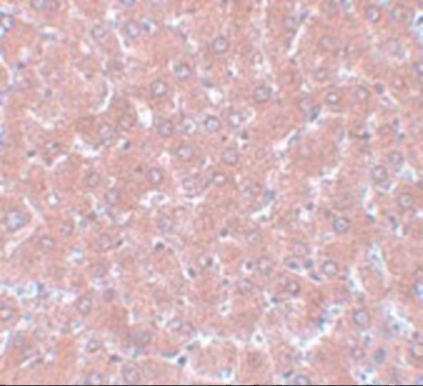
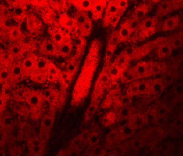
A14044 covers paraffin-section IHC and IF (catalog: IHC-P, IF), with images from rat liver tissue (catalog image captions) and listed reactivity to human, mouse and rat (catalog: reactivity).

Real IHC data Immunohistochemistry of NSA1 in rat liver tissue with NSA1 antibody at 5 μg/mL.
Anti-NSA1 WDR74 Antibody
Cat # A14044

A14044 has a rat liver IHC image at 5 μg/mL (catalog: IHC image caption). A14044 also has a rat liver IF image at 20 μg/mL (catalog: IF image caption).

Which to pick: Choose A14044 for tissue IHC because IHC-P is listed and its IHC image shows rat liver tissue (catalog: applications; IHC image caption); the fixative is unreported (catalog: IHC image caption). Choose A14044 for tissue IF because IF is listed and its IF image shows rat liver tissue (catalog: applications; IF image caption); ICC is not listed (catalog: applications). For cross-species work, A14044 lists human, mouse and rat reactivity (catalog: reactivity), but the supplied IHC and IF images show rat tissue only (catalog: image captions).

Each figure is that product's own IHC / IF validation image from its datasheet.

References

  1. UniProt Consortium. UniProt entry Q6RFH5 (WDR74_HUMAN, WD repeat-containing protein 74).
  2. Human Protein Atlas. WDR74 tissue IHC expression (reliability: Uncertain).
  3. Human Protein Atlas. WDR74 subcellular location (ICC-IF): Localized to the nucleoplasm and nucleoli..
  4. Human Protein Atlas. WDR74 antibody validation summary (2 antibodies).
  5. Identification of WDR74 and TNFRSF12A as biomarkers for early osteoarthritis using machine learning and immunohistochemistry. Frontiers in immunology 2025 — PMC11810735.
  6. A Pan-Cancer Analysis of the Oncogenic Role of WD Repeat Domain 74 in Multiple Tumors. Frontiers in genetics 2022 — PMC9086290.
  7. Ribosomal modifications are associated with mesenchymal fate selection in the neural crest lineage. Nature communications 2026 — PMC12976135.
  8. PubMed PMID:14702039 — UniProt-cited evidence.
  9. PubMed PMID:15489334 — UniProt-cited evidence.
  10. PubMed PMID:12429849 — UniProt-cited evidence.