YBX2 / Y-box-binding protein 2 · IHC design guide

Design Immunohistochemistry for YBX2

Plan YBX2 chromogenic IHC on paraffin sections using testis as a positive control and cytoplasmic staining in spermatocytes and spermatids as the expected pattern (HPA tissue IHC). For human tissue, start the IHC-P–validated catalog antibody A08610 at 10 μg/mL (datasheet A08610).

Evidence assembled Oct 2026 · For research use; verify linked source records and product datasheet before use
Immunohistochemistry protocol sheet for YBX2 (IHC for YBX2): expected localisation Cytoplasm in spermatids and spermatocytes (HPA tissue IHC), antibody A08610-2, validated IHC image, and IHC protocol steps
Printable YBX2 IHC protocol sheet — expected localisation Cytoplasm in spermatids and spermatocytes (HPA tissue IHC), antibody A08610-2, controls and protocol steps. Open the full YBX2 IHC guide →

YBX2 Immunohistochemistry Experimental Design Guide

Expected localisation, validated protocols, controls and antibodies — the at-a-glance facts below, then the full design guide.

Must know before staining
Expected localisation Cytoplasm in spermatids and spermatocytes (HPA tissue IHC)
Staining pattern Cytoplasmic staining in spermatids and spermatocytes (HPA tissue IHC)
Antigen retrieval EDTA pH 8.0 HIER, heat-mediated (datasheet A08610-2)
Positive control ⓘ Testis
Negative control ⓘ Adipose tissue+4 more · see all
Important caveats
Reasons your staining may differ from the expected pattern.
Fixation Keep fixation conditions consistent across sections. (standard IHC practice; not target-specific)
Caveat Staining depends on the germ-cell stages sampled (HPA tissue IHC)
Regulation Germ-cell stage-dependent expression (HPA tissue IHC)
Isoform / epitope No annotated isoforms or extracellular domain (UniProt)
Section 1

Recommended YBX2 IHC & IF Protocols

Compare the catalog antibody’s IHC-P protocol (datasheet A08610-2) with the published paraffin-section YBX2 workflow (PMC7893073).

Recommended immunohistochemistry (IHC-P) protocol parameters
SampleParaffin-embedded mouse testis tissue; fixative not specified (datasheet A08610-2)
FixationImage fixative and duration unreported (datasheet A08610-2); verify before use.
Sectioning4–5 µm sections on charged slides (standard)
DeparaffinisationXylene, graded ethanol series to water (standard)
Antigen retrievalHeat retrieval: EDTA pH 8.0 (datasheet A08610-2); 20 min, 95–100 °C (standard)
Peroxidase block3% H2O2, 10 min, room temperature (standard)
Blocking10% goat serum (datasheet A08610-2)
Primary antibodyRabbit anti-YBX2, 2-5 μg/ml (datasheet A08610-2)
Primary incubationOvernight at 4 °C (datasheet A08610-2)
DetectionHRP-conjugated secondary, DAB chromogen (datasheet A08610-2)
CounterstainHematoxylin, blue, dehydrate and mount (standard)
Expected resultYBX2-positive staining in elongated or late spermatids of testis (HPA tissue IHC: High). HPA tissue profile: Selective cytoplasmic expression in spermatids and spermatocytes of testis. No signal in the no-primary control.
💡Decision noteStart with heat-mediated EDTA pH 8.0 retrieval for the catalog antibody (datasheet A08610-2). The published excerpt does not specify retrieval (PMC7893073).
Section 2

What Is the Expected YBX2 Staining Pattern?

For paraffin-section IHC, expect selective cytoplasmic YBX2 staining in testicular germ cells, including spermatocytes and spermatids (HPA tissue IHC: Enhanced reliability). The strongest listed signal is in elongated or late spermatids (HPA tissue IHC: High). UniProt also lists YBX2 in the nucleus and cytoplasm and reports no transmembrane segment (UniProt Q9Y2T7: location and topology).

What am I looking at on my slide?
Strong cytoplasmic signal in elongated or late spermatids, with selective staining of spermatocytes.This matches the reported testis pattern; the late spermatid signal has a High staining level (HPA tissue IHC: Enhanced reliability; High in elongated or late spermatids). Compare cells within the same section before scoring overall intensity.
Predominantly membrane-like or extracellular staining, with little signal in testicular germ-cell cytoplasm.This conflicts with the observed cytoplasmic IHC pattern and the absence of a transmembrane segment (HPA tissue IHC: testis profile; UniProt Q9Y2T7: topology). Check the negative control and detection steps for artefact (general IHC practice). Nuclear signal alone needs a more cautious reading: UniProt lists nuclear YBX2, but HPA describes the testis IHC pattern as cytoplasmic (UniProt Q9Y2T7: location; HPA tissue IHC: testis profile).
Prominent staining in bronchial respiratory epithelium or other cells listed as undetected.That distribution disagrees with the supplied tissue observations (HPA tissue IHC: bronchial respiratory epithelial cells Not detected). Consider antibody cross-reactivity or endogenous detection activity; inspect reagent controls before assigning the signal to YBX2 (general IHC practice).
Diffuse colour across cells, stroma and slide areas without a selective germ-cell pattern.The distribution lacks the reported cytoplasmic selectivity in testis (HPA tissue IHC: testis profile). Review background in the negative control, then assess blocking, washes and detection reagent exposure (general IHC practice).
No signal in an adequately preserved testis section containing elongated or late spermatids.The expected High signal is absent (HPA tissue IHC: High in elongated or late spermatids). Check that those cells are present, then verify the antibody and detection run with appropriate controls before interpreting other tissues as negative (general IHC practice).
💡Expected YBX2 appearanceCall the result positive when strong cytoplasmic staining is selective for elongated or late spermatids, with the reported spermatocyte pattern; widespread epithelial or membrane-like staining is discordant and needs control review (HPA tissue IHC: testis profile and High late spermatids; UniProt Q9Y2T7: topology).
How each factor affects the staining
Cell type and reference tissueUse testis containing the relevant germ-cell stages to judge a run: HPA reports selective cytoplasmic staining in spermatocytes and spermatids, and High staining in elongated or late spermatids (HPA tissue IHC: testis profile). The listed negatives describe particular cells within tissues, rather than every cell in those sections (HPA tissue IHC: negative-cell entries).
CompartmentThe observed testis IHC pattern is cytoplasmic, while UniProt assigns YBX2 to both cytoplasm and nucleus (HPA tissue IHC: testis profile; UniProt Q9Y2T7: location). Interpret an unexpected nuclear pattern against tissue and reagent controls rather than treating nuclear location alone as proof of artefact (general IHC practice).
Topology and processingUniProt reports no transmembrane segment, signal peptide or propeptide, and lists one chain spanning residues 1–364 (UniProt Q9Y2T7: topology and processing). These annotations give no basis for expecting a membrane rim or a shed extracellular staining pattern.
Antibody validationThree listed antibodies have Enhanced IHC status: HPA053904, HPA056744 and CAB012332 (HPA antibodies: IHC validation). Enhanced supports the reported pattern through independent antibodies or orthogonal data; it does not establish that every staining event in a new section is specific (HPA antibodies: validation definition; general IHC practice).
IF/ICC: what localisation is established?The supplied HPA subcellular record has no main location or ICC-IF images, so it provides no observed IF/ICC pattern to use as a reference (HPA subcellular: location Not available; no ICC-IF images). UniProt lists cytoplasm and nucleus, but that annotation is not an IF/ICC validation result (UniProt Q9Y2T7: location).
Why is my staining missing, weak or wrong?
SituationLikely causeNext action
Testis is blank, including elongated or late spermatids.The known High-staining cell population has yielded no detectable signal (HPA tissue IHC: High in elongated or late spermatids). A failed antibody or detection step is possible (general IHC practice).Confirm the relevant cells are present, then review the run controls, antibody use and chromogenic detection steps (general IHC practice). Do not score the run's other sections as YBX2 negative until the positive control works.
Signal appears only along cell borders or outside cells.The pattern conflicts with selective cytoplasmic testis staining and YBX2's lack of a transmembrane segment (HPA tissue IHC: testis profile; UniProt Q9Y2T7: topology).Compare with a negative reagent control and inspect reagent distribution or precipitate before accepting the pattern (general IHC practice). Recheck whether germ-cell cytoplasm shows the expected signal (HPA tissue IHC: testis profile).
Bronchial respiratory epithelium stains prominently.HPA lists that cell type as Not detected (HPA tissue IHC: bronchus). Cross-reactivity or endogenous detection activity may explain discordant staining (general IHC practice).Review the negative reagent control and the detection system's endogenous-activity control, then compare the same run with testis (general IHC practice; HPA tissue IHC: testis profile).
Colour is diffuse and obscures germ-cell boundaries.Background can prevent recognition of the selective cytoplasmic pattern reported in testis (HPA tissue IHC: testis profile; general IHC practice).Inspect negative-control background and review blocking, washes and detection reagent exposure under the laboratory's IHC procedure (general IHC practice). Judge YBX2 staining only where cell boundaries remain interpretable.
Nuclear staining dominates in testis.UniProt lists nuclear YBX2, whereas the supplied testis IHC observation is selective and cytoplasmic (UniProt Q9Y2T7: location; HPA tissue IHC: testis profile). The evidence does not establish a nuclear-dominant testis IHC pattern.Compare compartment staining across germ-cell stages and controls before calling it specific; document the discordance with the HPA testis pattern (general IHC practice; HPA tissue IHC: testis profile).
An IF/ICC image is being used to validate the IHC result.The supplied HPA record has no ICC-IF images or assigned main subcellular location (HPA subcellular: no ICC-IF images; location Not available).Base this IHC interpretation on the observed testis tissue pattern and IHC controls (HPA tissue IHC: testis profile; general IHC practice). Assess IF/ICC on its own guide page.

Sample controls for YBX2 IHC & IF

🧪Run testis first and score elongated or late spermatids, which must stain (HPA: High in elongated or late spermatids). Use adipose tissue adipocytes as the negative comparison (HPA: Not detected in adipocytes); on the testis slide, assess other cells for background staining without assuming they are YBX2-negative.
Positive control tissue: Testis (Elongated or late spermatids, HPA High)
Negative control tissue: Adipose tissue (HPA Not detected)
ICC-IF cell lines (HPA subcellular resource): HPA carries no ICC-IF cell line for YBX2; derive a cell-line control from the positive tissue's cell type (Elongated or late spermatids) and confirm it by RNA or western blot first.
Technical controls: Include a no-primary (secondary-only) control, a concentration-matched rabbit IgG isotype control, and knockout tissue or antigen-peptide competition as a biological specificity control (selected IHC caption: rabbit primary antibody). For chromogenic testis sections, check endogenous peroxidase signal before interpreting DAB staining (selected IHC caption: peroxidase secondary and DAB detection).
⚠️Feasibility: No target-specific fixation window or fixation effect is reported, and the fixative in the selected paraffin-section caption is unreported (selected IHC caption: fixative not stated). The example uses heat retrieval in EDTA at pH 8.0; test whether that retrieval is needed under the chosen processing conditions (selected IHC caption: heat-mediated EDTA retrieval). The supplied evidence does not establish that frozen sections or IF/ICC are easier (HPA: no ICC-IF images); endogenous peroxidase can complicate DAB interpretation in testis sections (selected IHC caption: peroxidase and DAB detection).

HPA tissue IHC evidence for YBX2

Comprehensive Human Protein Atlas IHC scoring per tissue (reliability: Enhanced — High consistency between antibody staining and RNA expression data.). Rows are taken directly from the HPA tissue chart — click any row's HPA link to view the source.

Positive expression · recommended positive controls

TissueCell typeLevelEvidenceSource
Testis Elongated or late spermatids High Protein (IHC) HPA →

Undetected expression · recommended negative controls

TissueCell typeLevelEvidenceSource
Adipose tissue Adipocytes Not detected Protein (IHC) HPA →
Adrenal gland Glandular cells Not detected Protein (IHC) HPA →
Appendix Glandular cells Not detected Protein (IHC) HPA →
Bone marrow Hematopoietic cells Not detected Protein (IHC) HPA →
Breast Adipocytes Not detected Protein (IHC) HPA →
Section 3

Advanced YBX2 IHC Tips

Use cell type and compartment controls alongside the catalog antibody’s paraffin section protocol when assessing YBX2 staining.

What retrieval should I try first for weak YBX2 staining in paraffin sections?
Start with heat-mediated retrieval in EDTA at pH 8.0 (datasheet A08610-2). The selected mouse testis image used that retrieval, followed by 10% goat serum blocking and 2 μg/ml primary antibody overnight at 4°C (caption A08610-2). If staining is weak, first check that the retrieval bath reached its intended temperature and that sections stayed covered by buffer throughout heating (standard IHC practice). Compare any revised heating time against a testis control in the same run, keeping detection and development conditions matched (HPA: testis expression; standard IHC practice). Excessive retrieval can damage morphology and make compartment-level scoring unreliable (standard IHC practice).
How should I handle fixation when YBX2 staining varies between paraffin blocks?
Target-specific sensitivity to fixation is unknown from the supplied evidence; the selected paraffin-section caption does not state its fixative (caption A08610-2). Record each block’s fixative, fixation duration, processing history and section age before comparing staining across runs (standard IHC practice). Process a testis control alongside the study sections, then hold EDTA pH 8.0 retrieval and primary antibody at 2 μg/ml constant while investigating variability (caption A08610-2; HPA: testis expression). Assess preserved morphology and staining in the expected germ-cell compartments before adjusting detection or chromogen development (HPA: selective cytoplasmic testis staining; standard IHC practice). Do not assign a particular fixation effect to YBX2 without a controlled comparison.
Should YBX2 appear in nuclei, cytoplasm, or both in testis sections?
Expect the strongest interpretive support for cytoplasmic staining in spermatocytes and spermatids, with high expression reported in elongated or late spermatids (HPA: testis IHC). Nuclear localisation remains biologically possible because YBX2 is annotated in both cytoplasm and nucleus (UniProt Q9Y2T7: subcellular location). Its role in messenger ribonucleoprotein particles and maternal messenger RNA retention provides context for cytoplasmic signal (UniProt Q9Y2T7: function). Score nuclear and cytoplasmic staining separately, using cell morphology and a matched testis control to distinguish compartments (standard IHC practice; HPA: testis IHC). Diffuse nuclear staining across unrelated cell types warrants review of background and tissue preservation before interpretation (standard IHC practice).
Could isoforms or epitope masking explain discordant YBX2 staining?
The supplied record lists 0 isoforms and a single 1–364 amino-acid chain, so it does not support an isoform-specific explanation (UniProt Q9Y2T7: processing and isoforms). YBX2 has a cold-shock domain at residues 93–163 and reported phosphothreonines at 65 and 76 (UniProt Q9Y2T7: domains and modified residues). Without an antibody epitope map, those features cannot establish which tissue-processing step masks binding (supplied catalog evidence). Compare sections using the specified EDTA pH 8.0 retrieval and a matched testis control before changing conditions (datasheet A08610-2; HPA: testis expression). Document any retrieval adjustment alongside morphology and compartment-level staining (standard IHC practice).
How can I assess YBX2 by multiplex IF after chromogenic IHC?
Treat IF staining as a separate optimisation because the supplied antibody evidence describes paraffin-section chromogenic IHC, without an IF dilution or fixation condition (caption A08610-2). Multiplex YBX2 with a validated marker identifying the expected testicular germ-cell population, and include single-stain controls to check channel separation (HPA: testis IHC; standard IF practice). Choose fluorophores after measuring tissue autofluorescence, placing the weaker signal in a cleaner detection channel where possible (standard IF practice). YBX2 has no transmembrane segment and is annotated in cytoplasm and nucleus, so permeabilisation must allow access to intracellular epitopes (UniProt Q9Y2T7: topology and subcellular location). Optimise permeabilisation against morphology and background rather than transferring the IHC primary concentration directly (standard IF practice).
What should I check when YBX2 DAB staining appears widespread?
Compare the pattern with testis staining expected mainly in spermatocytes and spermatids; broad staining across unrelated cells merits investigation (HPA: testis IHC). The selected image used 10% goat serum blocking, 2 μg/ml primary antibody overnight at 4°C, and DAB detection (caption A08610-2). Run a primary-omission control and check endogenous peroxidase blocking to assess detection-related signal (standard IHC practice). Examine section edges, folds and damaged areas separately from intact tissue before changing antibody concentration (standard IHC practice). If background persists, compare shorter chromogen development and stronger blocking against a matched positive testis section, retaining the EDTA pH 8.0 retrieval baseline (datasheet A08610-2; standard IHC practice).
How should I quantify YBX2 staining across testis sections? ⚠ ANSWER MARKED FOR VERIFICATION
Define the germ-cell population before scoring, because the reference pattern is selective cytoplasmic staining in spermatocytes and spermatids (HPA: testis IHC). Report the percentage of positive cells and, when intensity is reproducible, an H-score from 0–300 for cytoplasmic signal (standard IHC practice). Score nuclear signal separately rather than combining compartments, since both localisations are annotated but the tissue reference emphasises cytoplasm (UniProt Q9Y2T7: subcellular location; HPA: testis IHC). Normalise positive-cell counts to the number of evaluable cells in the same defined population, or report positive-cell density per mm² of evaluable tissue (standard IHC practice). Apply one threshold and exclude damaged regions consistently across slides (standard IHC practice).
How can I distinguish true YBX2 staining from artefact?
Give greatest weight to cytoplasmic signal in the expected testicular germ cells, especially elongated or late spermatids (HPA: testis IHC). Nuclear signal can be plausible because YBX2 is also annotated in the nucleus, but evaluate its cell type and morphology independently (UniProt Q9Y2T7: subcellular location). Staining concentrated at section edges, within necrotic areas, or across unrelated cell populations should prompt an artefact review (standard IHC practice; HPA: selective testis pattern). Compare intact regions with a matched testis control and a primary-omission control, including a check for endogenous peroxidase signal (standard IHC practice). Interpret unexpected carcinoma staining cautiously because expression is reported in various carcinomas without establishing this slide’s staining specificity (UniProt Q9Y2T7: tissue specificity).
Boster reagents

Best YBX2 / Y-box-binding protein 2 IHC Antibodies

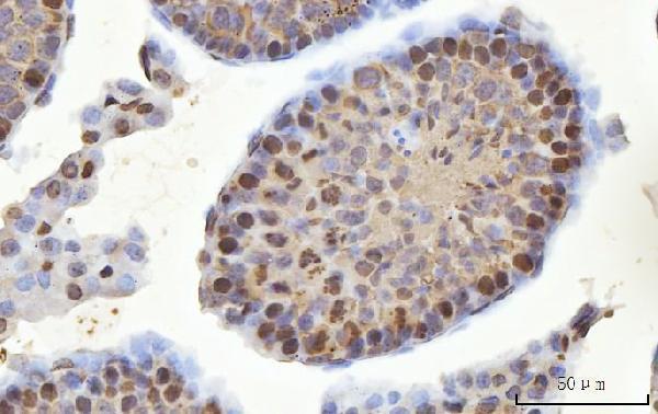
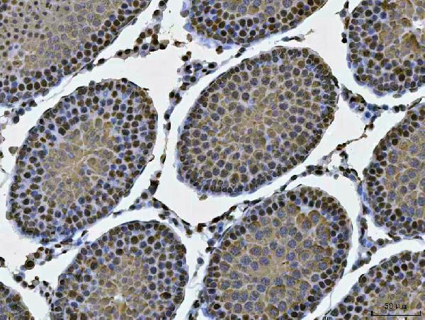
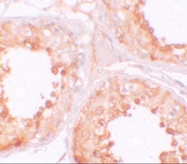
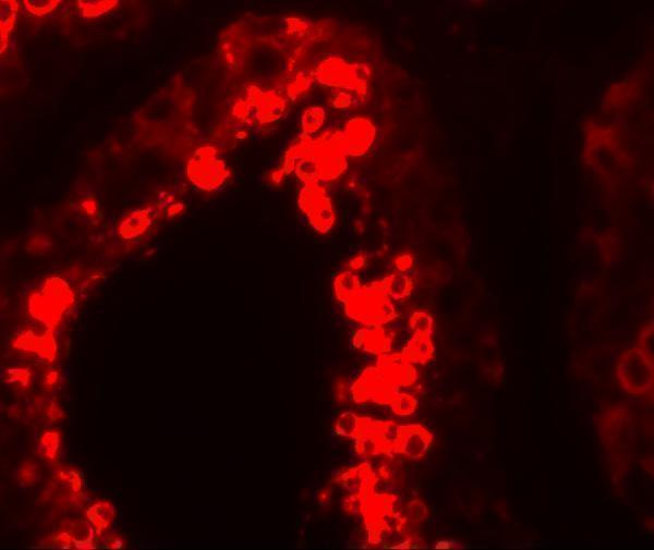
Both catalog antibodies have testis IHC images: human for A08610 (A08610 IHC image caption) and mouse and rat for A08610-2 (A08610-2 IHC image captions). A08610 also has a human testis IF image (A08610 IF image caption).

Real IHC data IHC analysis of MSY2/YBOX2/YBX2 using anti-MSY2/YBOX2/YBX2 antibody (A08610-2). MSY2/YBOX2/YBX2 was detected in a paraffin-embedded section of mouse testis tissue. Heat mediated antigen retrieval was performed in EDTA buffer (pH 8.0, epitope retrieval solution). The tissue section was blocked with 10% goat serum. The tissue section was then incubated with 2 μg/ml rabbit anti-MSY2/YBOX2/YBX2 Antibody (A08610-2) overnight at 4°C. Peroxidase Conjugated Goat Anti-rabbit IgG was used as secondary antibody and incubated for 30 minutes at 37°C. The tissue section was developed using HRP Conjugated Rabbit IgG Super Vision Assay Kit (Catalog # SV0002) with DAB as the chromogen.
Anti-MSY2/YBOX2/YBX2 Antibody ®
Cat # A08610-2
Real IHC data Immunohistochemistry of YBX2 in human testis tissue with YBX2 antibody at 10 μg/mL.
Anti-Y-box-binding protein 2 YBX2 Antibody
Cat # A08610

A08610 has IHC and IF images from human testis and lists human, mouse and rat reactivity (A08610 image captions; A08610 catalog applications and reactivity). A08610-2 has IHC images from mouse and rat testis and lists human, mouse and rat reactivity (A08610-2 IHC image captions; A08610-2 catalog reactivity).

Which to pick: For paraffin-section IHC in mouse or rat testis, choose A08610-2: its captions document paraffin sections, EDTA retrieval at pH 8.0, and 2 μg/ml primary antibody; the fixative is unreported (A08610-2 IHC image captions). For human testis IHC, choose A08610 at 10 μg/mL; its IHC caption does not report section processing or fixative (A08610 IHC image caption). For IF, choose A08610, which has a human testis IF image at 20 μg/mL; ICC is not documented, and its listed mouse and rat reactivity does not establish IHC or IF validation in those species (A08610 IF image caption; A08610 catalog applications and reactivity).

Each figure is that product's own IHC / IF validation image from its datasheet.

References

  1. UniProt Consortium. UniProt entry Q9Y2T7 (YBOX2_HUMAN, Y-box-binding protein 2).
  2. Human Protein Atlas. YBX2 tissue IHC expression (reliability: Enhanced).
  3. Human Protein Atlas. YBX2 subcellular location (ICC-IF): Highest expression in MCF-7: 12.3 nTPM.
  4. Human Protein Atlas. YBX2 antibody validation summary (3 antibodies).
  5. Analgesic exposure in pregnant rats affects fetal germ cell development with inter-generational reproductive consequences. Scientific reports 2016 — PMC4728385.
  6. YBX2 and cancer testis antigen 45 contribute to stemness, chemoresistance and a high degree of malignancy in human endometrial cancer. Scientific reports 2021 — PMC7893073.
  7. PAPOLB/TPAP regulates spermiogenesis independently of chromatoid body-associated factors. The Journal of reproduction and development 2018 — PMC5830355.
  8. Acute Dietary Zinc Deficiency Impairs Sperm Motility but Not Gonadal mRNA Methylation in Mice. Molecular reproduction and development 2026 — PMC13501185.
  9. PubMed PMID:10100484 — UniProt-cited evidence.
  10. PubMed PMID:15489334 — UniProt-cited evidence.
  11. PubMed PMID:16479255 — UniProt-cited evidence.