This website uses cookies to ensure you get the best experience on our website.


- Table of Contents
Plan YWHAH paraffin IHC around cytoplasmic staining and high signals in Purkinje cells and neuropil (HPA tissue IHC). Keep fixation consistent, and interpret staining cautiously because antibody staining has low concordance with RNA expression (HPA tissue IHC).

Expected localisation, validated protocols, controls and antibodies — the at-a-glance facts below, then the full design guide.
| Expected localisation | Cytoplasmic staining in several tissues (HPA tissue IHC) | |
| Staining pattern | Cytoplasmic staining; high in Purkinje cells and neuropil (HPA tissue IHC) | |
| Antigen retrieval | EDTA pH 8.0 HIER, heat-mediated (datasheet A04219-2) | |
| Positive control | Cerebellum+4 more · see all | |
| Negative control | Duodenum+4 more · see all |
| Fixation | Keep fixation consistent across sections (standard IHC practice; not target-specific). Selected-image fixative and duration unreported (datasheet A04219-2); verify before use. | |
| Caveat | Antibody staining has low concordance with RNA expression (HPA tissue IHC) | |
| Regulation | Mainly expressed in brain (UniProt) | |
| Isoform / epitope | No annotated isoforms; chain spans residues 2–246 (UniProt) |
The catalog antibody’s IHC-P protocol (datasheet: A04219-2) is accompanied by one published YWHAH tissue IHC protocol (PMC12549071: IHC methods).
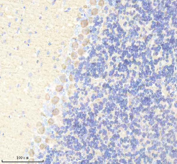
| Sample | Paraffin-embedded mouse cerebellum tissue; fixative not specified (datasheet A04219-2) |
| Fixation | Image fixative and duration unreported (datasheet A04219-2); verify before use. |
| Sectioning | 4–5 µm sections on charged slides (standard) |
| Deparaffinisation | Xylene, graded ethanol series to water (standard) |
| Antigen retrieval | Heat retrieval: EDTA pH 8.0 (datasheet A04219-2); 20 min, 95–100 °C (standard) |
| Peroxidase block | 3% H2O2, 10 min, room temperature (standard) |
| Blocking | 10% goat serum (datasheet A04219-2) |
| Primary antibody | Rabbit anti-YWHAH, 2-5 μg/ml (datasheet A04219-2) |
| Primary incubation | Overnight at 4 °C (datasheet A04219-2) |
| Detection | HRP-conjugated secondary, DAB chromogen (datasheet A04219-2) |
| Counterstain | Hematoxylin, blue, dehydrate and mount (standard) |
| Expected result | YWHAH-positive staining in purkinje cells of cerebellum (HPA tissue IHC: High). HPA tissue profile: Cytoplasmic expression in a number of tissues. No signal in the no-primary control. |
YWHAH should appear mainly cytoplasmic in tissue sections, with strong staining in cerebellar Purkinje cells and cerebral cortex neuropil (HPA tissue IHC: High in both). It has no transmembrane segment (UniProt Q04917 topology). Use these patterns as guides, not absolute pass criteria: HPA rates tissue IHC reliability Uncertain because antibody staining and RNA expression show low consistency (HPA tissue IHC: Uncertain).
| Strong cytoplasmic staining in Purkinje cells; strong signal in cerebral cortex neuropil. | This matches the reported high-signal sites (HPA tissue IHC: High in Purkinje cells and neuropil). Compare stained structures with the counterstain and tissue morphology. HPA’s Uncertain tissue IHC rating means a matching pattern supports interpretation but does not, by itself, establish antibody specificity (HPA tissue IHC: Uncertain). |
| Staining appears only at cell borders or as a dominant nuclear signal in tissue. | Recheck localisation and morphology: the reported tissue pattern is cytoplasmic (HPA tissue IHC: cytoplasmic expression), and YWHAH has no transmembrane segment (UniProt Q04917 topology). A nuclear signal alone is not proof of artefact, since additional nuclear locations are reported in ICC-IF (HPA subcellular: nucleoplasm and nucleoli rim). |
| Strong staining appears in a cell population reported as undetected, while expected positive cells are weak. | Possible explanations include cross-reactivity or endogenous detection activity; check the no-primary control and the location of signal within each cell. For example, HPA reports no detection in lymph node germinal center cells, but that observation is an uncertain IHC reference rather than an absolute negative standard (HPA tissue IHC: Not detected; Uncertain). |
| Colour is diffuse across the section, including spaces outside cells or unrelated structures. | This does not resolve the cytoplasmic pattern reported for YWHAH (HPA tissue IHC: cytoplasmic expression). In general IHC practice, excess primary or secondary reagent, inadequate blocking or washing, and detection-system background can obscure localisation. Compare with a no-primary control before assigning the diffuse colour to YWHAH. |
| Purkinje cells or cerebral cortex neuropil show no detectable stain. | These are useful positive reference structures because HPA reports High staining there (HPA tissue IHC: High in Purkinje cells and neuropil). First check that the intended structures are present, then review the antibody’s IHC-P conditions, retrieval, detection reagents and control results. HPA’s Uncertain rating prevents a negative result from being interpreted alone as target absence (HPA tissue IHC: Uncertain). |
| Tissue and cell selection | Brain is a useful starting point because UniProt reports expression mainly there, and HPA reports High staining in Purkinje cells and cortex neuropil (UniProt Q04917 tissue specificity; HPA tissue IHC: High). Medium staining in several other tissues and undetected staining in selected cell populations make comparisons cell-specific (HPA tissue IHC: Medium; Not detected). |
| Compartment and topology | Expect the reported cytoplasmic tissue pattern, while interpreting borders against cell morphology (HPA tissue IHC: cytoplasmic expression; UniProt Q04917 topology: no transmembrane segment). UniProt gives no subcellular annotation for YWHAH, so it does not independently validate a precise tissue compartment (UniProt Q04917 subcellular record: not annotated). |
| Strength of IHC evidence | HPA rates tissue IHC Uncertain because antibody staining has low consistency with RNA expression (HPA tissue IHC: Uncertain). The listed antibody CAB025918 also has Uncertain IHC status; HPA061603 has Supported ICC status, which is a separate application assessment (HPA antibodies: CAB025918 IHC Uncertain; HPA061603 ICC Supported). |
| IF/ICC question: can nuclear signal occur? | Yes. HPA reports mainly cytosolic localisation, with additional nucleoplasm and nucleoli-rim signal in ICC-IF (HPA subcellular: cytosol, nucleoplasm, nucleoli rim). Treat that as IF context, not a tissue IHC acceptance rule; HPA cautions that the subcellular summary uses antibodies targeting proteins from multiple genes (HPA subcellular: caution). |
| Situation | Likely cause | Next action |
|---|---|---|
| Expected positive brain structures are blank. | The relevant cells may be absent from the section, or an IHC workflow step may have failed; HPA reports High staining in Purkinje cells and cortex neuropil (HPA tissue IHC: High). | Confirm structures on the counterstain and compare a known-positive section processed in the same run. In general IHC practice, verify the antibody’s stated IHC-P conditions, retrieval, primary incubation and detection reagents before changing interpretation. |
| Signal is weak in a tissue chosen as a positive control. | The sampled cell population may differ from the reported positive population; HPA levels refer to specific cells, not whole organs (HPA tissue IHC: named cells and levels). | Identify the reported cells first. Prefer Purkinje cells or cortex neuropil for a high-signal comparison, and do not expect the same intensity from sites listed as Medium (HPA tissue IHC: High; Medium). |
| Background covers cells and extracellular areas. | General IHC causes include insufficient blocking or washing, concentrated reagents, or endogenous detection activity. A diffuse stain cannot be assigned to the reported cytoplasmic YWHAH pattern (HPA tissue IHC: cytoplasmic expression). | Inspect the no-primary control, then adjust blocking, washes or detection controls according to the assay chemistry. Reassess cell boundaries and intracellular signal after background is reduced. |
| Only nuclei stain in an IHC section. | Tissue IHC is reported as cytoplasmic, although ICC-IF also shows nuclear locations (HPA tissue IHC: cytoplasmic expression; HPA subcellular: additional nucleoplasm and nucleoli rim). | Check the counterstain, no-primary control and positive tissue comparison. Describe reproducible nuclear staining separately; do not score it as the expected tissue pattern solely on the basis of ICC-IF. |
| A reported undetected cell population stains strongly. | Cross-reactivity or endogenous detection activity is possible; HPA’s undetected calls are uncertain tissue IHC observations, not definitive absence (HPA tissue IHC: Not detected; Uncertain). | Confirm the cell identity and compartment, inspect the no-primary control, and compare the result with a reported positive population in the same run. Treat persistent discordance as unresolved specificity evidence. |
| Two tissue sections give conflicting intensity calls. | HPA’s tissue IHC profile has low agreement with RNA expression, and its calls distinguish particular cell populations (HPA tissue IHC: Uncertain; named cells and levels). | Compare the same cell type and compartment in matched staining runs; record section quality and control results. Keep the discrepancy visible in the interpretation instead of treating an HPA level as a fixed threshold. |




Comprehensive Human Protein Atlas IHC scoring per tissue (reliability: Uncertain — Low consistency between antibody staining and RNA expression data.). Rows are taken directly from the HPA tissue chart — click any row's HPA link to view the source.
| Tissue | Cell type | Level | Evidence | Source |
|---|---|---|---|---|
| Cerebellum | Purkinje cells | High | Protein (IHC) | HPA → |
| Cerebral cortex | Neuropil | High | Protein (IHC) | HPA → |
| Adipose tissue | Adipocytes | Medium | Protein (IHC) | HPA → |
| Appendix | Glandular cells | Medium | Protein (IHC) | HPA → |
| Bone marrow | Hematopoietic cells | Medium | Protein (IHC) | HPA → |
| Tissue | Cell type | Level | Evidence | Source |
|---|---|---|---|---|
| Duodenum | Glandular cells | Not detected | Protein (IHC) | HPA → |
| Esophagus | Squamous epithelial cells | Not detected | Protein (IHC) | HPA → |
| Lymph node | Germinal center cells | Not detected | Protein (IHC) | HPA → |
| Oral mucosa | Squamous epithelial cells | Not detected | Protein (IHC) | HPA → |
| Seminal vesicle | Glandular cells | Not detected | Protein (IHC) | HPA → |
Troubleshoot YWHAH staining in paraffin sections by comparing retrieval, controls, cell type and compartment before interpreting chromogenic signal.
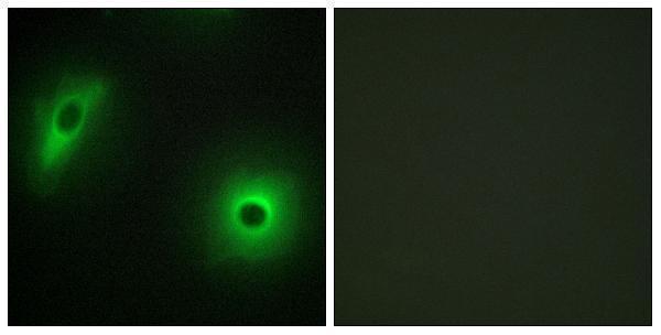
The catalog includes YWHAH antibodies with IHC images from paraffin sections of mouse and rat cerebellum and human stomach, plus IF images from cells and rat cerebellum (catalog image captions).
A04219-2 has IHC images from paraffin sections of mouse and rat cerebellum and human stomach, and IF images from cells and rat cerebellum (A04219-2 image captions). A04219 lists IHC and IF/ICC applications and has a peptide-blocked IF image from HeLa cells (A04219 catalog entry and image caption).
Which to pick: Choose A04219-2 for tissue IHC: its images document paraffin sections with EDTA retrieval at pH 8.0 and 2 μg/ml primary antibody; the fixative is unreported (A04219-2 IHC image captions). Choose A04219 for cell IF/ICC when the peptide-blocked HeLa IF image is useful evidence; its listed IF dilution is 1:200–1:1000 (A04219 catalog entry and image caption). For broader listed species reactivity, A04219-2 includes human, monkey, mouse and rat, although its IHC images document human, mouse and rat only (A04219-2 catalog entry and IHC image captions).