ZBTB21 / Zinc finger and BTB domain-containing protein 21 · IHC design guide

Design Immunohistochemistry for ZBTB21

Plan ZBTB21 IHC around the nuclear staining reported in several tissues (HPA tissue IHC). Use high staining in parathyroid glandular and placental decidual cells to guide controls, and optimize the catalog antibody within its 1:100–1:300 IHC dilution range (HPA tissue IHC; datasheet).

Evidence assembled Oct 2026 · For research use; verify linked source records and product datasheet before use
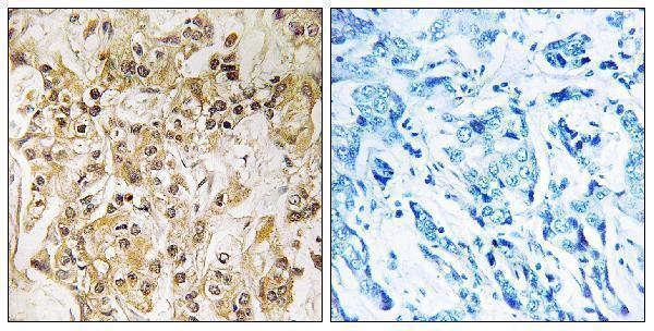
Immunohistochemistry protocol sheet for ZBTB21 (IHC for ZBTB21): expected localisation Nuclear staining in several tissues (HPA tissue IHC), antibody A30772, validated IHC image, and IHC protocol steps
Printable ZBTB21 IHC protocol sheet — expected localisation Nuclear staining in several tissues (HPA tissue IHC), antibody A30772, controls and protocol steps. Open the full ZBTB21 IHC guide →

ZBTB21 Immunohistochemistry Experimental Design Guide

Expected localisation, validated protocols, controls and antibodies — the at-a-glance facts below, then the full design guide.

Must know before staining
Expected localisation Nuclear staining in several tissues (HPA tissue IHC)
Staining pattern Nuclear signal across several tissue cell types (HPA tissue IHC)
Antigen retrieval Tris-EDTA pH 9.0 HIER, 95–98 °C, 20 min (rule: nuclear antigen)
Positive control ⓘ Nasopharynx+4 more · see all
Negative control ⓘ Caudate+4 more · see all
Important caveats
Reasons your staining may differ from the expected pattern.
Fixation Matched tissue-IHC evidence does not establish the fixation claim. Validate the specimen-specific method before use. (selected-SKU IHC image A30772); effects on ZBTB21 are unclear.
Caveat Staining–RNA agreement has medium consistency (HPA tissue IHC)
Regulation Bone marrow–enhanced RNA (HPA tissue RNA)
Isoform / epitope 2 isoforms; verify antibody epitope coverage (UniProt)
Section 1

Recommended ZBTB21 IHC & IF Protocols

The catalog antibody’s IHC-P protocol is accompanied by published ZBTB21 IHC methods for colorectal specimens (PMC13612771) and a lung adenocarcinoma tissue microarray (PMC13088348).

Recommended immunohistochemistry (IHC-P) protocol parameters
SampleParaffin-embedded human breast carcinoma tissue; fixative not specified (datasheet A30772)
FixationImage fixative and duration unreported (datasheet A30772); verify before use.
Sectioning4–5 µm sections on charged slides (standard)
DeparaffinisationXylene, graded ethanol series to water (standard)
Antigen retrievalHeat-induced epitope retrieval in Tris-EDTA buffer, pH 9.0, 20 min at 95–98 °C (standard rule: nuclear antigen)
Peroxidase block3% H2O2, 10 min, room temperature (standard)
Blocking10% normal serum of the secondary host, 30 min, room temperature (standard)
Primary antibodyRabbit anti-ZBTB21, 1:100 - 1:300 (datasheet A30772)
Primary incubationOvernight at 4 °C (standard)
DetectionHRP-polymer secondary, DAB chromogen 5–10 min (standard)
CounterstainHematoxylin, blue, dehydrate and mount (standard)
Expected resultZBTB21-positive staining in ciliated cells (cell body) of nasopharynx (HPA tissue IHC: High). HPA tissue profile: Nuclear expression in several tissues. No signal in the no-primary control.
💡Decision noteStart with Tris-EDTA pH 9.0 retrieval at 95–98 °C for 20 min (page retrieval); the colorectal study also used alkaline EDTA retrieval (PMC13612771).
Section 2

What Is the Expected ZBTB21 Staining Pattern?

ZBTB21 is a nuclear protein with no transmembrane segment (UniProt Q9ULJ3: localization and topology). For paraffin-section IHC, expect staining in nuclei of selected cell populations: ciliated cells in nasopharynx, parathyroid glandular cells, and placental decidual cells show high staining (HPA tissue IHC). HPA describes nuclear expression in several tissues and rates its tissue profile Enhanced, with medium consistency between staining and RNA data (HPA tissue IHC).

What am I looking at on my slide?
Distinct nuclear staining in nasopharyngeal ciliated cells, parathyroid glandular cells, or placental decidual cells.This fits the reported high cell-level staining and overall nuclear profile (HPA tissue IHC). Judge the named cells separately from surrounding tissue; a positive tissue does not mean every cell should stain.
Strong cytoplasmic-only, membranous, or extracellular staining with little nuclear signal.This conflicts with the nuclear localization and absence of a transmembrane segment (UniProt Q9ULJ3) and the mainly nucleoplasmic IF pattern (HPA ICC-IF). Treat it as a possible IHC artifact until controls and antibody specificity are checked.
Prominent staining in glia, cholangiocytes, or salivary glandular cells.These cell types were not detected in the listed HPA tissue samples (HPA tissue IHC). Check cell identification, cross-reactivity, and endogenous detection activity; the HPA result is a reference pattern, not proof that every specimen must be negative.
Diffuse color across nuclei, cytoplasm, and tissue spaces without clear cell boundaries.A widespread deposit is difficult to assign to ZBTB21's expected nuclear compartment (UniProt Q9ULJ3; HPA tissue IHC). In general chromogenic IHC practice, incomplete blocking, excessive reagent concentration, or inadequate washing can produce background.
No nuclear signal in a section containing a listed high cell population.That result disagrees with the HPA reference pattern for the identified cells (HPA tissue IHC). First verify that the cells are present and the detection run worked; the Enhanced profile still has medium staining-to-RNA consistency (HPA tissue IHC).
💡Expected ZBTB21 appearanceCall a convincing positive when the named cell population shows clear nuclear chromogen, with high staining expected in the three listed high-level populations (HPA tissue IHC); strong cytoplasmic-only or widespread cell-independent color is suspect against the nuclear reference (UniProt Q9ULJ3; HPA tissue IHC).
How each factor affects the staining
Cell population and tissueHPA reports high staining in three listed cell populations, medium in several others, low in others, and no detection in selected cells (HPA tissue IHC). Score the identified cell type rather than a whole-section average.
Antibody evidenceHPA024655 has Enhanced IHC validation; HPA031757 and HPA031758 have Enhanced ICC validation but no listed IHC status (HPA antibodies). Keep assay-specific validation separate when weighing a disputed IHC result.
Isoforms and antibody epitopeTwo ZBTB21 isoforms are listed (UniProt Q9ULJ3). Their relevant epitopes and relative tissue abundance are unspecified here, so this record cannot predict whether a given antibody detects both or explain an intensity difference.
IF/ICC Q: What compartment should appear?A: Mainly the nucleoplasm (HPA ICC-IF: enhanced). This supports a localization cross-check for IHC, while the IF images do not set a chromogenic intensity threshold or supply an IHC protocol.
Why is my staining missing, weak or wrong?
SituationLikely causeNext action
Listed positive cells are present, but their nuclei are unstained.The result differs from the reported high staining in those cells (HPA tissue IHC); failed detection or unsuitable assay conditions are possible general IHC causes.Confirm cell identity and a working positive control. As general IHC practice, review retrieval, primary dilution, incubation, and detection against the antibody's IHC-P instructions; no ZBTB21-specific retrieval effect is established here.
Color is strongest outside nuclei.A cytoplasmic-only or membranous pattern conflicts with nuclear localization (UniProt Q9ULJ3) and the HPA nuclear tissue profile (HPA tissue IHC).Inspect the nuclear counterstain and morphology, then compare a validated positive tissue and detection controls. Reassess antibody specificity if the outside-nucleus pattern persists.
Glia or cholangiocytes stain strongly.HPA lists these cells as not detected in its sampled tissues (HPA tissue IHC); cell misidentification, cross-reactivity, or endogenous detection activity may account for the discrepancy.Confirm the cell type and review appropriate negative and detection-only controls. Interpret the HPA comparison at cell level, since its sampled result does not establish a universal absence.
Chromogen covers broad areas or the whole section.Diffuse deposit obscures the expected nuclear distribution (UniProt Q9ULJ3; HPA tissue IHC). Excess primary or detection reagent, incomplete blocking, or insufficient washing are general IHC possibilities.Use general IHC controls to locate the background source; optimize blocking, washes, and reagent concentration, then reassess whether signal resolves into identifiable nuclei.
A low-level population appears unstained.HPA lists alveolar cells, intestinal glandular cells, cardiomyocytes, and some other populations as low (HPA tissue IHC). Weak staining can be hard to distinguish from background in a chromogenic assay.Assess a listed high-level population in the same run before calling the assay unsuccessful. Record low-level cells separately, and avoid treating a barely visible result as a reliable negative.
Different antibodies give different nuclear intensities.The record lists two isoforms (UniProt Q9ULJ3), but provides no epitope mapping or isoform-specific tissue pattern. HPA also reports different IHC and ICC validation statuses across its antibodies (HPA antibodies).Compare like-for-like IHC conditions and the same named cell population. Check each antibody's documented IHC validation and epitope information before attributing the difference to ZBTB21 biology.

Sample controls for ZBTB21 IHC & IF

🧪Run parathyroid gland first; its glandular cells should stain (HPA: High in parathyroid glandular cells). Use salivary gland glandular cells as the biological tissue negative (HPA: Not detected in salivary gland glandular cells); cells without specific staining on the positive slide should remain at background, but the supplied evidence does not establish a named internal negative cell type.
Positive control tissue: Nasopharynx (Ciliated cells (cell body), HPA High)
Negative control tissue: Caudate (HPA Not detected)
ICC-IF cell lines (HPA subcellular resource): HPA ICC-IF images show ZBTB21 in A-431, U-251MG, U2OS, HEK293, MCF-7, KOLF2.1J, with annotated localisation: Nucleoplasm (enhanced) (HPA subcellular).
Technical controls: Include a no-primary, secondary-only control and an isotype control matched to the primary antibody’s host species, immunoglobulin class, and clonality; confirm specificity with a ZBTB21 knockout or a peptide block (A30772 tissue-IHC caption: staining was blocked with synthesized peptide). For chromogenic IHC, block endogenous peroxidase and check the parathyroid section for background signal under the chosen detection system.
⚠️Feasibility: A target-specific fixation window and retrieval dependency are unreported in the supplied evidence; optimize antigen retrieval for paraffin sections using the positive and negative controls. The selected A30772 paraffin-section caption does not report a fixative. Frozen sections and IF cannot be judged easier from these data; IF/ICC images show mainly nucleoplasmic localization (HPA: Nucleoplasm enhanced), and no parathyroid-specific artefact is established by the supplied evidence.

HPA tissue IHC evidence for ZBTB21

Comprehensive Human Protein Atlas IHC scoring per tissue (reliability: Enhanced — Medium consistency between antibody staining and RNA expression data.). Rows are taken directly from the HPA tissue chart — click any row's HPA link to view the source.

Positive expression · recommended positive controls

TissueCell typeLevelEvidenceSource
Nasopharynx Ciliated cells (cell body) High Protein (IHC) HPA →
Parathyroid gland Glandular cells High Protein (IHC) HPA →
Placenta Decidual cells High Protein (IHC) HPA →
Stomach Glandular cells Medium Protein (IHC) HPA →
Adipose tissue Adipocytes Medium Protein (IHC) HPA →

Undetected expression · recommended negative controls

TissueCell typeLevelEvidenceSource
Caudate Glial cells Not detected Protein (IHC) HPA →
Hippocampus Glial cells Not detected Protein (IHC) HPA →
Liver Cholangiocytes Not detected Protein (IHC) HPA →
Ovary Follicle cells Not detected Protein (IHC) HPA →
Salivary gland Glandular cells Not detected Protein (IHC) HPA →
Section 3

Advanced ZBTB21 IHC Tips

Troubleshoot ZBTB21 staining in paraffin sections by checking retrieval, nuclear localisation, controls and cell-specific scoring before interpreting signal (UniProt Q9ULJ3; HPA tissue IHC).

How should I retrieve ZBTB21 in paraffin sections when nuclear staining is weak?
Start with Tris-EDTA pH 9.0 heat-induced epitope retrieval at 95–98 °C for 20 min (page retrieval specification). Allow sections to cool in retrieval buffer, then compare staining with a known positive section in the same run (standard IHC practice; HPA: nuclear expression in several tissues). If nuclei remain weak, check heating uniformity and primary-antibody incubation before testing a different retrieval condition on matched sections (standard IHC practice). Excessively harsh retrieval can damage tissue morphology and make nuclear scoring unreliable, so assess staining and morphology together (standard IHC practice). The selected catalog image establishes peptide-blockable staining in paraffin-embedded breast carcinoma, but its caption gives no retrieval conditions (caption: A30772).
Could fixation explain inconsistent ZBTB21 staining between paraffin blocks?
Target-specific ZBTB21 sensitivity to fixation is unknown from the supplied evidence; the selected paraffin-section caption does not state the fixative (caption: A30772). Record each block’s fixative, fixation duration and processing history, then compare sections processed and stained together (standard IHC practice). Uneven or prolonged fixation can alter epitope accessibility in IHC generally, so vary retrieval on matched sections while monitoring tissue morphology (standard IHC practice). Include a reference section in each run to separate block effects from staining-run variation (standard IHC practice). Do not attribute a weak result to a particular ZBTB21 modification or tissue pattern without a direct comparison (UniProt Q9ULJ3: modified residues; HPA: tissue IHC profile).
What should I check if ZBTB21 staining appears predominantly cytoplasmic?
Prioritise nuclear staining when evaluating ZBTB21: UniProt places it in the nucleus, and HPA reports mainly nucleoplasmic localisation (UniProt Q9ULJ3: subcellular location; HPA: subcellular summary). Score nuclei separately from cytoplasm, using a counterstain to define cell boundaries and nuclear integrity (standard IHC practice). Diffuse cytoplasmic chromogen without a corresponding nuclear signal warrants checks of antibody concentration, secondary-only background and tissue pigment (standard IHC practice). The protein has no transmembrane segment, so a membrane-restricted pattern would also require independent verification (UniProt Q9ULJ3: topology). Compare the suspect section with a positive tissue and peptide-blocked control where available; the selected catalog image includes a peptide-blocked panel (caption: A30772).
Can an epitope or isoform difference account for patchy nuclear staining?
ZBTB21 has 2 listed isoforms and a BTB domain at residues 30–96, but the supplied evidence does not map the catalog antibody’s epitope (UniProt Q9ULJ3: isoforms and domains; supplied catalog evidence). Do not assign a staining difference to either isoform or to a specific phosphorylation site without epitope mapping and a direct test (UniProt Q9ULJ3: isoforms and modified residues). If staining is patchy, first compare intact nuclei, retrieval consistency and section quality across the slide (standard IHC practice). An antibody raised against a shared sequence could detect both isoforms, whereas an isoform-specific sequence could change coverage; confirm the actual immunogen before making either claim (standard antibody practice).
How can I investigate an IHC localisation discrepancy with multiplex IF?
Use IF as a separate localisation check, pairing ZBTB21 with a validated marker for the expected cell type and a nuclear counterstain (standard IF practice; HPA: high staining in nasopharyngeal ciliated cells and parathyroid glandular cells). HPA reports mainly nucleoplasmic ZBTB21 and lists ICC/IF images, so assess overlap within identified nuclei rather than whole-cell fluorescence alone (HPA: subcellular summary and ICC/IF image list). Choose a far-red fluorophore when tissue autofluorescence is prominent, and inspect unstained and single-label controls before interpreting multiplex overlap (standard IF practice). Because ZBTB21 lacks a transmembrane segment and is nuclear, permeabilise sufficiently for antibody access to the nuclear epitope while preserving nuclear morphology (UniProt Q9ULJ3: topology and location; standard IF practice).
How do I distinguish nonspecific chromogen from genuine ZBTB21 staining?
Examine a no-primary control and an appropriate negative tissue alongside the test section to locate reagent and tissue background (standard IHC practice). Block endogenous peroxidase before chromogenic detection, and inspect pigment or precipitate before treating brown deposits as specific DAB signal (standard IHC practice). Interpret the selected catalog peptide-blocked panel as evidence that its paraffin-section staining can be competed by peptide, while recognising that the caption does not establish every tissue’s specificity (caption: A30772). HPA reports ZBTB21 as mainly nucleoplasmic; widespread extracellular deposits or uniform staining across unrelated compartments should prompt troubleshooting (HPA: subcellular summary; standard IHC practice). Reduce overly concentrated primary antibody or detection reagent only after comparing controls processed in the same run (standard IHC practice).
How should I quantify ZBTB21 IHC across samples with different cell composition? ⚠ ANSWER MARKED FOR VERIFICATION
Define the cell population and count only interpretable nuclei within matched tissue regions, excluding folds, necrosis and damaged edges (standard IHC practice; HPA: nuclear expression in several tissues). Report both the percentage of positive nuclei and intensity categories; an H-score sums percentage at each intensity multiplied by its 0–3 score, giving a 0–300 range (standard IHC scoring practice). Normalise positive counts to eligible cells of the same type, or report density per mm² of the defined region when cell counts are impractical (standard IHC practice). Keep retrieval, detection, exposure and scoring thresholds consistent across groups (standard IHC practice). HPA’s variable cell-type staining makes a whole-section average sensitive to differences in tissue composition (HPA: tissue IHC profile).
When should I question an apparently positive ZBTB21 IHC result?
A convincing result should show signal in intact nuclei of the evaluated cells, consistent with nuclear and mainly nucleoplasmic localisation (UniProt Q9ULJ3: location; HPA: subcellular summary). Check cell identity before generalising across a tissue: HPA reports high staining in parathyroid glandular cells but no detected staining in salivary glandular cells (HPA: tissue IHC profile). Suspect artefact when signal is confined to section edges, necrotic areas or damaged cells, or appears in the no-primary control (standard IHC practice). Endogenous peroxidase activity can also create misleading chromogen if blocking is inadequate (standard IHC practice). The catalog peptide-blocked breast carcinoma image supports that specific comparison, while broader biological interpretation needs matched controls and cell-level scoring (caption: A30772; standard IHC practice).
Boster reagents

Best ZBTB21 / Zinc finger and BTB domain-containing protein 21 IHC Antibodies

A30772 has a human paraffin-section IHC image (image caption). IF/ICC applications and Human/Mouse reactivity are listed (catalog); no IF image is supplied (catalog).

Real IHC data Immunohistochemistry analysis of paraffin-embedded human breast carcinoma tissue, using ZNF295 Antibody. The picture on the right is blocked with the synthesized peptide.
Anti-ZNF295 ZBTB21 Antibody
Cat # A30772

A30772 is listed for IHC, IF and ICC in Human and Mouse samples (catalog). Its IHC image shows a paraffin-embedded human breast carcinoma section with peptide blocking (image caption); no IF image is supplied (catalog).

Which to pick: For tissue IHC, choose A30772: this rabbit polyclonal antibody lists an IHC dilution of 1:100–1:300 (catalog), and its image shows a paraffin-embedded human section (image caption); the fixative is unreported (image caption). For IF/ICC, A30772 lists both applications and an IF dilution of 1:50 (catalog). For cross-species work, A30772 lists Human and Mouse reactivity (catalog), while its depicted IHC evidence is limited to human tissue (image caption).

Each figure is that product's own IHC / IF validation image from its datasheet.

References

  1. UniProt Consortium. UniProt entry Q9ULJ3 (ZBT21_HUMAN, Zinc finger and BTB domain-containing protein 21).
  2. Human Protein Atlas. ZBTB21 tissue IHC expression (reliability: Enhanced).
  3. Human Protein Atlas. ZBTB21 subcellular location (ICC-IF): Mainly localized to the nucleoplasm..
  4. Human Protein Atlas. ZBTB21 antibody validation summary (3 antibodies).
  5. Integrative single-cell and multi-omics analysis of ZBTB21-mediated serine metabolism in colorectal cancer: from metabolic reprogramming to immune microenvironment modulation. Cancer immunology, immunotherapy : CII 2026 — PMC13612771.
  6. ZBTB21 Is a Dual Suppressor of Pyroptosis and MHC-I Antigen Presentation That Promotes Tumor Immune Evasion. Advanced science (Weinheim, Baden-Wurttemberg, Germany) 2026 — PMC13088348.
  7. PubMed PMID:15629158 — UniProt-cited evidence.
  8. PubMed PMID:10574462 — UniProt-cited evidence.
  9. PubMed PMID:10830953 — UniProt-cited evidence.