This website uses cookies to ensure you get the best experience on our website.


- Table of Contents
Plan chromogenic paraffin IHC around the predominantly nuclear ZBTB7B pattern, with bone marrow hematopoietic cells and adipocytes as high-staining and undetected comparators (HPA tissue IHC). Catalog antibody M06831 lists a human IHC dilution of 1:200–1:1000 (datasheet).

Expected localisation, validated protocols, controls and antibodies — the at-a-glance facts below, then the full design guide.
| Expected localisation | Predominantly nuclear staining (HPA tissue IHC) | |
| Staining pattern | Nuclear staining in most tissue cell types (HPA tissue IHC) | |
| Antigen retrieval | Tris-EDTA pH 9.0 HIER, 95–98 °C, 20 min (rule: nuclear antigen) | |
| Positive control | Adrenal gland+4 more · see all | |
| Negative control | Adipose tissue+4 more · see all |
| Fixation | Keep fixation consistent across sections (standard IHC practice; not target-specific) | |
| Caveat | Antibody staining shows low consistency with RNA data (HPA tissue IHC) | |
| Regulation | Low tissue specificity at RNA level (HPA tissue RNA) | |
| Isoform / epitope | Two isoforms; epitope coverage is unclear (UniProt) |
The catalog antibody’s IHC-P protocol is followed by two published ZBTB7B IHC methods (PMC12686175; PMC9235105).
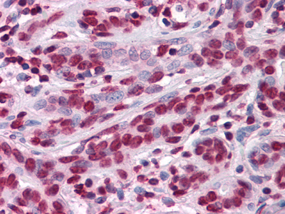
| Sample | Paraffin-embedded Human Breast tissues; fixative not specified (datasheet M06831) |
| Fixation | Image fixative and duration unreported (datasheet M06831); verify before use. |
| Sectioning | 4–5 µm sections on charged slides (standard) |
| Deparaffinisation | Xylene, graded ethanol series to water (standard) |
| Antigen retrieval | Heat-induced epitope retrieval in Tris-EDTA buffer, pH 9.0, 20 min at 95–98 °C (standard rule: nuclear antigen) |
| Peroxidase block | 3% H2O2, 10 min, room temperature (standard) |
| Blocking | 10% normal serum of the secondary host, 30 min, room temperature (standard) |
| Primary antibody | Mouse monoclonal (clone 7C12) anti-ZBTB7B, 1:200-1:1000 (datasheet M06831) |
| Primary incubation | Overnight at 4 °C (standard) |
| Detection | HRP-polymer secondary, DAB chromogen 5–10 min (standard) |
| Counterstain | Hematoxylin, blue, dehydrate and mount (standard) |
| Expected result | ZBTB7B-positive staining in glandular cells of adrenal gland (HPA tissue IHC: High). HPA tissue profile: Nuclear expression in most tissues. No signal in the no-primary control. |
ZBTB7B is a nuclear protein with no transmembrane segment (UniProt O15156 topology; UniProt O15156 subcellular location). In paraffin section IHC, expect nuclear staining in selected cells across many tissues, including glandular cells in colon and hematopoietic cells in bone marrow (HPA: nuclear expression in most tissues; HPA: High in colon glandular cells and bone marrow hematopoietic cells). Interpret tissue comparisons cautiously: HPA rates tissue IHC Enhanced but reports low consistency with RNA data and pending external verification (HPA tissue IHC reliability).
| Distinct nuclear chromogen in colon glandular cells or bone marrow hematopoietic cells, with nuclei identifiable on the counterstain (HPA: High in both cell populations). | This matches the reported compartment and positive cell populations (UniProt O15156: nucleus; HPA: nuclear expression in most tissues). Compare signal with a negative control before calling a sample positive (general IHC practice); the HPA tissue pattern awaits external verification (HPA tissue IHC reliability). |
| Predominantly cytoplasmic, membrane rim, or extracellular color while nuclei remain clear. | Those compartments conflict with the reported nuclear location and absence of a transmembrane segment (UniProt O15156 topology and subcellular location; HPA: nuclear expression). Review counterstain alignment and the negative control; a strong wrong-compartment signal warrants investigation as possible nonspecific staining (general IHC practice). |
| Strong color in hepatocytes, adipocytes, or glomerular cells, especially without nuclear restriction. | HPA reports ZBTB7B as Not detected in these respective cell populations (HPA: liver, adipose tissue, kidney). Check the cell identification and compare a negative control for antibody cross-reactivity or endogenous chromogenic activity (general IHC practice). A single unexpected sample does not by itself establish target expression. |
| Even haze across nuclei, cytoplasm, and empty slide areas obscures tissue boundaries. | A field-wide deposit does not resemble the reported nuclear pattern (HPA: nuclear expression in most tissues). Inspect the negative control and review blocking, washes, detection reagent, and chromogen development as general IHC background checks (general IHC practice); do not score the haze as ZBTB7B-positive cells. |
| No nuclear signal in a colon section's glandular cells or bone marrow section's hematopoietic cells. | This conflicts with HPA's High observations in those populations (HPA: colon; HPA: bone marrow), but tissue IHC has low consistency with RNA and awaits external verification (HPA tissue IHC reliability). Confirm that the intended cells are present, then check assay controls and routine IHC processing (general IHC practice). |
| Subcellular location and topology | ZBTB7B is nuclear and has no transmembrane segment (UniProt O15156). Score chromogen against the counterstained nuclei; a membrane pattern lacks support from the supplied localisation evidence (UniProt O15156; HPA: nuclear expression in most tissues). |
| Cell population within a tissue | High staining is reported for colon glandular cells and cerebellar Purkinje cells, while glial cells in caudate and hepatocytes are Not detected (HPA tissue IHC). Identify the specific cell population before assigning a tissue-wide result; HPA's entries do not make every cell in a positive tissue positive. |
| Interpretive confidence | Two rabbit polyclonal antibodies, HPA006811 and HPA025820, have IHC Enhanced status (HPA antibodies). The tissue profile still has low consistency between antibody staining and RNA data and is pending external verification (HPA tissue IHC reliability). Treat a matching stain as supportive evidence, not independent confirmation of specificity. |
| Isoforms and modifications | UniProt lists two isoforms and five modified residues, including phosphorylation and acetylation (UniProt O15156). The supplied records give no antibody epitope or isoform selectivity, so they cannot predict which isoform or modification a nuclear IHC signal represents. |
| Negative reference cells | Adipocytes, cardiomyocytes, and hepatocytes are reported Not detected in their listed tissues (HPA: adipose tissue, heart muscle, liver). These observations can help flag unexpected staining, but HPA's low antibody–RNA consistency limits their use as definitive biological negatives (HPA tissue IHC reliability). |
| IF/ICC Q&A: Where should signal appear? | In ICC-IF, HPA reports enhanced nucleoplasmic localisation and lists images from A-431, U-251MG, and U2OS (HPA subcellular; HPA antibodies: ICC Enhanced). This supports a nuclear interpretation for IF/ICC; the supplied record provides no IF/ICC protocol or dilution. |
| Situation | Likely cause | Next action |
|---|---|---|
| Positive tissue has no visible nuclear chromogen. | The expected cells may be absent from the section, or the IHC run may have failed; HPA reports High staining only for specified populations (HPA: colon glandular cells; HPA: bone marrow hematopoietic cells). | Locate the specified cells on the counterstain and check a concurrent positive control, antibody application, retrieval step, and detection reagents (general IHC practice). Do not infer ZBTB7B-specific fixation sensitivity; none is supplied. |
| Signal is mainly cytoplasmic or outlines cell membranes. | The compartment disagrees with nuclear localisation and the lack of a transmembrane segment (UniProt O15156; HPA: nuclear expression). Nonspecific staining or cell-boundary misreading is possible (general IHC practice). | Compare nuclear counterstain alignment and a negative control, then assess whether signal persists after routine antibody and detection checks (general IHC practice). Avoid scoring the wrong-compartment color as a positive nuclear result. |
| Hepatocytes, cardiomyocytes, or adipocytes show convincing color. | Those cells are reported Not detected in the corresponding HPA tissue entries (HPA: liver, heart muscle, adipose tissue). Cross-reactivity or endogenous chromogenic activity is a possible technical explanation (general IHC practice). | Verify cell identity and nuclear restriction, inspect a negative control, and check endogenous-activity blocking used by the chromogenic assay (general IHC practice). Report the discrepancy alongside HPA's limited tissue-profile confidence (HPA tissue IHC reliability). |
| Diffuse color covers tissue and surrounding blank areas. | Field-wide background is inconsistent with a cell-associated nuclear pattern (HPA: nuclear expression in most tissues). Incomplete blocking, washing, or overdevelopment can raise general IHC background (general IHC practice). | Check the negative control and blank areas, then review routine blocking, washes, and chromogen development (general IHC practice). Reassess nuclei only after background is low enough to distinguish cells from deposit. |
| A faint, patchy nuclear pattern is difficult to score. | Expression differs by cell population: HPA reports High in colon glandular cells but Low in lymph node germinal center cells (HPA tissue IHC). Variable cellular composition can complicate interpretation (general IHC practice). | Score the identified population and compartment, compare it with the same-run controls, and record heterogeneity without upgrading faint cells to High (general IHC practice; HPA: population-specific levels). |
| Two stained sections disagree about which cells are positive. | The HPA tissue profile has low antibody–RNA consistency despite IHC Enhanced antibody status (HPA tissue IHC reliability; HPA antibodies). Differences in section composition or assay performance also merit review (general IHC practice). | Match the same cell populations and nuclear scoring criteria, review controls and processing records, and describe the disagreement explicitly (general IHC practice). Avoid treating either section alone as external verification of the HPA pattern. |

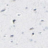
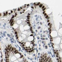
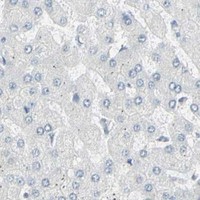


Comprehensive Human Protein Atlas IHC scoring per tissue (reliability: Enhanced — Low consistency between antibody staining and RNA expression data. Pending external verification.). Rows are taken directly from the HPA tissue chart — click any row's HPA link to view the source.
| Tissue | Cell type | Level | Evidence | Source |
|---|---|---|---|---|
| Adrenal gland | Glandular cells | High | Protein (IHC) | HPA → |
| Appendix | Glandular cells | High | Protein (IHC) | HPA → |
| Bone marrow | Hematopoietic cells | High | Protein (IHC) | HPA → |
| Bronchus | Respiratory epithelial cells | High | Protein (IHC) | HPA → |
| Cerebellum | Purkinje cells | High | Protein (IHC) | HPA → |
| Tissue | Cell type | Level | Evidence | Source |
|---|---|---|---|---|
| Adipose tissue | Adipocytes | Not detected | Protein (IHC) | HPA → |
| Caudate | Glial cells | Not detected | Protein (IHC) | HPA → |
| Cerebral cortex | Endothelial cells | Not detected | Protein (IHC) | HPA → |
| Epididymis | Glandular cells | Not detected | Protein (IHC) | HPA → |
| Heart muscle | Cardiomyocytes | Not detected | Protein (IHC) | HPA → |
Troubleshoot ZBTB7B staining by checking nuclear localisation, cell identity, processing conditions and control sections (UniProt O15156: nucleus; HPA: nuclear expression in most tissues).
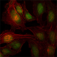
Two human-reactive ZBTB7B antibodies have image data: paraffin-section IHC of human breast tissue (M06831 IHC caption) and IF of HeLa cells (A06831 IF caption).
M06831 has an IHC image of paraffin-embedded human breast tissue with AEC staining (M06831 IHC caption). A06831 has a confocal IF image of HeLa cells (A06831 IF caption).
Which to pick: Choose mouse monoclonal M06831 for tissue IHC: its application list includes IHC, and its own image shows paraffin-embedded human breast tissue (M06831 catalog; M06831 IHC caption); the fixative is unreported (M06831 IHC caption). Choose rabbit polyclonal A06831 for IF/ICC in human cells: its application list includes IF, and its own image shows PFA-fixed HeLa cells (A06831 catalog; A06831 IF caption). Neither is documented for another species; both list Human reactivity only (M06831 catalog; A06831 catalog).