This website uses cookies to ensure you get the best experience on our website.


- Table of Contents
Plan chromogenic ZFP36L1 IHC on paraffin sections using an IHC-validated antibody at 1:100–1:300 (A05651 datasheet). Urothelial cells show high staining, but HPA rates the tissue IHC evidence uncertain (HPA tissue IHC).

Expected localisation, validated protocols, controls and antibodies — the at-a-glance facts below, then the full design guide.
| Expected localisation | Cytoplasmic tissue staining (HPA tissue IHC); nuclear localisation is possible (UniProt) | |
| Staining pattern | Cytoplasmic across tissues; high in urothelial cells (HPA tissue IHC) | |
| Antigen retrieval | Tris-EDTA pH 9.0 HIER, 95–98 °C, 20 min (rule: nuclear antigen) | |
| Positive control | Urinary bladder+4 more · see all | |
| Negative control | Adipose tissue+4 more · see all |
| Fixation | Keep fixation consistent across paraffin sections. (standard IHC practice; not target-specific) | |
| Caveat | Antibody staining has low concordance with RNA data (HPA tissue IHC) | |
| Regulation | Basal epidermis exceeds suprabasal layers (UniProt) | |
| Isoform / epitope | 0 annotated isoforms; one 1–338 chain (UniProt) |
The catalog antibody protocol is accompanied by three published ZFP36L1 IHC protocols covering lung sections, SCLC tumors, and nasopharyngeal carcinoma samples (PMC4192124; PMC9411550; PMC8406720).
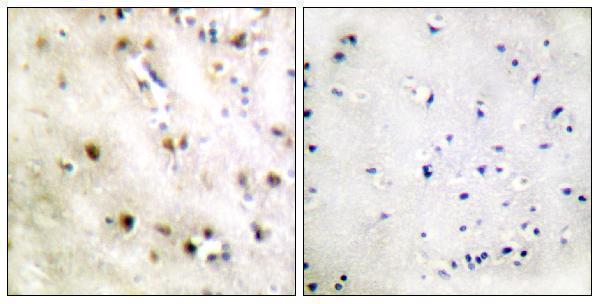
| Sample | Paraffin-embedded human brain tissue; fixative not specified (datasheet A05651) |
| Fixation | Image fixative and duration unreported (datasheet A05651); verify before use. |
| Sectioning | 4–5 µm sections on charged slides (standard) |
| Deparaffinisation | Xylene, graded ethanol series to water (standard) |
| Antigen retrieval | Heat-induced epitope retrieval in Tris-EDTA buffer, pH 9.0, 20 min at 95–98 °C (standard rule: nuclear antigen) |
| Peroxidase block | 3% H2O2, 10 min, room temperature (standard) |
| Blocking | 10% normal serum of the secondary host, 30 min, room temperature (standard) |
| Primary antibody | Rabbit anti-ZFP36L1, 1:100-1:300 (datasheet A05651) |
| Primary incubation | Overnight at 4 °C (standard) |
| Detection | HRP-polymer secondary, DAB chromogen 5–10 min (standard) |
| Counterstain | Hematoxylin, blue, dehydrate and mount (standard) |
| Expected result | ZFP36L1-positive staining in urothelial cells of urinary bladder (HPA tissue IHC: High). HPA tissue profile: Cytoplasmic expression in several different tissue types. No signal in the no-primary control. |
ZFP36L1 is a soluble protein that localizes to the cytoplasm, nucleus and cytoplasmic granules (UniProt Q07352: localization and topology). In paraffin section IHC, expect predominantly cytoplasmic staining in selected epithelial and glandular cells, including strongly stained urothelial cells (HPA: tissue IHC). Treat this pattern as provisional: the HPA tissue profile and its listed antibody, HPA001301, are both rated Uncertain (HPA: reliability; HPA: antibody validation).
| Clear cytoplasmic staining in urothelial cells, with weaker staining in some glandular cells (HPA: High in urothelial cells; Medium in listed glandular cells). | This matches the reported tissue pattern. Compare cell types within each section rather than treating every unstained cell as a failed assay: HPA reports low RNA tissue specificity but variable protein staining (HPA: tissue IHC). |
| Predominantly cell-surface or extracellular staining, without a convincing intracellular pattern (UniProt Q07352: localization and topology). | That distribution is inconsistent with the reported nucleus and cytoplasm localization and absence of a transmembrane segment (UniProt Q07352). Consider nonspecific staining or chromogen deposit; inspect morphology and a no-primary control (standard IHC practice). |
| Strong staining in adipocytes while nearby expected-positive cells are weak or negative (HPA: adipocytes Not detected; urothelial cells High). | Question antibody specificity or endogenous detection activity, especially if a no-primary control also stains (standard IHC practice). The adipocyte comparison is a warning, not definitive proof: HPA rates its tissue IHC evidence Uncertain (HPA: reliability). |
| Uniform brown haze across cells and stroma, or deposit outside recognizable cells (standard IHC practice). | This obscures the cell-specific cytoplasmic pattern reported by HPA (HPA: tissue IHC). Review blocking, washes, detection reagent and no-primary control before assigning biological intensity scores (standard IHC practice). |
| No staining in a section containing well-preserved urothelial cells (HPA: urothelial cells High). | Check a concurrently processed positive-control section, antibody application and detection performance (standard IHC practice). A negative result alone cannot distinguish technical failure from sample variation, especially with HPA's Uncertain reliability rating (HPA: tissue IHC). |
| Reference tissue and cell type (HPA: tissue IHC) | Urothelial cells provide the strongest listed IHC reference (High); breast, epididymis, fallopian tube and gallbladder glandular cells are listed as Medium (HPA: tissue IHC). Score the named cells, not the entire tissue uniformly. |
| Evidence strength (HPA: reliability; HPA: antibody validation) | The tissue profile and HPA001301 IHC assessment are Uncertain, with low consistency between antibody staining and RNA data (HPA: tissue IHC; HPA: HPA001301). Use the pattern as a comparison point, not a validated diagnostic threshold. |
| Intracellular distribution (UniProt Q07352: localization) | ZFP36L1 can shuttle between nucleus and cytoplasm and occur in stress granules and P-bodies (UniProt Q07352). Some nuclear or punctate signal can be plausible; compartment alone cannot establish specificity without suitable controls (standard IHC practice). |
| Epidermal comparison (UniProt Q07352: tissue specificity) | UniProt reports expression mainly in the basal epidermal layer and weakly in suprabasal layers, including protein-level evidence in keratinocytes (UniProt Q07352). This is a distinct reference from HPA's listed tissue IHC levels. |
| Topology and processing (UniProt Q07352: topology and processing) | No transmembrane segment, signal peptide or propeptide is reported; the annotated chain spans residues 1–338 (UniProt Q07352). These facts support an intracellular expectation but do not identify the antibody epitope or establish retrieval behavior. |
| IF/ICC cross-check? (HPA: subcellular; UniProt Q07352: localization) | HPA supplies no main ICC-IF location or cell-line images here, so it provides no image-based IF pattern to compare with this IHC result (HPA: subcellular). Use UniProt localization as context; consult the separate IF/ICC guide for assay design. |
| Situation | Likely cause | Next action |
|---|---|---|
| Urothelial cells show no signal (HPA: urothelial cells High). | The assay may have failed, or this specimen may differ from HPA's reported pattern; HPA rates its IHC evidence Uncertain (HPA: reliability). | Review section preservation, antibody and detection steps, and a concurrently processed positive control; adjust antigen retrieval only as a general IHC optimization, without assuming a ZFP36L1-specific fixation effect (standard IHC practice). |
| Only nuclei stain intensely while the cytoplasm is blank (UniProt Q07352: localization; HPA: cytoplasmic tissue profile). | Nuclear localization is possible, but a nucleus-only pattern departs from HPA's reported predominantly cytoplasmic IHC profile (UniProt Q07352; HPA: tissue IHC). | Recheck cellular boundaries and compare positive and no-primary controls; record nuclear and cytoplasmic scores separately before interpreting the shift (standard IHC practice). |
| Membranes or luminal material dominate the stain (UniProt Q07352: topology and localization). | The distribution conflicts with the reported intracellular localization and lack of a transmembrane segment (UniProt Q07352); nonspecific deposit is a possibility (standard IHC practice). | Inspect the deposit at higher magnification and compare a no-primary control and adjacent tissue morphology (standard IHC practice). Do not score extracellular deposit as ZFP36L1-positive cells. |
| Adipocytes stain strongly (HPA: adipocytes Not detected). | This differs from HPA's listed adipocyte observation, though that reference has Uncertain reliability (HPA: tissue IHC). Antibody cross-reactivity or endogenous detection activity are possible (standard IHC practice). | Compare no-primary and reagent controls, then review staining in a listed positive cell type on the same run (standard IHC practice; HPA: tissue IHC). |
| Brown haze prevents cell-by-cell scoring (standard IHC practice). | Background from the detection workflow can mask the cytoplasmic, cell-associated pattern reported by HPA (standard IHC practice; HPA: tissue IHC). | Inspect no-primary staining; review blocking, reagent concentration, incubation and wash steps according to the assay controls (standard IHC practice). Score only after cell boundaries are readable. |
| Small cytoplasmic dots are interpreted as a definitive positive (UniProt Q07352: cytoplasmic granules). | P-body or stress-granule localization is biologically plausible, but dot morphology alone does not identify either structure or prove antibody specificity (UniProt Q07352: localization; standard IHC practice). | Document the dots alongside diffuse cytoplasmic staining and controls; reserve granule identity for a separately validated colocalization assessment (standard IHC practice). |
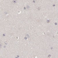

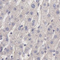

Comprehensive Human Protein Atlas IHC scoring per tissue (reliability: Uncertain — Low consistency between antibody staining and RNA expression data. Pending external verification.). Rows are taken directly from the HPA tissue chart — click any row's HPA link to view the source.
| Tissue | Cell type | Level | Evidence | Source |
|---|---|---|---|---|
| Urinary bladder | Urothelial cells | High | Protein (IHC) | HPA → |
| Breast | Glandular cells | Medium | Protein (IHC) | HPA → |
| Epididymis | Glandular cells | Medium | Protein (IHC) | HPA → |
| Fallopian tube | Glandular cells | Medium | Protein (IHC) | HPA → |
| Gallbladder | Glandular cells | Medium | Protein (IHC) | HPA → |
| Tissue | Cell type | Level | Evidence | Source |
|---|---|---|---|---|
| Adipose tissue | Adipocytes | Not detected | Protein (IHC) | HPA → |
| Bone marrow | Hematopoietic cells | Not detected | Protein (IHC) | HPA → |
| Caudate | Glial cells | Not detected | Protein (IHC) | HPA → |
| Cerebellum | Cells in granular layer | Not detected | Protein (IHC) | HPA → |
| Cerebral cortex | Endothelial cells | Not detected | Protein (IHC) | HPA → |
Troubleshoot ZFP36L1 staining in paraffin section IHC by checking retrieval, compartment, controls and cell resolved scoring before interpreting chromogenic signal.
Both anti-ZFP36L1 antibodies have IHC images from human paraffin sections; both list human, mouse and rat reactivity. Only A05651 lists IF, without an IF image (catalog applications; IHC image captions).
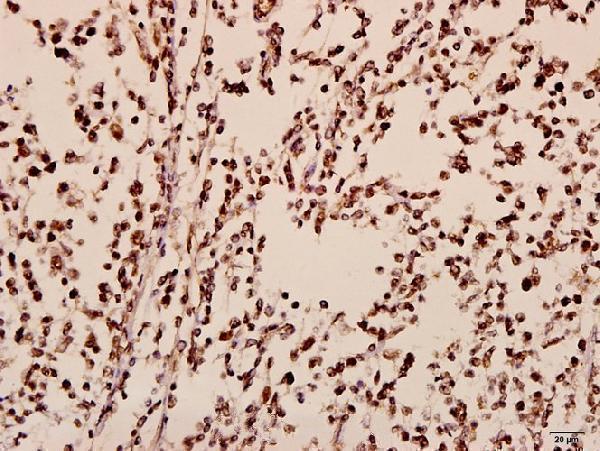
A05651 has an IHC image from human paraffin-embedded brain tissue, including a peptide-blocked comparison; its catalog lists IHC and IF (A05651 image caption; catalog applications). A05651-1 has an IHC image from human paraffin-embedded tonsil carcinoma tissue at 1:50 and lists IHC only (A05651-1 image caption; catalog applications).
Which to pick: For tissue IHC, choose either according to the sample: A05651 has a human brain paraffin-section image, while A05651-1 has a human tonsil carcinoma paraffin-section image and is listed as polyclonal (each SKU’s image caption; A05651-1 catalog description). For IF, choose A05651 because IF is listed at 1:50; ICC validation and an IF image are unreported (A05651 catalog applications, dilution and image list). Both list human, mouse and rat reactivity, but their supplied IHC images show human tissue only; neither caption reports the fixative (catalog reactivity; each SKU’s IHC image caption).