This website uses cookies to ensure you get the best experience on our website.


- Table of Contents
Plan chromogenic ZNF397 IHC on paraffin sections with the catalog antibody at 1:100–1:300 (datasheet A13857). Assess cytoplasmic staining (HPA tissue IHC) with controls, and interpret tissue patterns cautiously because staining and RNA expression show low consistency (HPA tissue IHC).

Expected localisation, validated protocols, controls and antibodies — the at-a-glance facts below, then the full design guide.
| Expected localisation | Mostly cytoplasmic in tissue IHC (HPA tissue IHC); nuclear location annotated (UniProt) | |
| Staining pattern | Cytoplasmic staining in most tissues (HPA tissue IHC) | |
| Antigen retrieval | Tris-EDTA pH 9.0 HIER, heat-mediated (datasheet A13857) | |
| Positive control | Bone marrow+4 more · see all | |
| Negative control | Oral mucosa+1 more · see all |
| Fixation | Keep fixation consistent across paraffin sections (standard IHC practice; not target-specific); Matched tissue-IHC evidence does not establish the fixation claim. Validate the specimen-specific method before use. (selected-SKU IHC image A13857) | |
| Caveat | Staining and RNA expression show low consistency (HPA tissue IHC) | |
| Regulation | Strongly expressed in testis (UniProt) | |
| Isoform / epitope | Three isoforms; antibody epitope coverage is unspecified (UniProt) |
Compare the catalog antibody’s IHC-P protocol (datasheet A13857) with published ZNF397 IHC in mouse xenograft tumors (PMC11285331).
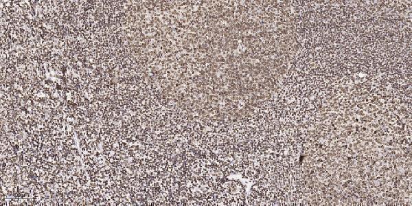
| Sample | Paraffin-embedded human tonsil tissue; fixative not specified (datasheet A13857) |
| Fixation | Image fixative and duration unreported (datasheet A13857); verify before use. |
| Sectioning | 4–5 µm sections on charged slides (standard) |
| Deparaffinisation | Xylene, graded ethanol series to water (standard) |
| Antigen retrieval | Heat retrieval: Tris-EDTA pH 9.0 (datasheet A13857); 20 min, 95–100 °C (standard) |
| Peroxidase block | 3% H2O2, 10 min, room temperature (standard) |
| Blocking | 10% normal serum of the secondary host, 30 min, room temperature (standard) |
| Primary antibody | Rabbit anti-ZNF397, 1:100-1:300 (datasheet A13857) |
| Primary incubation | Overnight at 4 °C (standard) |
| Detection | HRP-polymer secondary, DAB chromogen 5–10 min (standard) |
| Counterstain | Hematoxylin, blue, dehydrate and mount (standard) |
| Expected result | ZNF397-positive staining in hematopoietic cells of bone marrow (HPA tissue IHC: High). HPA tissue profile: Cytoplasmic expression in most tissues. No signal in the no-primary control. |
In paraffin-section IHC, ZNF397 is reported mainly as cytoplasmic staining across tissues, with high staining in selected glandular and hematopoietic cells and in cerebral-cortex neuropil (HPA tissue IHC). Nuclear staining is biologically plausible because ZNF397 localizes to both nucleus and cytoplasm (UniProt Q8NF99). It has no transmembrane segment (UniProt Q8NF99 topology). Treat the tissue pattern as provisional: HPA rates its IHC reliability Uncertain because antibody staining and RNA expression have low consistency (HPA tissue IHC).
| Cytoplasmic staining is strong in bone-marrow hematopoietic cells or breast and duodenal glandular cells (HPA: High). | This matches reported positive cell populations and the predominant tissue-IHC compartment (HPA tissue IHC). Compare stained cells with nearby unstained structures; a matching pattern supports interpretation but does not establish specificity because tissue-IHC reliability is Uncertain (HPA tissue IHC). |
| Staining is confined to nuclei, with little cytoplasmic signal in a tissue expected to show cytoplasmic staining (HPA tissue IHC). | This departs from the reported tissue-IHC profile (HPA tissue IHC). Nuclear localization remains possible (UniProt Q8NF99), and nucleoli are an approved ICC-IF location (HPA subcellular). Review controls and morphology before calling nuclear-only IHC either genuine or artefactual. |
| Strong chromogen appears in oral-mucosa squamous epithelium or skeletal-muscle myocytes (HPA: Not detected). | These cell types are reported as unstained in tissue IHC (HPA tissue IHC). Check for cross-reactivity and endogenous detection activity (general IHC practice). HPA's Uncertain reliability prevents treating either reported negative as a definitive biological absence (HPA tissue IHC). |
| Diffuse colour covers cells and extracellular spaces without a readable cellular pattern. | This is background until controls show otherwise (general IHC practice). Reassess blocking, washing, antibody concentration and chromogen development (general IHC practice); diffuse colour cannot establish ZNF397 localization, even where HPA reports positive staining (HPA tissue IHC). |
| A bone-marrow section shows no hematopoietic-cell signal, including with the positive control (HPA: High in hematopoietic cells). | First investigate assay performance and whether the expected cell population is present (general IHC practice). HPA reports high staining there, but its tissue-IHC reliability is Uncertain; one negative section alone cannot resolve ZNF397 expression (HPA tissue IHC). |
| Compartment and topology | Nucleus and cytoplasm are listed locations, and no transmembrane segment is annotated (UniProt Q8NF99). Tissue IHC is predominantly cytoplasmic, while ICC-IF adds nucleoli, plasma membrane, cytosol and microtubules (HPA tissue IHC; HPA subcellular). Interpret each application against its own observed pattern. |
| Tissue and cell selection | High IHC staining is reported in bone-marrow hematopoietic cells, several glandular populations, endometrial stromal cells and cerebral-cortex neuropil (HPA tissue IHC). Oral-mucosa squamous cells and skeletal-muscle myocytes are reported Not detected (HPA tissue IHC). These are provisional comparison sites because reliability is Uncertain (HPA tissue IHC). |
| Expression and staining discordance | UniProt reports moderate expression in skeletal muscle, whereas HPA reports myocyte IHC as Not detected (UniProt Q8NF99 tissue specificity; HPA tissue IHC). HPA also describes low tissue RNA specificity and low staining–RNA consistency (HPA tissue IHC). Do not infer protein absence or assay specificity from one readout. |
| Antibody evidence | The listed antibody HPA026087 is rated IHC Uncertain and ICC Supported; the overall tissue-IHC profile is also Uncertain (HPA antibodies; HPA tissue IHC). Record which antibody and controls produced a pattern before comparing results. An ICC rating does not upgrade tissue-IHC confidence (HPA antibodies). |
| Isoforms and epitope | Three isoforms are annotated, and the SCAN box spans residues 50–132 (UniProt Q8NF99). No antibody epitope or isoform coverage is supplied here, so staining cannot be assigned to one isoform or explained by differential recognition. Verify those properties from antibody-specific evidence before making an isoform claim. |
| IF/ICC Q: What localization should an IF image show? | A: HPA reports approved nucleolar, plasma-membrane and cytosolic locations, plus microtubules, with images from A-431, U-251MG and U2OS (HPA subcellular). HPA026087 is ICC Supported (HPA antibodies). This observation is context for the separate IF/ICC guide, not a tissue-IHC scoring rule. |
| Situation | Likely cause | Next action |
|---|---|---|
| No cytoplasmic signal in bone-marrow hematopoietic cells (HPA: High). | A failed staining run or an unrepresentative section is possible (general IHC practice); HPA's IHC call is Uncertain (HPA tissue IHC). | Confirm the cells are present, inspect a concurrently stained control, and review retrieval and detection settings (general IHC practice). |
| Strong staining in oral-mucosa squamous cells (HPA: Not detected). | Cross-reactivity or endogenous detection activity is possible (general IHC practice); the HPA negative is provisional (HPA tissue IHC: Uncertain). | Check a no-primary control and the distribution of colour, then compare with a reported positive cell population (general IHC practice; HPA tissue IHC). |
| Nuclear-only staining in a tissue with expected cytoplasmic staining (HPA tissue IHC). | The pattern differs from tissue IHC, although nuclear ZNF397 is annotated (UniProt Q8NF99; HPA tissue IHC). | Inspect morphology and controls; repeat with independently validated evidence before interpreting a nuclear-only tissue pattern (general IHC practice). |
| Diffuse colour obscures cell boundaries. | Excess antibody, insufficient washing or prolonged chromogen development can raise background (general IHC practice). | Review the no-primary control and adjust staining conditions systematically, preserving a readable cellular pattern (general IHC practice). |
| Colour persists when primary antibody is omitted. | Endogenous detection activity or nonspecific detection-reagent binding is possible (general IHC practice). | Address the activity appropriate to the detection chemistry and repeat the no-primary control (general IHC practice). |
| Skeletal-muscle myocytes stain despite the HPA Not detected call (HPA tissue IHC). | The finding conflicts with HPA IHC yet UniProt reports moderate skeletal-muscle expression; neither resolves antibody specificity (UniProt Q8NF99; HPA tissue IHC). | Check cell identification and controls, then seek independent validation before accepting or rejecting the signal (general IHC practice; HPA tissue IHC: Uncertain). |


Comprehensive Human Protein Atlas IHC scoring per tissue (reliability: Uncertain — Low consistency between antibody staining and RNA expression data. Pending external verification.). Rows are taken directly from the HPA tissue chart — click any row's HPA link to view the source.
| Tissue | Cell type | Level | Evidence | Source |
|---|---|---|---|---|
| Bone marrow | Hematopoietic cells | High | Protein (IHC) | HPA → |
| Breast | Glandular cells | High | Protein (IHC) | HPA → |
| Cerebral cortex | Neuropil | High | Protein (IHC) | HPA → |
| Duodenum | Glandular cells | High | Protein (IHC) | HPA → |
| Endometrium | Cells in endometrial stroma | High | Protein (IHC) | HPA → |
Use matched controls and compartment-specific scoring when assessing ZNF397 in paraffin sections; reported tissue staining has uncertain reliability (HPA tissue IHC).
A13857 is a human-reactive anti-ZNF397 antibody with paraffin-section IHC data (catalog; IHC figure caption). IF is listed as an application, but no IF figure is supplied (catalog).
A13857 is listed for human IHC and IF (catalog: applications and reactivity). Its IHC figure shows paraffin-embedded human tonsil; the catalog supplies no IF figure (IHC figure caption; catalog).
Which to pick: For tissue IHC, choose A13857: its own figure documents paraffin-embedded human tonsil, Tris-EDTA retrieval at pH 9.0, and a 1:200 primary dilution; the fixative is unreported (A13857 IHC figure caption). For IF/ICC, A13857 lists IF at 1:200–1:1000, but the payload provides no IF figure or ICC validation (A13857 catalog). No cross-species option is documented because A13857 lists human reactivity only (A13857 catalog).