ZNRF2 / E3 ubiquitin-protein ligase ZNRF2 · IHC design guide

Design Immunohistochemistry for ZNRF2

Plan ZNRF2 staining in paraffin sections using the reported cytoplasmic tissue pattern (HPA tissue IHC) and the catalog antibody at 1:100–1:300 (datasheet A11505). Compare high-staining neuronal or glandular cells with muscle cells reported as undetected (HPA tissue IHC).

Evidence assembled Oct 2026 · For research use; verify linked source records and product datasheet before use
Immunohistochemistry protocol sheet for ZNRF2 (IHC for ZNRF2): expected localisation General cytoplasmic staining (HPA tissue IHC), antibody A11505, validated IHC image, and IHC protocol steps
Printable ZNRF2 IHC protocol sheet — expected localisation General cytoplasmic staining (HPA tissue IHC), antibody A11505, controls and protocol steps. Open the full ZNRF2 IHC guide →

ZNRF2 Immunohistochemistry Experimental Design Guide

Expected localisation, validated protocols, controls and antibodies — the at-a-glance facts below, then the full design guide.

Must know before staining
Expected localisation General cytoplasmic staining (HPA tissue IHC)
Staining pattern Cytoplasmic staining in neuronal and glandular cells (HPA tissue IHC)
Antigen retrieval Tris-EDTA pH 9.0 HIER, heat-mediated (datasheet A11505)
Positive control ⓘ Appendix+4 more · see all
Negative control ⓘ Heart muscle+2 more · see all
Important caveats
Reasons your staining may differ from the expected pattern.
Fixation Keep fixation consistent across sections (standard IHC practice; not target-specific)
Caveat Antibody staining and RNA show medium consistency (HPA tissue IHC)
Regulation Higher brain expression in development (UniProt)
Isoform / epitope No isoforms annotated; chain spans residues 2–242 (UniProt)
Section 1

Recommended ZNRF2 IHC & IF Protocols

The catalog antibody has an IHC-P protocol (datasheet A11505). One published ZNRF2 IHC protocol uses paraffin-embedded human glioma and non-tumoral brain sections (PMC11829375).

Recommended immunohistochemistry (IHC-P) protocol parameters
SampleParaffin-embedded human Squamous cell carcinoma of lung tissue; fixative not specified (datasheet A11505)
FixationImage fixative and duration unreported (datasheet A11505); verify before use.
Sectioning4–5 µm sections on charged slides (standard)
DeparaffinisationXylene, graded ethanol series to water (standard)
Antigen retrievalHeat retrieval: Tris-EDTA pH 9.0 (datasheet A11505); 20 min, 95–100 °C (standard)
Peroxidase block3% H2O2, 10 min, room temperature (standard)
Blocking10% normal serum of the secondary host, 30 min, room temperature (standard)
Primary antibodyRabbit anti-ZNRF2, 1:100 - 1:300 (datasheet A11505)
Primary incubationOvernight at 4 °C (standard)
DetectionHRP-polymer secondary, DAB chromogen 5–10 min (standard)
CounterstainHematoxylin, blue, dehydrate and mount (standard)
Expected resultZNRF2-positive staining in glandular cells of appendix (HPA tissue IHC: High). HPA tissue profile: General cytoplasmic expression. No signal in the no-primary control.
💡Decision noteStart with heat-mediated Tris-EDTA pH 9.0 retrieval (datasheet A11505); the published glioma protocol does not specify retrieval conditions (PMC11829375).
Section 2

What Is the Expected ZNRF2 Staining Pattern?

In paraffin-section IHC, expect chiefly cytoplasmic staining in selected glandular cells, neurons, Purkinje cells and kidney tubule cells (HPA: general cytoplasmic expression; High in these cells). Endosome and lysosome membranes, presynaptic membrane and cytoplasm are annotated locations despite no transmembrane segment (UniProt Q8NHG8 topology). Treat the pattern as a guide: HPA rates tissue IHC Approved, with medium consistency between staining and RNA data (HPA: tissue IHC reliability).

What am I looking at on my slide?
Clear cytoplasmic staining in kidney tubules, with much less signal in adjacent unstained structures.This fits a documented positive cell population (HPA: High in kidney tubule cells). Judge the cell type and compartment together; a positive tissue does not imply every cell should stain (HPA: tissue IHC profile).
Cytoplasmic staining in caudate or cerebral-cortex neurons, or cerebellar Purkinje cells.These are documented high-staining populations (HPA: High in neuronal and Purkinje cells). Vesicle-associated signal is biologically plausible, but puncta are not required for a valid chromogenic tissue result (UniProt Q8NHG8 localisation; HPA: general cytoplasmic expression).
Predominantly nuclear chromogen with little cytoplasmic signal in an IHC section.Recheck specificity and image interpretation: the tissue IHC profile is generally cytoplasmic (HPA: tissue IHC profile). Nuclear ICC/IF localisation is separately reported as uncertain, so nuclear signal alone does not establish a correct tissue IHC pattern (HPA: subcellular).
Strong staining throughout cardiomyocytes, skeletal myocytes or smooth muscle cells.This conflicts with the listed tissue IHC negatives (HPA: Not detected in these cell types). Consider cross-reactivity or endogenous detection activity; compare with omission and detection controls before assigning the signal to ZNRF2 (general IHC practice).
Uniform brown haze across cells, extracellular areas and blank slide regions.A non-cell-selective deposit cannot be read as the reported cytoplasmic pattern (HPA: tissue IHC profile). Background from detection reagents or inadequate blocking is a general IHC possibility; compare controls and inspect tissue-free areas (general IHC practice).
💡Expected ZNRF2 appearanceA convincing positive shows stronger cytoplasmic chromogen in a documented high-staining population, such as kidney tubule cells or Purkinje cells (HPA: High); broad muscle staining or dominant nuclear-only IHC signal warrants investigation (HPA: muscle Not detected; general cytoplasmic profile).
How each factor affects the staining
Cell compartment and membrane associationZNRF2 is annotated in cytoplasm and on endosome, lysosome and presynaptic membranes, with no transmembrane segment (UniProt Q8NHG8 topology). Cytoplasmic tissue staining is therefore coherent; a membrane-only outline is not a required IHC endpoint (HPA: general cytoplasmic expression).
Choice of positive and low-signal comparatorsAppendix and duodenum glandular cells, kidney tubules, selected neurons and Purkinje cells are High (HPA: tissue IHC). Muscle cell types are Not detected, while some other glandular populations are Low; choose comparators at the cell-type level (HPA: tissue IHC).
Strength of tissue evidenceThe tissue IHC profile is Approved but has medium consistency with RNA expression (HPA: tissue IHC reliability). The listed antibody HPA072244 is IHC Approved, not reported here as IHC Enhanced (HPA: antibody validation); unexpected staining needs controls and cautious interpretation.
Protein forms and epitope limitsUniProt lists a 242-residue protein, chain 2–242, no annotated isoforms, and several phosphoserines (UniProt Q8NHG8 processing and modified residues). No antibody epitope or target-specific retrieval response is supplied; these annotations cannot predict staining intensity or justify a retrieval setting.
IF/ICC Q: What localisation should I expect?A: HPA reports approved vesicles, uncertain nucleoplasm and uncertain nucleoli fibrillar center in ICC/IF images from RT-4, SH-SY5Y and U2OS (HPA: subcellular). This separate cell-based observation does not replace the generally cytoplasmic tissue IHC profile (HPA: tissue IHC).
Why is my staining missing, weak or wrong?
SituationLikely causeNext action
No signal in kidney tubules or Purkinje cells.These are documented high-staining cells (HPA: tissue IHC); a blank result may reflect a failed IHC run or unsuitable assay settings (general IHC practice).Confirm tissue identity and preservation, then check primary antibody, detection reagents and a run control; empirically assess retrieval and dilution without assuming a ZNRF2-specific optimum (general IHC practice).
Strong staining in cardiomyocytes or skeletal myocytes.Both are listed as Not detected (HPA: tissue IHC). Cross-reactivity or endogenous detection activity is possible (general IHC practice).Compare primary-omission and detection controls; inspect whether the deposit follows tissue structures, then reassess antibody concentration and blocking (general IHC practice).
Nuclear signal dominates a paraffin-section result.The reported tissue profile is generally cytoplasmic (HPA: tissue IHC); nucleoplasm is an uncertain ICC/IF location (HPA: subcellular).Check counterstain versus chromogen, compare a documented positive cell population, and verify specificity with controls before interpreting nuclear staining as ZNRF2 (general IHC practice).
Brown haze covers positive and negative areas alike.Indiscriminate deposit obscures the cell-selective cytoplasmic pattern (HPA: tissue IHC profile); detection background is a general IHC possibility.Inspect tissue-free regions and omission controls, then adjust blocking, washes or detection development using the assay's controls (general IHC practice).
Signal appears only at section edges or in damaged tissue.Uneven processing or reagent coverage can create a spatial staining artifact (general IHC practice). The distribution does not establish the HPA cell-type pattern (HPA: tissue IHC).Score intact interior tissue, check section adhesion and reagent coverage, and repeat with a documented positive cell population on the same run (general IHC practice).
Weak staining in one expected-positive tissue, but the run control works.HPA reports high staining in specific cell types, not uniform staining across each organ; its tissue IHC reliability has medium RNA concordance (HPA: tissue IHC).Verify the relevant cells are present, compare another listed high-staining population, and record intensity by cell type. Treat a single weak field as inconclusive (HPA: tissue IHC; general IHC practice).

Sample controls for ZNRF2 IHC & IF

🧪Run cerebellum first and confirm staining in Purkinje cells (HPA: High in cerebellar Purkinje cells). Run heart muscle as the negative tissue, where cardiomyocytes are not detected (HPA: Not detected in heart cardiomyocytes); neighboring non-Purkinje cells are candidate internal negatives only if they show little or no specific chromogen on the same slide (standard IHC practice).
Positive control tissue: Appendix (Glandular cells, HPA High)
Negative control tissue: Heart muscle (HPA Not detected)
ICC-IF cell lines (HPA subcellular resource): HPA ICC-IF images show ZNRF2 in RT-4, SH-SY5Y, U2OS, with annotated localisation: Nucleoplasm (uncertain), Vesicles (approved) (HPA subcellular).
Technical controls: Include no-primary (secondary-only) and host-species- and clonality-matched isotype controls, plus ZNRF2 knockout tissue or a validated immunizing-peptide block as a biological specificity control (standard IHC practice). Block endogenous peroxidase for chromogenic detection and assess pigment-related background in cerebellum (standard IHC practice).
⚠️Feasibility: A target-specific fixation window or fixation effect is unreported, and the selected A11505 paraffin-section caption does not report its fixative (selected A11505 tissue-IHC caption). That caption uses Tris-EDTA at pH 9.0 for antigen retrieval, but whether retrieval is required for cerebellum is unreported (selected A11505 tissue-IHC caption). Frozen sections or IF cannot be judged easier from the supplied evidence; neuronal pigment can complicate interpretation, particularly with fluorescence (standard IHC/IF practice).

HPA tissue IHC evidence for ZNRF2

Comprehensive Human Protein Atlas IHC scoring per tissue (reliability: Approved — Medium consistency between antibody staining and RNA expression data.). Rows are taken directly from the HPA tissue chart — click any row's HPA link to view the source.

Positive expression · recommended positive controls

TissueCell typeLevelEvidenceSource
Appendix Glandular cells High Protein (IHC) HPA →
Caudate Neuronal cells High Protein (IHC) HPA →
Cerebellum Purkinje cells High Protein (IHC) HPA →
Cerebral cortex Neuronal cells High Protein (IHC) HPA →
Duodenum Glandular cells High Protein (IHC) HPA →

Undetected expression · recommended negative controls

TissueCell typeLevelEvidenceSource
Heart muscle Cardiomyocytes Not detected Protein (IHC) HPA →
Skeletal muscle Myocytes Not detected Protein (IHC) HPA →
Smooth muscle Smooth muscle cells Not detected Protein (IHC) HPA →
Section 3

Advanced ZNRF2 IHC Tips

Troubleshoot ZNRF2 staining in paraffin sections using the catalog antibody’s IHC conditions, tissue staining patterns, and reported subcellular locations.

Which retrieval condition should I try first for weak ZNRF2 staining?
Use heat-mediated retrieval in Tris-EDTA, pH 9.0, for paraffin sections (datasheet A11505). The selected tissue image used this condition with primary antibody at 1:200 overnight at 4°C (caption A11505). If staining remains weak, compare retrieval duration on adjacent sections while keeping antibody incubation and chromogenic development constant (standard IHC practice). Include a tissue compartment with reported high staining, such as kidney tubule cells (HPA: High in kidney tubule cells), to distinguish a failed run from a weak study specimen. Assess morphology alongside signal, because excessive heating can damage sections and make apparent improvement difficult to interpret (standard IHC practice).
Could fixation explain weak or uneven ZNRF2 staining?
Target-specific fixation sensitivity is unknown; the selected paraffin-section caption does not state a fixative (caption A11505). Record the actual fixative and fixation duration for each specimen, then compare sections processed in the same batch before attributing differences to ZNRF2 biology (standard IHC practice). Keep retrieval at Tris-EDTA, pH 9.0, and the primary incubation at 1:200 overnight at 4°C during that comparison (datasheet A11505; caption A11505). Uneven staining across a section warrants checking tissue preservation, section attachment, and retrieval uniformity (standard IHC practice). Do not treat a staining difference alone as evidence that ZNRF2 is especially fixation sensitive.
Should ZNRF2 look diffuse, punctate, or nuclear in chromogenic sections?
Expect a predominantly cytoplasmic tissue staining pattern (HPA tissue IHC: general cytoplasmic expression), and assess punctate signal against that baseline rather than requiring it in every cell. Endosome and lysosome membranes, the presynaptic cell membrane, and cytoplasm are reported locations (UniProt Q8NHG8: subcellular location). Vesicles are an approved location in cell imaging, whereas nucleoplasm and the nucleolar fibrillar center are uncertain locations (HPA subcellular). If nuclear staining dominates a chromogenic section, check whether the cytoplasmic pattern and appropriate positive cells are also present before calling it ZNRF2. Compare adjacent sections and a no-primary control to evaluate staining consistency and nonspecific chromogen deposition (standard IHC practice).
How should epitope uncertainty affect my interpretation of ZNRF2 staining?
The supplied record lists 0 annotated isoforms and no transmembrane segment (UniProt Q8NHG8: isoforms and topology). It also lists multiple modified residues, including phosphoserines at positions 19, 21, 25, 82, and 89 (UniProt Q8NHG8: modified residues). Without an epitope map for this antibody, do not infer that a staining change measures one phosphorylation state or a particular membrane-facing epitope. When comparing specimens, use the same retrieval at Tris-EDTA, pH 9.0, and the same antibody conditions (datasheet A11505; caption A11505). Document compartment and cell type as well as intensity, then seek independent validation for any modification-specific interpretation (standard IHC practice).
How can IF help examine the cell identity and location of ZNRF2 signal?
For a separate IF experiment, multiplex ZNRF2 with a validated marker for the expected cell type, such as a neuronal marker when examining caudate neuronal cells (HPA tissue IHC: High in caudate neuronal cells). Choose spectrally separated fluorophores and consider a far-red channel if specimen autofluorescence obscures shorter wavelengths (standard IF practice). Use a mild permeabilisation pilot for intracellular epitopes, but establish epitope accessibility from the antibody documentation because ZNRF2 has no transmembrane segment and is reported at membranes and in cytoplasm (UniProt Q8NHG8: topology and subcellular location). Include single-label and no-primary controls to assess bleed-through and background (standard IF practice). Treat this as an IF troubleshooting comparison, not as a validated IF protocol for the catalog antibody.
What should I check when ZNRF2 chromogenic staining is widespread?
Start with a no-primary control to assess secondary reagent and detection-system background, and include an endogenous peroxidase block before peroxidase-based DAB detection (standard IHC practice). The selected image used primary antibody at 1:200 overnight at 4°C and secondary antibody at 1:200 for 45 minutes at room temperature (caption A11505). If background persists, compare shorter development or a more dilute primary antibody on adjacent sections without changing retrieval simultaneously (standard IHC practice). Check whether chromogen follows tissue edges, necrotic areas, pigment, or the same pattern in the no-primary control (standard IHC practice). Use HPA-reported unstained myocytes as a contextual comparator, not an absolute specificity test (HPA: Not detected in skeletal muscle myocytes).
How should I score ZNRF2 staining across tissue samples? ⚠ ANSWER MARKED FOR VERIFICATION
Define the cell population and viable tissue area before scoring, because ZNRF2 staining varies by cell type in tissue images (HPA tissue IHC: High in kidney tubule cells; Not detected in cardiomyocytes). For intensity scoring, use an H-score from 0–300, calculated as the sum of each intensity grade multiplied by its percentage of cells (standard IHC practice). Alternatively, report the percentage of positive cells or positive-cell density per mm² of viable tissue when those measures fit the study question (standard IHC practice). Keep thresholds, counterstain, imaging, and analysis settings consistent across batches (standard IHC practice). Normalise to the number of eligible cells or their viable area, and report compartments separately if their staining patterns differ.
How can I distinguish plausible ZNRF2 signal from staining artefact?
Give greatest weight to reproducible cytoplasmic staining in the expected cell population, consistent with the reported general tissue pattern (HPA tissue IHC: general cytoplasmic expression). Kidney tubule cells and caudate neuronal cells have reported high staining, while cardiomyocytes are reported as not detected (HPA tissue IHC: cell-specific staining levels). Puncta may merit closer inspection because endosome and lysosome membranes are reported locations, but nuclear predominance needs caution because nucleoplasm localisation is uncertain (UniProt Q8NHG8: subcellular location; HPA subcellular). Exclude tissue-edge staining, necrotic deposits, and signal present in a no-primary control before scoring (standard IHC practice). Check endogenous peroxidase background with an appropriate block when using peroxidase-based DAB detection (standard IHC practice).
Boster reagents

Best ZNRF2 / E3 ubiquitin-protein ligase ZNRF2 IHC Antibodies

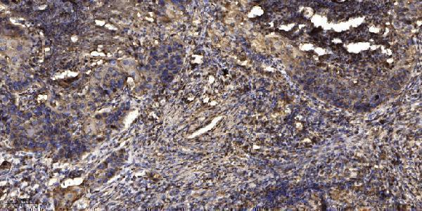
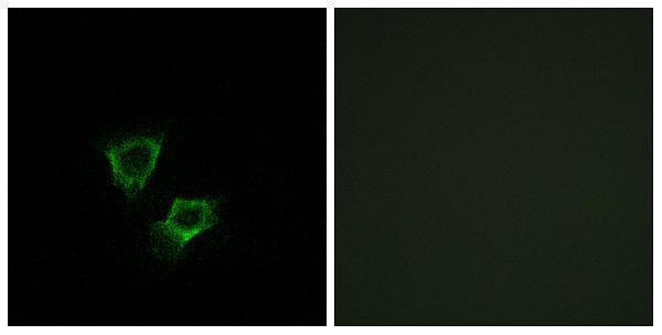
A11505 has documented IHC in paraffin-embedded human lung carcinoma and IF in A549 cells (A11505 image captions); both SKUs list human and mouse reactivity, while M11505 also lists rat (catalog: reactivity).

Real IHC data Immunohistochemical analysis of paraffin-embedded human Squamous cell carcinoma of lung. 1, Antibody was diluted at 1:200(4° overnight). 2, Tris-EDTA,pH9.0 was used for antigen retrieval. 3,Secondary antibody was diluted at 1:200(room temperature, 45min).
Anti-ZNRF2 Antibody
Cat # A11505

A11505 lists IHC and IF/ICC, with images of paraffin-embedded human lung squamous cell carcinoma and A549 cells, respectively (catalog: A11505 applications and image captions). M11505 lists IHC and human, mouse, and rat reactivity, but provides no IHC image caption (catalog: M11505).

Which to pick: For tissue IHC, choose A11505: its own caption documents paraffin-embedded human lung squamous cell carcinoma; the fixative is unreported (A11505 IHC caption). For IF/ICC, choose A11505 because both applications are listed and its IF image shows A549 cells (catalog: A11505 applications and IF caption). For rat-inclusive IHC, M11505 is the rabbit monoclonal option with listed human, mouse, and rat reactivity, though it has no IHC image caption (catalog: M11505).

Each figure is that product's own IHC / IF validation image from its datasheet.

References

  1. UniProt Consortium. UniProt entry Q8NHG8 (ZNRF2_HUMAN, E3 ubiquitin-protein ligase ZNRF2).
  2. Human Protein Atlas. ZNRF2 tissue IHC expression (reliability: Approved).
  3. Human Protein Atlas. ZNRF2 subcellular location (ICC-IF): Mainly localized to the nucleoplasm and vesicles. In addition localized to the nucleoli fibrillar center..
  4. Human Protein Atlas. ZNRF2 antibody validation summary (1 antibodies).
  5. A pancancer analysis of the oncogenic role of ZNRF2 in human tumours. Journal of cellular and molecular medicine 2023 — PMC10623518.
  6. Role of hsa‑miR‑105 during the pathogenesis of paclitaxel resistance and its clinical implication in ovarian cancer. Oncology reports 2021 — PMC8025119.
  7. ZNRF2 is essential for gliomagenesis through orchestrating glycolysis and acts as a promising therapeutic target in glioma. Journal of translational medicine 2025 — PMC11829375.
  8. PubMed PMID:14561866 — UniProt-cited evidence.
  9. PubMed PMID:17081983 — UniProt-cited evidence.
  10. PubMed PMID:16215985 — UniProt-cited evidence.