Click image to see more details
Product Info Summary
| SKU: | A01257 |
|---|---|
| Size: | 100 μg/vial |
| Reactive Species: | Human, Mouse, Rat |
| Host: | Rabbit |
| Application: | ELISA, Flow Cytometry, WB |
Customers Who Bought This Also Bought

Product info
Product Name
Anti-BAFF/TNFSF13B Antibody Picoband™
View all BAFF/BLyS/TNFSF13B Antibodies
SKU/Catalog Number
A01257
Size
100 μg/vial
Form
Lyophilized
Description
Boster Bio Anti-BAFF/TNFSF13B Antibody Picoband™ catalog # A01257. Tested in ELISA, Flow Cytometry, WB applications. This antibody reacts with Human, Mouse, Rat.
Storage & Handling
Store at -20˚C for one year from date of receipt. After reconstitution, at 4˚C for one month. It can also be aliquotted and stored frozen at -20˚C for six months. Avoid repeated freeze-thaw cycles.
Cite This Product
Anti-BAFF/TNFSF13B Antibody Picoband™ (Boster Biological Technology, Pleasanton CA, USA, Catalog # A01257)
Host
Rabbit
Contents
Each vial contains 4mg Trehalose, 0.9mg NaCl, 0.2mg Na2HPO4, 0.05mg NaN3.
Clonality
Polyclonal
Isotype
Rabbit IgG
Immunogen
E.coli-derived human BAFF/TNFSF13B recombinant protein (Position: A134-T277).
*Blocking peptide can be purchased. Costs vary based on immunogen length. Contact us for pricing.
Cross-reactivity
No cross-reactivity with other proteins.
Reactive Species
A01257 is reactive to TNFSF13B in Human, Mouse, Rat
Applications
A01257 is guaranteed for ELISA, Flow Cytometry, WB Boster Guarantee
Observed Molecular Weight
34 kDa
Calculated molecular weight
31.223kDa
Background of BAFF/BLyS/TNFSF13B
BAFF was regularly detected by enzyme-linked immunosorbent assay in brain tissue lysates and in normal spinal fluid, and in astrocytes by double fluorescence microscopy. BAFF was localized in astrocytes close to BAFF-R-expressing immune cells. BAFF receptors were strongly expressed in situ in primary central nervous system (CNS) lymphomas.1 The TNF superfamily member B cell-activating factor (BAFF) plays an important role in humoral immunity and in autoimmune diseases, including RA.Local BAFF gene targeting inhibited proinflammatory cytokine expression, suppressed generation of plasma cells and Th17 cells, and markedly ameliorated joint pathology. The B cell activating factor BAFF (BlyS/TALL-1/zTNF4) is a tumor necrosis factor (TNF)-related ligand that promotes B cell survival and binds to three receptors (BCMA, TACI, and the recently described BAFF-R). Human BAFF was mapped to chromosome 13q32-34. The standard used in this kit is recombinant soluble human BAFF (A134-L295) with the molecular mass of 19.6KDa.
Antibody Validation
Boster validates all antibodies on WB, IHC, ICC, Immunofluorescence, and ELISA with known positive control and negative samples to ensure specificity and high affinity, including thorough antibody incubations.
Innovating Scientists Reward
If you are the first to review this product, or if you have results for a special sample, species or application this product is not validated in, share your results with us and receive product credits you can use towards any Boster products! Applicable to all scientists worldwide.
Submit A Review
Assay dilution & Images
Reconsitution
Add 0.2ml of distilled water will yield a concentration of 500ug/ml.
Assay Dilutions Recommendation
The recommendations below provide a starting point for assay optimization. The actual working concentration varies and should be decided by the user.
Western blot, 0.25-0.5μg/ml
Flow Cytometry, 1-3μg/1x106 cells
Direct ELISA, 0.1-0.5μg/ml
Validation Images & Assay Conditions
Click image to see more details
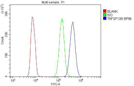
Figure 1. Flow Cytometry analysis of HL-60 cells using anti-TNFSF13B antibody (A01257).
Overlay histogram showing HL-60 cells stained with A01257 (Blue line).The cells were blocked with 10% normal goat serum. And then incubated with rabbit anti-TNFSF13B Antibody (A01257,1μg/1x106 cells) for 30 min at 20°C. DyLight®488 conjugated goat anti-rabbit IgG (BA1127, 5-10μg/1x106 cells) was used as secondary antibody for 30 minutes at 20°C. Isotype control antibody (Green line) was rabbit IgG (1μg/1x106) used under the same conditions. Unlabelled sample (Red line) was also used as a control.

Click image to see more details
Figure 2. Flow Cytometry analysis of U87 cells using anti-TNFSF13B antibody (A01257).
Overlay histogram showing U87 cells stained with A01257 (Blue line).The cells were blocked with 10% normal goat serum. And then incubated with rabbit anti-TNFSF13B Antibody (A01257,1μg/1x106 cells) for 30 min at 20°C. DyLight®488 conjugated goat anti-rabbit IgG (BA1127, 5-10μg/1x106 cells) was used as secondary antibody for 30 minutes at 20°C. Isotype control antibody (Green line) was rabbit IgG (1μg/1x106) used under the same conditions. Unlabelled sample (Red line) was also used as a control.

Click image to see more details
Figure 3. Western blot analysis of TNFSF13B using anti-TNFSF13B antibody (A01257).
Electrophoresis was performed on a 5-20% SDS-PAGE gel at 70V (Stacking gel) / 90V (Resolving gel) for 2-3 hours. The sample well of each lane was loaded with 50ug of sample under reducing conditions.
Lane 1: human U-937 whole cell lysates,
Lane 2: human Caco-2 whole cell lysates,
Lane 3: human PANC-1 whole cell lysates,
Lane 4: human CCRF-CEM whole cell lysates,
Lane 5: human MDA-MB-231 whole cell lysates.
After Electrophoresis, proteins were transferred to a Nitrocellulose membrane at 150mA for 50-90 minutes. Blocked the membrane with 5% Non-fat Milk/ TBS for 1.5 hour at RT. The membrane was incubated with rabbit anti-TNFSF13B antigen affinity purified polyclonal antibody (Catalog # A01257) at 0.5 μg/mL overnight at 4°C, then washed with TBS-0.1%Tween 3 times with 5 minutes each and probed with a goat anti-rabbit IgG-HRP secondary antibody at a dilution of 1:10000 for 1.5 hour at RT. The signal is developed using an Enhanced Chemiluminescent detection (ECL) kit (Catalog # EK1002) with Tanon 5200 system. A specific band was detected for TNFSF13B at approximately 34KD. The expected band size for TNFSF13B is at 34KD.

Click image to see more details
Figure 4. Western blot analysis of TNFSF13B using anti-TNFSF13B antibody (A01257).
Electrophoresis was performed on a 5-20% SDS-PAGE gel at 70V (Stacking gel) / 90V (Resolving gel) for 2-3 hours. The sample well of each lane was loaded with 50ug of sample under reducing conditions.
Lane 1: rat testicular issue lysates,
Lane 2: mouse testicular issue lysates.
After Electrophoresis, proteins were transferred to a Nitrocellulose membrane at 150mA for 50-90 minutes. Blocked the membrane with 5% Non-fat Milk/ TBS for 1.5 hour at RT. The membrane was incubated with rabbit anti-TNFSF13B antigen affinity purified polyclonal antibody (Catalog # A01257) at 0.5 μg/mL overnight at 4°C, then washed with TBS-0.1%Tween 3 times with 5 minutes each and probed with a goat anti-rabbit IgG-HRP secondary antibody at a dilution of 1:10000 for 1.5 hour at RT. The signal is developed using an Enhanced Chemiluminescent detection (ECL) kit (Catalog # EK1002) with Tanon 5200 system. A specific band was detected for TNFSF13B at approximately 34KD. The expected band size for TNFSF13B is at 34KD.
Protein Target Info & Infographic
Gene/Protein Information For TNFSF13B (Source: Uniprot.org, NCBI)
Gene Name
TNFSF13B
Full Name
Tumor necrosis factor ligand superfamily member 13B
Weight
31.223kDa
Superfamily
tumor necrosis factor family
Alternative Names
ApoL related ligand TALL-1; B lymphocyte stimulator; BAFF; BAFFB-cell activating factor; B-cell-activating factor; BLyS; BLYSB-lymphocyte stimulator; CD257 antigen; CD257; Dendritic cell-derived TNF-like molecule; DTL; TALL1; TALL-1delta BAFF; TALL1Delta4 BAFF; THANK; TNF- and APOL-related leukocyte expressed ligand 1; TNF and ApoL-related leukocyte expressed ligand 1; TNF homolog that activates apoptosis; TNFSF13B; TNFSF20; tumor necrosis factor (ligand) superfamily, member 13b; tumor necrosis factor (ligand) superfamily, member 20; tumor necrosis factor ligand superfamily member 13B; tumor n TNFSF13B BAFF, BLYS, CD257, DTL, TALL-1, TALL1, THANK, TNFSF20, TNLG7A, ZTNF4 TNF superfamily member 13b tumor necrosis factor ligand superfamily member 13B|ApoL related ligand TALL-1|B-cell-activating factor|B-lymphocyte stimulator|Delta4 BAFF|TNF and ApoL-related leukocyte expressed ligand 1|TNF homolog that activates apoptosis|delta BAFF|dendritic cell-derived TNF-like molecule|epididymis secretory sperm binding protein|tumor necrosis factor (ligand) superfamily, member 13b|tumor necrosis factor (ligand) superfamily, member 20|tumor necrosis factor ligand 7A|tumor necrosis factor superfamily member 13b|tumor necrosis factor-like protein ZTNF4
*If product is indicated to react with multiple species, protein info is based on the gene entry specified above in "Species".For more info on TNFSF13B, check out the TNFSF13B Infographic

We have 30,000+ of these available, one for each gene! Check them out.
In this infographic, you will see the following information for TNFSF13B: database IDs, superfamily, protein function, synonyms, molecular weight, chromosomal locations, tissues of expression, subcellular locations, post-translational modifications, and related diseases, research areas & pathways. If you want to see more information included, or would like to contribute to it and be acknowledged, please contact [email protected].
Specific Publications For Anti-BAFF/TNFSF13B Antibody Picoband™ (A01257)
Hello CJ!
A01257 has been cited in 1 publications:
*The publications in this section are manually curated by our staff scientists. They may differ from Bioz's machine gathered results. Both are accurate. If you find a publication citing this product but is missing from this list, please let us know we will issue you a thank-you coupon.
Expression of B-cell activating factor in acute lymphoblastic leukemia patients
Recommended Resources
Here are featured tools and databases that you might find useful.
- Boster's Pathways Library
- Protein Databases
- Bioscience Research Protocol Resources
- Data Processing & Analysis Software
- Photo Editing Software
- Scientific Literature Resources
- Research Paper Management Tools
- Molecular Biology Software
- Primer Design Tools
- Bioinformatics Tools
- Phylogenetic Tree Analysis
Customer Reviews
Have you used Anti-BAFF/TNFSF13B Antibody Picoband™?
Submit a review and receive an Amazon gift card.
- $30 for a review with an image
Be the first to review Anti-BAFF/TNFSF13B Antibody Picoband™
*The first user to submit a review for a product is eligible for Boster's Innovating Scientists Reward, which gives product credits. This is in addition to the gift card reward.
Customer Q&As
Have a question?
Find answers in Q&As, reviews.
Can't find your answer?
Submit your question
