Click image to see more details
-

-
-
-
-
+2
Product Info Summary
| SKU: | PA1021-2 |
|---|---|
| Size: | 100 μg/vial |
| Reactive Species: | Human, Rat |
| Host: | Rabbit |
| Application: | Flow Cytometry, IHC, WB |
Customers Who Bought This Also Bought


Product info
Product Name
Anti-CD44 antigen CD44 Antibody
SKU/Catalog Number
PA1021-2
Size
100 μg/vial
Form
Lyophilized
Description
Boster Bio Anti-CD44 antigen CD44 Antibody catalog # PA1021-2. Tested in Flow Cytometry, IHC, WB applications. This antibody reacts with Human, Rat.
Storage & Handling
Store at -20˚C for one year from date of receipt. After reconstitution, at 4˚C for one month. It can also be aliquotted and stored frozen at -20˚C for six months. Avoid repeated freeze-thaw cycles.
Cite This Product
Anti-CD44 antigen CD44 Antibody (Boster Biological Technology, Pleasanton CA, USA, Catalog # PA1021-2)
Host
Rabbit
Contents
Each vial contains 4 mg Trehalose, 0.9 mg NaCl and 0.2 mg Na2HPO4.
Clonality
Polyclonal
Isotype
Rabbit IgG
Immunogen
A synthetic peptide corresponding to a sequence at the C-terminus of human CD44, different from the related mouse and rat sequences by one amino acid.
*Blocking peptide can be purchased. Costs vary based on immunogen length. Contact us for pricing.
Cross-reactivity
No cross-reactivity with other proteins
Reactive Species
PA1021-2 is reactive to CD44 in Human, Rat
Reconstitution
Add 0.2ml of distilled water will yield a concentration of 500ug/ml.
Observed Molecular Weight
82 kDa
Calculated molecular weight
81538 MW
Background of CD44
The CD44 gene, which is a transmembrane protein, is expressed as a family of molecular isoforms generated from alternative RNA splicing and posttranslational modifications. The gene, which contains 19 exons spanning some 50 kb of genomic DNA, is a widely expressed integral membrane protein that acts as a receptor for hyaluronan (HA) and is important to cell-extracellular matrix interaction. CD44 binding with HA can play an important role in cellular aggregation and tumor cell growth. CD44 is necessary for limb development and functions in a novel growth factor presentation mechanism. A specific CD44 splice variant is crucial for the proliferation of these mesenchymal cells. CD44 glycoproteins are involved in leukocyte extravasation but also in the regulation of growth factor activation, stability, and signaling. Moreover, it plays a pivotal role in arteriogenesis.
Antibody Validation
Boster validates all antibodies on WB, IHC, ICC, Immunofluorescence, and ELISA with known positive control and negative samples to ensure specificity and high affinity, including thorough antibody incubations.
Application & Images
Applications
PA1021-2 is guaranteed for Flow Cytometry, IHC, WB Boster Guarantee
Assay Dilutions Recommendation
The recommendations below provide a starting point for assay optimization. The actual working concentration varies and should be decided by the user.
Western blot, 0.1-0.5μg/ml, Human, Rat
Immunohistochemistry (Paraffin-embedded Section), 2-5μg/ml, Human, Rat, By Heat
Flow Cytometry (Fixed), 1-3 μg/1x106 cells, Human
Positive Control
WB: human Hela whole cell, rat PC-12 whole cell
IHC: human laryngeal squamous cell carcinoma tissue, human rectal cancer tissue, rat lung tissue
FCM: HL-60 cell
Validation Images & Assay Conditions
Click image to see more details
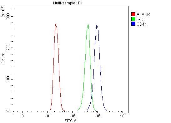
Figure 5. Flow Cytometry analysis of HL-60 cells using anti-CD44 antibody (PA1021-2).
Overlay histogram showing HL-60 cells stained with PA1021-2 (Blue line). To facilitate intracellular staining, cells were fixed with 4% paraformaldehyde and permeabilized with permeabilization buffer. The cells were blocked with 10% normal goat serum. And then incubated with rabbit anti-CD44 Antibody (PA1021-2, 1 μg/1x106 cells) for 30 min at 20°C. DyLight®488 conjugated goat anti-rabbit IgG (BA1127, 5-10 μg/1x106 cells) was used as secondary antibody for 30 minutes at 20°C. Isotype control antibody (Green line) was rabbit IgG (1 μg/1x106) used under the same conditions. Unlabelled sample without incubation with primary antibody and secondary antibody (Red line) was used as a blank control.

Click image to see more details
Figure 1. Western blot analysis of CD44 using anti-CD44 antibody (PA1021-2).
Electrophoresis was performed on a 5-20% SDS-PAGE gel at 70V (Stacking gel) / 90V (Resolving gel) for 2-3 hours. The sample well of each lane was loaded with 30 ug of sample under reducing conditions.
Lane 1: human Hela whole cell lysates,
Lane 2: rat PC-12 whole cell lysates.
After electrophoresis, proteins were transferred to a nitrocellulose membrane at 150 mA for 50-90 minutes. Blocked the membrane with 5% non-fat milk/TBS for 1.5 hour at RT. The membrane was incubated with rabbit anti-CD44 antigen affinity purified polyclonal antibody (Catalog # PA1021-2) at 0.5 μg/mL overnight at 4°C, then washed with TBS-0.1%Tween 3 times with 5 minutes each and probed with a goat anti-rabbit IgG-HRP secondary antibody at a dilution of 1:5000 for 1.5 hour at RT. The signal is developed using an Enhanced Chemiluminescent detection (ECL) kit (Catalog # EK1002) with Tanon 5200 system. A specific band was detected for CD44 at approximately 82 kDa. The expected band size for CD44 is at 82 kDa.

Click image to see more details
Figure 6. Western blot analysis of CD44 using anti-CD44 antigen CD44 antibody (PA1021-2).
Electrophoresis was performed on a 5-20% SDS-PAGE gel at 70V (Stacking gel) / 90V (Resolving gel) for 2-3 hours. The sample well of each lane was loaded with 30 ug of sample under reducing conditions.
After electrophoresis, proteins were transferred to a nitrocellulose membrane at 150 mA for 50-90 minutes. Blocked the membrane with 5% milk in PBS/0.05% Tween-20 (5% milk/PBS/Tw) for 1.5 hour at RT. The membrane was incubated with rabbit anti-CD44 antigen CD44 antibody (PA1021-2) at 1 ug/ml in 5% milk/BPS/Tw overnight at 4°C, then washed with TBS-0.1%Tween 3 times with 5 minutes each and probed with a goat anti-rabbit IgG-HRP secondary antibody at at 1:3,000 in 5% milk/PBS/Tw for 1.5 hour at RT. The signal is developed using a chemiluminescence: West Pico from Thermo Scientific. A specific band was detected for CD44 at approximately 81 kDa. The expected band size for CD44 is at 81 kDa.

Click image to see more details
Figure 2. IHC analysis of CD44 using anti-CD44 antibody (PA1021-2).
CD44 was detected in a paraffin-embedded section of human laryngeal squamous cell carcinoma tissue. Heat mediated antigen retrieval was performed in EDTA buffer (pH 8.0, epitope retrieval solution). The tissue section was blocked with 10% goat serum. The tissue section was then incubated with 2 μg/ml rabbit anti-CD44 Antibody (PA1021-2) overnight at 4°C. Peroxidase Conjugated Goat Anti-rabbit IgG was used as secondary antibody and incubated for 30 minutes at 37°C. The tissue section was developed using HRP Conjugated Rabbit IgG Super Vision Assay Kit (Catalog # SV0002) with DAB as the chromogen.

Click image to see more details
Figure 3. IHC analysis of CD44 using anti-CD44 antibody (PA1021-2).
CD44 was detected in a paraffin-embedded section of human rectal cancer tissue. Heat mediated antigen retrieval was performed in EDTA buffer (pH 8.0, epitope retrieval solution). The tissue section was blocked with 10% goat serum. The tissue section was then incubated with 2 μg/ml rabbit anti-CD44 Antibody (PA1021-2) overnight at 4°C. Peroxidase Conjugated Goat Anti-rabbit IgG was used as secondary antibody and incubated for 30 minutes at 37°C. The tissue section was developed using HRP Conjugated Rabbit IgG Super Vision Assay Kit (Catalog # SV0002) with DAB as the chromogen.

Click image to see more details
Figure 4. IHC analysis of CD44 using anti-CD44 antibody (PA1021-2).
CD44 was detected in a paraffin-embedded section of rat lung tissue. Heat mediated antigen retrieval was performed in EDTA buffer (pH 8.0, epitope retrieval solution). The tissue section was blocked with 10% goat serum. The tissue section was then incubated with 2 μg/ml rabbit anti-CD44 Antibody (PA1021-2) overnight at 4°C. Peroxidase Conjugated Goat Anti-rabbit IgG was used as secondary antibody and incubated for 30 minutes at 37°C. The tissue section was developed using HRP Conjugated Rabbit IgG Super Vision Assay Kit (Catalog # SV0002) with DAB as the chromogen.
Protein Target Info & Infographic
Gene/Protein Information For CD44 (Source: Uniprot.org, NCBI)
Gene Name
CD44
Full Name
CD44 antigen
Weight
81538 MW
Alternative Names
CD44 antigen;CDw44;Epican;Extracellular matrix receptor III;ECMR-III;GP90 lymphocyte homing/adhesion receptor;HUTCH-I;Heparan sulfate proteoglycan;Hermes antigen;Hyaluronate receptor;Phagocytic glycoprotein 1;PGP-1;Phagocytic glycoprotein I;PGP-I;CD44;CD44;LHR, MDU2, MDU3, MIC4; CD44 CDW44, CSPG8, ECMR-III, HCELL, HUTCH-I, IN, LHR, MC56, MDU2, MDU3, MIC4, Pgp1 CD44 molecule (Indian blood group) CD44 |GP90 lymphocyte homing/adhesion receptor|Hermes |Indian blood group |cell surface glycoprotein CD44|chondroitin sulfate proteoglycan 8|epican|extracellular matrix receptor III|hematopoietic cell E- and L-selectin ligand|heparan sulfate proteoglycan|homing function and Indian blood group system|hyaluronate receptor|phagocytic glycoprotein 1|soluble CD44
*If product is indicated to react with multiple species, protein info is based on the gene entry specified above in "Species".For more info on CD44, check out the CD44 Infographic

We have 30,000+ of these available, one for each gene! Check them out.
In this infographic, you will see the following information for CD44: database IDs, superfamily, protein function, synonyms, molecular weight, chromosomal locations, tissues of expression, subcellular locations, post-translational modifications, and related diseases, research areas & pathways. If you want to see more information included, or would like to contribute to it and be acknowledged, please contact [email protected].
Specific Publications For Anti-CD44 antigen CD44 Antibody (PA1021-2)
Hello CJ!
PA1021-2 has been cited in 35 publications:
*The publications in this section are manually curated by our staff scientists. They may differ from Bioz's machine gathered results. Both are accurate. If you find a publication citing this product but is missing from this list, please let us know we will issue you a thank-you coupon.
Clinical prognostic significance of cancer stem cell markers in patients with papillary thyroid carcinoma
CD44 promotes the migration of bone marrow-derived mesenchymal stem cells toward glioma
Rhus coriaria L. (Sumac) Demonstrates Oncostatic Activity in the Therapeutic and Preventive Model of Breast Carcinoma
Kubatka P,Kello M,Kajo K,Samec M,Jasek K,Vybohova D,Uramova S,Liskova A,Sadlonova V,Koklesova L,Murin R,Adamkov M,Smejkal K,Svajdlenka E,Solar P,Samuel SM,Kassayova M,Kwon TK,Zubor P,Pec M,Danko J,Büsselberg D,Mojzis J.Chemopreventive and Therapeutic Efficacy of Cinnamomum zeylanicum L. Bark in Experimental Breast Carcinoma: Mechanistic In Vivo and In Vitro Analyses.Molecules.2020 Mar 19;25(6):1399.doi:10.3390/molecules25061399.PMID:32204409;PMCID:PMC7144360.
Species: Mouse,Rat
PA1021-2 usage in article: APP:IHC, SAMPLE:MAMMARY GLAND TISSUE, DILUTION:NA
Wu,H.,Jiang,X.,Li,Y.,Zhou,Y.,Zhang,T.,Zhi,P.,Gao,J.,Engineering Stem Cell Derived Biomimetic Vesicles for Versatility and Effective Targeted Delivery.Adv. Funct.Mater.2020, 30, 2006169.https://doi.org/10.1002/adfm.202006169
Species: Rat,Mouse
PA1021-2 usage in article: APP:WB, SAMPLE:MSC, DILUTION:NA
Wang,Xiaoyu & Wang,Linlin & Qi,Lili & Bian,Huimiao & Yan,Xing & Wang,Yan & Li,Xiaodong & Cho,Kenka & Wu,Guojiang & Jiang,Baohong.(2020).Protective Effects of Ginsenoside Rg1 on Acute Myocardial Infarction. Journal of Pharmacy and Pharmacology Research.04.
Species: Rat
PA1021-2 usage in article: APP:IHC, SAMPLE:HEART TISSUE, DILUTION:1:100
Zhang GF,Wu JC,Wang HY,Jiang WD,Qiu L. Overexpression of microRNA-205-5p exerts suppressive effects on stem cell drug resistance in gallbladder cancer by down-regulating PRKCE. Biosci Rep.2020 Sep 30;40(9): BSR20194509.doi:10.1042/BSR20194509.PMID: 328698
Species: Human,Mouse
PA1021-2 usage in article: APP:FLOW CYTOMETRY, SAMPLE:GBC STEM CELLS, DILUTION:NA
Kubatka P,Kello M,Kajo K,Samec M,Liskova A,Jasek K,Koklesova L,Kuruc T,Adamkov M,Smejkal K,Svajdlenka E,Solar P,Pec M,Büsselberg D,Sadlonova V,Mojzis J.Rhus coriaria L.(Sumac) Demonstrates Oncostatic Activity in the Therapeutic and Preventive Model of Bre
Species: Human,Mouse,Rat
PA1021-2 usage in article: APP:IHC, SAMPLE:MAMMARY TISSUE, DILUTION:NA
Antineoplastic effects of clove buds (Syzygium aromaticum L.) in the model of breast carcinoma
Rat nasal respiratory mucosa-derived ectomesenchymal stem cells differentiate into Schwann-like cells promoting the differentiation of PC12 cells and %u2026
Recommended Resources
Here are featured tools and databases that you might find useful.
- Boster's Pathways Library
- Protein Databases
- Bioscience Research Protocol Resources
- Data Processing & Analysis Software
- Photo Editing Software
- Scientific Literature Resources
- Research Paper Management Tools
- Molecular Biology Software
- Primer Design Tools
- Bioinformatics Tools
- Phylogenetic Tree Analysis
Customer Reviews
Have you used Anti-CD44 antigen CD44 Antibody?
Submit a review and receive an Amazon gift card.
- $30 for a review with an image
1 Reviews For Anti-CD44 antigen CD44 Antibody
0
Anti-C-Terminal CD44 Antibody for Western Blotting
Excellent

Source: Biocompare.com
| Application | Western Blot |
|---|---|
| Sample | Tumor cell lysate |
| Primary Incubation | Overnight at 8-10 degrees Celcius, with rocking, 1 ug/ml in 5% milk/BPS/Tw |
| Blocking Agent | 5% milk in PBS/0.05% Tween-20 (5% milk/PBS/Tw) |
| Secondary Incubation | Goat anti-Rabbit antibody conjugated with HRP at 1:3,000 in 5% milk/PBS/Tw |
| Tertiary Incubation | HRP-bound secondary antibodies were detected by WestPico from ThermoScientific/Tw |
| Detection | Chemiluminescence: West Pico from Thermo Scientific |
| Results Summary | The antibody recognizes the expected ~80 kDa full-length CD44 and its low mol. wt. fragments containing C-terminal domain. The antibody is highly specific, produces "clean", definitive results; does not produce any non-specific bands. The antibody is sensitive and detects CD44 band when total protein per lane is loaded at 10-20 ug. The antibody is stable and could be re-used for blotting several times when stored in the original 5% milk/PBS/Tw solution at -20℃. |
"The antibody was used to detect the full length and cleaved fragments of human transmembrane protein CD44. The rabbit antibody PA1021-2 is sensitive, i.e. detects CD44 protein bands under reducing conditions and also when tested material is loaded at low total protein per lane. The antibody is highly specific, i.e. does not recognize any bands of unknown nature on the membrane."
Elena Deryugina
Verified customer
Submitted 2024-07-25
Customer Q&As
Have a question?
Find answers in Q&As, reviews.
Can't find your answer?
Submit your question
16 Customer Q&As for Anti-CD44 antigen CD44 Antibody
Question
I have a question about product PA1021-2, anti-CD44 antibody. I was wondering if it would be possible to conjugate this antibody with biotin. I would need it to be without BSA or sodium azide. I am planning on using a buffer exchange of sodium azide with PBS only. Would there be problems for me to conjugate the antibody and store it in -20 degrees in small aliquots?
Verified Customer
Verified customer
Asked: 2020-04-14
Answer
We do not advise storing this antibody with PBS buffer only in -20 degrees. If you want to store it in -20 degrees it is best to add some cryoprotectant like glycerol. If you want carrier free PA1021-2 anti-CD44 antibody, we can provide it to you in a special formula with trehalose and/or glycerol. These molecules will not interfere with conjugation chemistry and provide a good level of protection for the antibody from degradation. Please be sure to specify this in your purchase order.
Boster Scientific Support
Answered: 2020-04-14
Question
I was wanting to use your anti-CD44 antibody for ICC for rat spinal cord on frozen tissues, but I want to know if it has been tested for this particular application. Has this antibody been tested and is this antibody a good choice for rat spinal cord identification?
Verified Customer
Verified customer
Asked: 2020-02-27
Answer
As indicated on the product datasheet, PA1021-2 anti-CD44 antibody has been tested for IHC, ICC, WB on human, mouse, rat tissues. We have an innovator award program that if you test this antibody and show it works in rat spinal cord in IHC-frozen, you can get your next antibody for free.
Boster Scientific Support
Answered: 2020-02-27
Question
We have been able to see staining in rat foreskin. Do you have any suggestions? Is anti-CD44 antibody supposed to stain foreskin positively?
Verified Customer
Verified customer
Asked: 2020-01-14
Answer
According to literature foreskin does express CD44. According to Uniprot.org, CD44 is expressed in parotid gland, reticulocyte, myeloid leukemia cell, keratinocyte, lymphoblast, mammary carcinoma, articular cartilage, colon adenocarcinoma retinal pigment epithelium, spinal cord, pancreas retinal pigment epithelium, glial tumor, peripheral blood, foreskin, lung, plasma, cervix carcinoma, t-cell, liver, leukemic t-cell, among other tissues. Regarding which tissues have CD44 expression, here are a few articles citing expression in various tissues:
Cervix carcinoma, Pubmed ID: 17081983, 18669648, 18691976, 20068231, 23186163
Foreskin, Pubmed ID: 2007624
Glial tumor, Pubmed ID: 7527301
Keratinocyte, Pubmed ID: 1281868
Leukemic T-cell, Pubmed ID: 19690332
Liver, Pubmed ID: 19159218, 24275569
Lung, Pubmed ID: 1717145, 7528188
Lymphoblast, Pubmed ID: 1465456, 1922057
Mammary carcinoma, Pubmed ID: 8352881
Myeloid leukemia cell, Pubmed ID: 2056274
Pancreas, and Retinal pigment epithelium, Pubmed ID: 15489334
Peripheral blood, Pubmed ID: 7508992
Plasma, Pubmed ID: 16335952
Reticulocyte, Pubmed ID: 1840487
Spinal cord, Pubmed ID: 17974005
T-cell, Pubmed ID: 19367720
Boster Scientific Support
Answered: 2020-01-14
Question
My boss were happy with the WB result of your anti-CD44 antibody. However we have observed positive staining in cervix carcinoma cell membrane using this antibody. Is that expected? Could you tell me where is CD44 supposed to be expressed?
Verified Customer
Verified customer
Asked: 2020-01-06
Answer
From what I have seen in literature, cervix carcinoma does express CD44. Generally CD44 expresses in cell membrane. Regarding which tissues have CD44 expression, here are a few articles citing expression in various tissues:
Cervix carcinoma, Pubmed ID: 17081983, 18669648, 18691976, 20068231, 23186163
Foreskin, Pubmed ID: 2007624
Glial tumor, Pubmed ID: 7527301
Keratinocyte, Pubmed ID: 1281868
Leukemic T-cell, Pubmed ID: 19690332
Liver, Pubmed ID: 19159218, 24275569
Lung, Pubmed ID: 1717145, 7528188
Lymphoblast, Pubmed ID: 1465456, 1922057
Mammary carcinoma, Pubmed ID: 8352881
Myeloid leukemia cell, Pubmed ID: 2056274
Pancreas, and Retinal pigment epithelium, Pubmed ID: 15489334
Peripheral blood, Pubmed ID: 7508992
Plasma, Pubmed ID: 16335952
Reticulocyte, Pubmed ID: 1840487
Spinal cord, Pubmed ID: 17974005
T-cell, Pubmed ID: 19367720
Boster Scientific Support
Answered: 2020-01-06
Question
I was wanting to use to test anti-CD44 antibody PA1021-2 on rat spinal cord for research purposes, then I may be interested in using anti-CD44 antibody PA1021-2 for diagnostic purposes as well. Is the antibody suitable for diagnostic purposes?
Verified Customer
Verified customer
Asked: 2019-08-29
Answer
The products we sell, including anti-CD44 antibody PA1021-2, are only intended for research use. They would not be suitable for use in diagnostic work. If you have the means to develop a product into diagnostic use, and are interested in collaborating with us and develop our product into an IVD product, please contact us for more discussions.
Boster Scientific Support
Answered: 2019-08-29
Question
We are currently using anti-CD44 antibody PA1021-2 for rat tissue, and we are satisfied with the IHC results. The species of reactivity given in the datasheet says human, mouse, rat. Is it possible that the antibody can work on pig tissues as well?
Verified Customer
Verified customer
Asked: 2019-08-02
Answer
The anti-CD44 antibody (PA1021-2) has not been tested for cross reactivity specifically with pig tissues, though there is a good chance of cross reactivity. We have an innovator award program that if you test this antibody and show it works in pig you can get your next antibody for free. Please contact me if I can help you with anything.
Boster Scientific Support
Answered: 2019-08-02
Question
I am interested in using your anti-CD44 antibody for t cell activation studies. Has this antibody been tested with western blotting on hela whole cell lysate? We would like to see some validation images before ordering.
Verified Customer
Verified customer
Asked: 2019-07-17
Answer
We appreciate your inquiry. This PA1021-2 anti-CD44 antibody is validated on intestinal cancer tissue, tissue lysate, rat ovary tissue, testis tissue, hela whole cell lysate, hepa whole cell lysate. It is guaranteed to work for IHC, ICC, WB in human, mouse, rat. Our Boster guarantee will cover your intended experiment even if the sample type has not been be directly tested.
Boster Scientific Support
Answered: 2019-07-17
Question
Is a blocking peptide available for product anti-CD44 antibody (PA1021-2)?
Verified Customer
Verified customer
Asked: 2019-04-17
Answer
We do provide the blocking peptide for product anti-CD44 antibody (PA1021-2). If you would like to place an order for it please contact [email protected] and make a special request.
Boster Scientific Support
Answered: 2019-04-17
Question
I see that the anti-CD44 antibody PA1021-2 works with ICC, what is the protocol used to produce the result images on the product page?
J. Wu
Verified customer
Asked: 2019-01-09
Answer
You can find protocols for ICC on the "support/technical resources" section of our navigation menu. If you have any further questions, please send an email to [email protected]
Boster Scientific Support
Answered: 2019-01-09
Question
Would anti-CD44 antibody PA1021-2 work for ICC with spinal cord?
A. Li
Verified customer
Asked: 2018-04-17
Answer
According to the expression profile of spinal cord, CD44 is highly expressed in spinal cord. So, it is likely that anti-CD44 antibody PA1021-2 will work for ICC with spinal cord.
Boster Scientific Support
Answered: 2018-04-17
Question
See below the WB image, lot number and protocol we used for spinal cord using anti-CD44 antibody PA1021-2. Please let me know if you require anything else.
Verified Customer
Verified customer
Asked: 2017-11-17
Answer
Thank you very much for the data. Our lab team are working to resolve this as quickly as possible, and we appreciate your patience and understanding! You have provided everything we needed. Please let me know if there is anything you need in the meantime.
Boster Scientific Support
Answered: 2017-11-17
Question
Is this PA1021-2 anti-CD44 antibody reactive to the isotypes of CD44?
Verified Customer
Verified customer
Asked: 2017-07-28
Answer
The immunogen of PA1021-2 anti-CD44 antibody is A synthetic peptide corresponding to a sequence at the C-terminus of human CD44(728-742aa DETRNLQNVDMKIGV), different from the related mouse and rat sequences by one amino acid. Could you tell me which isotype you are interested in so I can help see if the immunogen is part of this isotype?
Boster Scientific Support
Answered: 2017-07-28
Question
We ordered your anti-CD44 antibody for IHC on t-cell in the past. I am using mouse, and We are going to use the antibody for ICC next. I was wanting to use examining t-cell as well as lymphoblast in our next experiment. Do you have any suggestion on which antibody would work the best for ICC?
Verified Customer
Verified customer
Asked: 2017-07-28
Answer
I have checked the website and datasheets of our anti-CD44 antibody and it seems that PA1021-2 has been validated on mouse in both IHC and ICC. Thus PA1021-2 should work for your application. Our Boster satisfaction guarantee will cover this product for ICC in mouse even if the specific tissue type has not been validated. We do have a comprehensive range of products for ICC detection and you can check out our website bosterbio.com to find out more information about them.
Boster Scientific Support
Answered: 2017-07-28
Question
Will PA1021-2 anti-CD44 antibody work on parafin embedded sections? If so, which fixation method do you recommend we use (PFA, paraformaldehyde, other)?
Verified Customer
Verified customer
Asked: 2017-05-23
Answer
As indicated on the product datasheet, PA1021-2 anti-CD44 antibody as been validated on ICC. It is best to use PFA for fixation because it has better tissue penetration ability. PFA needs to be prepared fresh before use. Long term stored PFA turns into formalin, as the PFA molecules congregate and become formalin.
Boster Scientific Support
Answered: 2017-05-23
Question
We appreciate helping with my inquiry over the phone. Here are the WB image, lot number and protocol we used for spinal cord using anti-CD44 antibody PA1021-2. Let me know if you need anything else.
A. Mitchell
Verified customer
Asked: 2016-01-12
Answer
I appreciate the data. You have provided everything we needed. Our lab team are working to resolve your inquiry as quickly as possible, and we appreciate your patience and understanding! Please let me know if there is anything you need in the meantime.
Boster Scientific Support
Answered: 2016-01-12
Question
Do you have a BSA free version of anti-CD44 antibody PA1021-2 available?
R. Kulkarni
Verified customer
Asked: 2014-02-11
Answer
I appreciate your recent telephone inquiry. I can confirm that some lots of this anti-CD44 antibody PA1021-2 are BSA free. For now, these lots are available and we can make a BSA free formula for you free of charge. It will take 3 extra days to prepare. If you require this antibody BSA free again in future, please do not hesitate to contact me and I will be pleased to check which lots we have in stock that are BSA free.
Boster Scientific Support
Answered: 2014-02-11
