Click image to see more details
-

-
-
-
-
+1
Product Info Summary
| SKU: | A02296-3 |
|---|---|
| Size: | 100 μg/vial |
| Reactive Species: | Human, Mouse, Rat |
| Host: | Rabbit |
| Application: | IF, IHC, ICC, WB |
Customers Who Bought This Also Bought


Product info
Product Name
Anti-EEA1 Antibody Picoband™
SKU/Catalog Number
A02296-3
Size
100 μg/vial
Form
Lyophilized
Description
Boster Bio Anti-EEA1 Antibody Picoband™ catalog # A02296-3. Tested in IF, IHC, ICC, WB applications. This antibody reacts with Human, Mouse, Rat.
Storage & Handling
Store at -20˚C for one year from date of receipt. After reconstitution, at 4˚C for one month. It can also be aliquotted and stored frozen at -20˚C for six months. Avoid repeated freeze-thaw cycles.
Cite This Product
Anti-EEA1 Antibody Picoband™ (Boster Biological Technology, Pleasanton CA, USA, Catalog # A02296-3)
Host
Rabbit
Contents
Each vial contains 4mg Trehalose, 0.9mg NaCl, 0.2mg Na2HPO4, 0.01mg NaN3.
Clonality
Polyclonal
Isotype
Rabbit IgG
Immunogen
A synthetic peptide corresponding to a sequence at the C-terminus of human EEA1, identical to the related mouse and rat sequences.
*Blocking peptide can be purchased. Costs vary based on immunogen length. Contact us for pricing.
Cross-reactivity
No cross-reactivity with other proteins.
Reactive Species
A02296-3 is reactive to EEA1 in Human, Mouse, Rat
Applications
A02296-3 is guaranteed for IF, IHC, ICC, WB Boster Guarantee
Observed Molecular Weight
170 kDa
Calculated molecular weight
162.466kDa
Background of EEA1
The gene EEA1 encodes for the 1400 amino acid protein, Early Endosome Antigen 1. It localizes exclusively to early endosomes and has an important role in endosomal trafficking. EEA1 binds directly to the phospholipid phosphatidylinositol 3-phosphate through its C-terminal FYVE domain and forms ahomodimer through a coiled coil. Furthermore, EEA1 acts as a tethering molecule that couples vesicle docking with SNAREs such as N-ethylmaleimide sensitive fusion protein, bringing the endosomes physically closer and ultimately resulting in the fusion and delivery of endosomal cargo.
Antibody Validation
Boster validates all antibodies on WB, IHC, ICC, Immunofluorescence, and ELISA with known positive control and negative samples to ensure specificity and high affinity, including thorough antibody incubations.
Innovating Scientists Reward
If you are the first to review this product, or if you have results for a special sample, species or application this product is not validated in, share your results with us and receive product credits you can use towards any Boster products! Applicable to all scientists worldwide.
Submit A Review
Assay dilution & Images
Reconsitution
Add 0.2ml of distilled water will yield a concentration of 500ug/ml.
Assay Dilutions Recommendation
The recommendations below provide a starting point for assay optimization. The actual working concentration varies and should be decided by the user.
Western blot, 0.1-0.5μg/ml
Immunohistochemistry, 2-5 μg/ml
Immunocytochemistry/Immunofluorescence, 5μg/ml
Validation Images & Assay Conditions
Click image to see more details
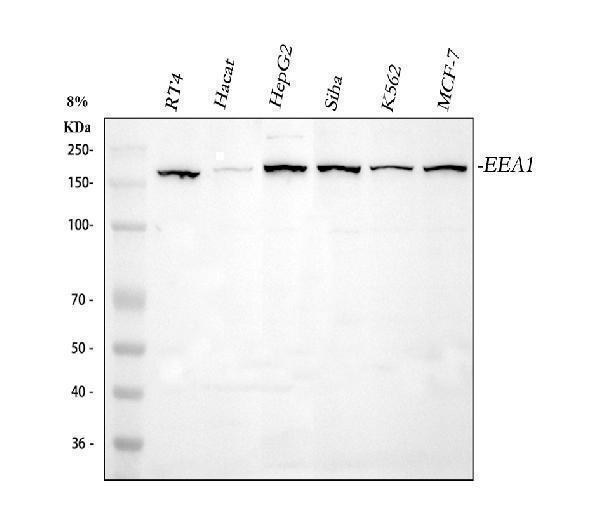
Figure 1. Western blot analysis of EEA1 using anti-EEA1 antibody (A02296-3).
Electrophoresis was performed on a 5-20% SDS-PAGE gel at 70V (Stacking gel) / 90V (Resolving gel) for 2-3 hours. The sample well of each lane was loaded with 30 ug of sample under reducing conditions.
Lane 1: human RT4 whole cell lysates,
Lane 2: human Hacat whole cell lysates,
Lane 3: human HepG2 whole cell lysates,
Lane 4: human SiHa whole cell lysates,
Lane 5: humna K562 whole cell lysates,
Lane 6: humna MCF-7 whole cell lysates.
After electrophoresis, proteins were transferred to a nitrocellulose membrane at 150 mA for 50-90 minutes. Blocked the membrane with 5% non-fat milk/TBS for 1.5 hour at RT. The membrane was incubated with rabbit anti-EEA1 antigen affinity purified polyclonal antibody (Catalog # A02296-3) at 0.5 μg/mL overnight at 4°C, then washed with TBS-0.1%Tween 3 times with 5 minutes each and probed with a goat anti-rabbit IgG-HRP secondary antibody at a dilution of 1:5000 for 1.5 hour at RT. The signal is developed using an Enhanced Chemiluminescent detection (ECL) kit (Catalog # EK1002) with Tanon 5200 system. A specific band was detected for EEA1 at approximately 162 kDa. The expected band size for EEA1 is at 162 kDa.

Click image to see more details
Figure 2. Western blot analysis of EEA1 using anti-EEA1 antibody (A02296-3).
Electrophoresis was performed on a 5-20% SDS-PAGE gel at 70V (Stacking gel) / 90V (Resolving gel) for 2-3 hours. The sample well of each lane was loaded with 30 ug of sample under reducing conditions.
Lane 1: rat brain tissue lysates,
Lane 2: rat lung tissue lysates,
Lane 3: mouse brain tissue lysates,
Lane 4: mouse kidney tissue lysates,
Lane 5: mouse lung tissue lysates.
After electrophoresis, proteins were transferred to a nitrocellulose membrane at 150 mA for 50-90 minutes. Blocked the membrane with 5% non-fat milk/TBS for 1.5 hour at RT. The membrane was incubated with rabbit anti-EEA1 antigen affinity purified polyclonal antibody (Catalog # A02296-3) at 0.5 μg/mL overnight at 4°C, then washed with TBS-0.1%Tween 3 times with 5 minutes each and probed with a goat anti-rabbit IgG-HRP secondary antibody at a dilution of 1:5000 for 1.5 hour at RT. The signal is developed using an Enhanced Chemiluminescent detection (ECL) kit (Catalog # EK1002) with Tanon 5200 system. A specific band was detected for EEA1 at approximately 162 kDa. The expected band size for EEA1 is at 162 kDa.

Click image to see more details
Figure 3. IHC analysis of EEA1 using anti-EEA1 antibody (A02296-3).
EEA1 was detected in a paraffin-embedded section of human breast cancer tissue. Heat mediated antigen retrieval was performed in EDTA buffer (pH 8.0, epitope retrieval solution). The tissue section was blocked with 10% goat serum. The tissue section was then incubated with 2 μg/ml rabbit anti-EEA1 Antibody (A02296-3) overnight at 4°C. Peroxidase Conjugated Goat Anti-rabbit IgG was used as secondary antibody and incubated for 30 minutes at 37°C. The tissue section was developed using HRP Conjugated Rabbit IgG Super Vision Assay Kit (Catalog # SV0002) with DAB as the chromogen.

Click image to see more details
Figure 4. IHC analysis of EEA1 using anti-EEA1 antibody (A02296-3).
EEA1 was detected in a paraffin-embedded section of rat brain tissue. Heat mediated antigen retrieval was performed in EDTA buffer (pH 8.0, epitope retrieval solution). The tissue section was blocked with 10% goat serum. The tissue section was then incubated with 2 μg/ml rabbit anti-EEA1 Antibody (A02296-3) overnight at 4°C. Peroxidase Conjugated Goat Anti-rabbit IgG was used as secondary antibody and incubated for 30 minutes at 37°C. The tissue section was developed using HRP Conjugated Rabbit IgG Super Vision Assay Kit (Catalog # SV0002) with DAB as the chromogen.

Click image to see more details
Figure 5. IF analysis of EEA1 using anti- EEA1 antibody (A02296-3).
EEA1 was detected in immunocytochemical section of A549 cells. Enzyme antigen retrieval was performed using IHC enzyme antigen retrieval reagent (AR0022) for 15 mins. The cells were blocked with 10% goat serum. And then incubated with 5μg/mL rabbit anti- EEA1 Antibody (A02296-3) overnight at 4°C. DyLight®550 Conjugated Goat Anti-Rabbit IgG (BA1135) was used as secondary antibody at 1:500 dilution and incubated for 30 minutes at 37°C. The section was counterstained with DAPI. Visualize using a fluorescence microscope and filter sets appropriate for the label used.
Protein Target Info & Infographic
Gene/Protein Information For EEA1 (Source: Uniprot.org, NCBI)
Gene Name
EEA1
Full Name
Early endosome antigen 1
Weight
162.466kDa
Alternative Names
early endosome antigen 1; early endosome antigen 1, 162kD; early endosome-associated protein; EEA1; Endosome-associated protein p162; MST105; MSTP105; ZFYVE2; ZFYVE2MST105; Zinc finger FYVE domain-containing protein 2 EEA1 MST105, MSTP105, ZFYVE2 early endosome antigen 1 early endosome antigen 1|early endosome antigen 1, 162kD|early endosome-associated protein|endosome-associated protein p162|zinc finger FYVE domain-containing protein 2
*If product is indicated to react with multiple species, protein info is based on the gene entry specified above in "Species".For more info on EEA1, check out the EEA1 Infographic

We have 30,000+ of these available, one for each gene! Check them out.
In this infographic, you will see the following information for EEA1: database IDs, superfamily, protein function, synonyms, molecular weight, chromosomal locations, tissues of expression, subcellular locations, post-translational modifications, and related diseases, research areas & pathways. If you want to see more information included, or would like to contribute to it and be acknowledged, please contact [email protected].
Specific Publications For Anti-EEA1 Antibody Picoband™ (A02296-3)
Hello CJ!
A02296-3 has been cited in 1 publications:
*The publications in this section are manually curated by our staff scientists. They may differ from Bioz's machine gathered results. Both are accurate. If you find a publication citing this product but is missing from this list, please let us know we will issue you a thank-you coupon.
Wang,Y.,Pang,J.,Wang,Q.,Yan,L.,Wang,L.,Xing,Z.,Wang,C.,Zhang,J., Dong,L.,Delivering Antisense Oligonucleotides across the Blood‐Brain Barrier by Tumor Cell‐Derived Small Apoptotic Bodies.Adv.Sci.2021,2004929.https://doi.org/10.1002/advs.202004929
Species: Mouse
A02296-3 usage in article: APP:TRANSCYTOSIS ANALYSIS, SAMPLE: B16F10 CELL, DILUTION:NA
Recommended Resources
Here are featured tools and databases that you might find useful.
- Boster's Pathways Library
- Protein Databases
- Bioscience Research Protocol Resources
- Data Processing & Analysis Software
- Photo Editing Software
- Scientific Literature Resources
- Research Paper Management Tools
- Molecular Biology Software
- Primer Design Tools
- Bioinformatics Tools
- Phylogenetic Tree Analysis
Customer Reviews
Have you used Anti-EEA1 Antibody Picoband™?
Submit a review and receive an Amazon gift card.
- $30 for a review with an image
Be the first to review Anti-EEA1 Antibody Picoband™
*The first user to submit a review for a product is eligible for Boster's Innovating Scientists Reward, which gives product credits. This is in addition to the gift card reward.
Customer Q&As
Have a question?
Find answers in Q&As, reviews.
Can't find your answer?
Submit your question
5 Customer Q&As for Anti-EEA1 Antibody Picoband™
Question
We are currently using anti-EEA1 antibody A02296-3 for mouse tissue, and we are happy with the WB results. The species of reactivity given in the datasheet says human, mouse, rat. Is it likely that the antibody can work on monkey tissues as well?
Verified Customer
Verified customer
Asked: 2020-04-09
Answer
The anti-EEA1 antibody (A02296-3) has not been validated for cross reactivity specifically with monkey tissues, but there is a good chance of cross reactivity. We have an innovator award program that if you test this antibody and show it works in monkey you can get your next antibody for free. Please contact me if I can help you with anything.
Boster Scientific Support
Answered: 2020-04-09
Question
We ordered your anti-EEA1 antibody for Flow Cytometry on cervix carcinoma erythroleukemia in a previous experiment. I am using human, and We intend to use the antibody for WB next. My lab would like examining cervix carcinoma erythroleukemia as well as biceps brachii in our next experiment. Could you please give me some suggestion on which antibody would work the best for WB?
Verified Customer
Verified customer
Asked: 2019-10-04
Answer
I have checked the website and datasheets of our anti-EEA1 antibody and it appears that A02296-3 has been tested on human in both Flow Cytometry and WB. Thus A02296-3 should work for your application. Our Boster satisfaction guarantee will cover this product for WB in human even if the specific tissue type has not been validated. We do have a comprehensive range of products for WB detection and you can check out our website bosterbio.com to find out more information about them.
Boster Scientific Support
Answered: 2019-10-04
Question
We have seen staining in rat liver. What should we do? Is anti-EEA1 antibody supposed to stain liver positively?
Verified Customer
Verified customer
Asked: 2019-07-26
Answer
From what I have seen in literature liver does express EEA1. From what I have seen in Uniprot.org, EEA1 is expressed in biceps brachii, cervix carcinoma, fetal brain cortex, cervix carcinoma erythroleukemia, liver, among other tissues. Regarding which tissues have EEA1 expression, here are a few articles citing expression in various tissues:
Cervix carcinoma, Pubmed ID: 7768953, 20068231
Cervix carcinoma, and Erythroleukemia, Pubmed ID: 23186163
Fetal brain cortex, Pubmed ID: 9697774
Liver, Pubmed ID: 24275569
Boster Scientific Support
Answered: 2019-07-26
Question
My boss were content with the WB result of your anti-EEA1 antibody. However we have been able to see positive staining in fetal brain cortex cytoplasm. early endosome membrane using this antibody. Is that expected? Could you tell me where is EEA1 supposed to be expressed?
A. Evans
Verified customer
Asked: 2019-07-23
Answer
From literature, fetal brain cortex does express EEA1. Generally EEA1 expresses in cytoplasm. early endosome membrane. Regarding which tissues have EEA1 expression, here are a few articles citing expression in various tissues:
Cervix carcinoma, Pubmed ID: 7768953, 20068231
Cervix carcinoma, and Erythroleukemia, Pubmed ID: 23186163
Fetal brain cortex, Pubmed ID: 9697774
Liver, Pubmed ID: 24275569
Boster Scientific Support
Answered: 2019-07-23
Question
you antibody using your anti-EEA1 antibody for early endosome to late endosome transport studies. Has this antibody been tested with western blotting on nih3t3 cell lysate? We would like to see some validation images before ordering.
Verified Customer
Verified customer
Asked: 2019-04-23
Answer
Thank you for your inquiry. This A02296-3 anti-EEA1 antibody is validated on rat thymus tissue, spleen tissue, tissue lysate, brain tissue, lung tissue, mouse spleen tissue, nih3t3 cell lysate, human placenta tissue, hela cell lysate, a549 cell lysate. It is guaranteed to work for Flow Cytometry, WB in human, mouse, rat. Our Boster guarantee will cover your intended experiment even if the sample type has not been be directly tested.
Boster Scientific Support
Answered: 2019-04-23
