Click image to see more details
-

-
-
-
-
+4
Product Info Summary
| SKU: | A00723 |
|---|---|
| Size: | 100 μg/vial |
| Reactive Species: | Human, Monkey, Mouse, Rat |
| Host: | Rabbit |
| Application: | IHC, WB |
Customers Who Bought This Also Bought


Product info
Product Name
Anti-FOXP1 Antibody Picoband™
SKU/Catalog Number
A00723
Size
100 μg/vial
Form
Lyophilized
Description
Boster Bio Anti-FOXP1 Antibody Picoband™ catalog # A00723. Tested in IHC, WB applications. This antibody reacts with Human, Monkey, Mouse, Rat.
Storage & Handling
Store at -20˚C for one year from date of receipt. After reconstitution, at 4˚C for one month. It can also be aliquotted and stored frozen at -20˚C for six months. Avoid repeated freeze-thaw cycles.
Cite This Product
Anti-FOXP1 Antibody Picoband™ (Boster Biological Technology, Pleasanton CA, USA, Catalog # A00723)
Host
Rabbit
Contents
Each vial contains 4 mg Trehalose, 0.9 mg NaCl and 0.2 mg Na2HPO4.
Clonality
Polyclonal
Isotype
Rabbit IgG
Immunogen
A synthetic peptide corresponding to a sequence at the C-terminus of human FOXP1, identical to the related mouse and rat sequences.
*Blocking peptide can be purchased. Costs vary based on immunogen length. Contact us for pricing.
Cross-reactivity
No cross-reactivity with other proteins.
Reactive Species
A00723 is reactive to FOXP1 in Human, Monkey, Mouse, Rat
Applications
A00723 is guaranteed for IHC, WB Boster Guarantee
Observed Molecular Weight
82 kDa
Calculated molecular weight
75.317kDa
Background of FOXP1
FOXP1 ("forkhead box P1") is a gene that is necessary for the proper development of the brain and lung in mammals. It is a member of the large FOX family of transcription factors. This gene belongs to subfamily P of the forkhead box (FOX) transcription factor family. Forkhead box transcription factors play important roles in the regulation of tissue- and cell type-specific gene transcription during both development and adulthood. Forkhead box P1 protein contains both DNA-binding- and protein-protein binding-domains. This gene may act as a tumor suppressor as it is lost in several tumor types and maps to a chromosomal region (3p14.1) reported to contain a tumor suppressor gene (s). Alternative splicing results in multiple transcript variants encoding different isoforms. It was shown that the embryonic stem cell (ESC)-specific isoform of FOXP1 stimulates the expression of transcription factor genes required for pluripotency, including OCT4, NANOG, NR5A2, and GDF3, while concomitantly repressing genes required for ESC differentiation. This isoform also promotes the maintenance of ESC pluripotency and contributes to efficient reprogramming of somatic cells into induced pluripotent stem cells. These results reveal a pivotal role for an Alternative splicing event in the regulation of pluripotency through the control of critical ESC-specific transcriptional programs (2).
Antibody Validation
Boster validates all antibodies on WB, IHC, ICC, Immunofluorescence, and ELISA with known positive control and negative samples to ensure specificity and high affinity, including thorough antibody incubations.
Innovating Scientists Reward
If you are the first to review this product, or if you have results for a special sample, species or application this product is not validated in, share your results with us and receive product credits you can use towards any Boster products! Applicable to all scientists worldwide.
Submit A Review
Assay dilution & Images
Reconsitution
Add 0.2ml of distilled water will yield a concentration of 500ug/ml.
Assay Dilutions Recommendation
The recommendations below provide a starting point for assay optimization. The actual working concentration varies and should be decided by the user.
Western blot, 0.25-0.5μg/ml, Human, Monkey
Immunohistochemistry (Paraffin-embedded Section), 1-2μg/ml, Human, Mouse, Rat
Validation Images & Assay Conditions
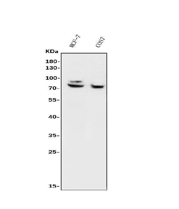
Click image to see more details
Figure 1. Western blot analysis of FOXP1 using anti-FOXP1 antibody (A00723).
Electrophoresis was performed on a 5-20% SDS-PAGE gel at 70V (Stacking gel) / 90V (Resolving gel) for 2-3 hours. The sample well of each lane was loaded with 30ug of sample under reducing conditions.
Lane 1: human MCF-7whole cell lysates,
Lane 2: monkey COS-7 whole cell lysates.
After Electrophoresis, proteins were transferred to a Nitrocellulose membrane at 150mA for 50-90 minutes. Blocked the membrane with 5% Non-fat Milk/ TBS for 1.5 hour at RT. The membrane was incubated with rabbit anti-FOXP1 antigen affinity purified polyclonal antibody (Catalog # A00723) at 0.5 μg/mL overnight at 4°C, then washed with TBS-0.1%Tween 3 times with 5 minutes each and probed with a goat anti-rabbit IgG-HRP secondary antibody at a dilution of 1:5000 for 1.5 hour at RT. The signal is developed using an Enhanced Chemiluminescent detection (ECL) kit (Catalog # EK1002) with Tanon 5200 system. A specific band was detected for FOXP1 at approximately 82KD. The expected band size for FOXP1 is at 82KD.

Click image to see more details
Figure 2. IHC analysis of FOXP1 using anti-FOXP1 antibody (A00723).
FOXP1 was detected in paraffin-embedded section of human mammary cancer tissue. Heat mediated antigen retrieval was performed in EDTA buffer (pH8.0, epitope retrieval solution). The tissue section was blocked with 10% goat serum. The tissue section was then incubated with 2μg/ml rabbit anti-FOXP1 Antibody (A00723) overnight at 4°C. Biotinylated goat anti-rabbit IgG was used as secondary antibody and incubated for 30 minutes at 37°C. The tissue section was developed using Strepavidin-Biotin-Complex (SABC) (Catalog # SA1022) with DAB as the chromogen.

Click image to see more details
Figure 3. IHC analysis of FOXP1 using anti-FOXP1 antibody (A00723).
FOXP1 was detected in paraffin-embedded section of human mammary cancer tissue. Heat mediated antigen retrieval was performed in EDTA buffer (pH8.0, epitope retrieval solution). The tissue section was blocked with 10% goat serum. The tissue section was then incubated with 2μg/ml rabbit anti-FOXP1 Antibody (A00723) overnight at 4°C. Biotinylated goat anti-rabbit IgG was used as secondary antibody and incubated for 30 minutes at 37°C. The tissue section was developed using Strepavidin-Biotin-Complex (SABC) (Catalog # SA1022) with DAB as the chromogen.

Click image to see more details
Figure 4. IHC analysis of FOXP1 using anti-FOXP1 antibody (A00723).
FOXP1 was detected in paraffin-embedded section of human rectal cancer tissue. Heat mediated antigen retrieval was performed in EDTA buffer (pH8.0, epitope retrieval solution). The tissue section was blocked with 10% goat serum. The tissue section was then incubated with 2μg/ml rabbit anti-FOXP1 Antibody (A00723) overnight at 4°C. Biotinylated goat anti-rabbit IgG was used as secondary antibody and incubated for 30 minutes at 37°C. The tissue section was developed using Strepavidin-Biotin-Complex (SABC) (Catalog # SA1022) with DAB as the chromogen.

Click image to see more details
Figure 5. IHC analysis of FOXP1 using anti-FOXP1 antibody (A00723).
FOXP1 was detected in paraffin-embedded section of human rectal cancer tissue. Heat mediated antigen retrieval was performed in EDTA buffer (pH8.0, epitope retrieval solution). The tissue section was blocked with 10% goat serum. The tissue section was then incubated with 2μg/ml rabbit anti-FOXP1 Antibody (A00723) overnight at 4°C. Biotinylated goat anti-rabbit IgG was used as secondary antibody and incubated for 30 minutes at 37°C. The tissue section was developed using Strepavidin-Biotin-Complex (SABC) (Catalog # SA1022) with DAB as the chromogen.

Click image to see more details
Figure 6. IHC analysis of FOXP1 using anti-FOXP1 antibody (A00723).
FOXP1 was detected in paraffin-embedded section of mouse brain tissue. Heat mediated antigen retrieval was performed in EDTA buffer (pH8.0, epitope retrieval solution). The tissue section was blocked with 10% goat serum. The tissue section was then incubated with 2μg/ml rabbit anti-FOXP1 Antibody (A00723) overnight at 4°C. Biotinylated goat anti-rabbit IgG was used as secondary antibody and incubated for 30 minutes at 37°C. The tissue section was developed using Strepavidin-Biotin-Complex (SABC) (Catalog # SA1022) with DAB as the chromogen.

Click image to see more details
Figure 7. IHC analysis of FOXP1 using anti-FOXP1 antibody (A00723).
FOXP1 was detected in paraffin-embedded section of mouse brain tissue. Heat mediated antigen retrieval was performed in EDTA buffer (pH8.0, epitope retrieval solution). The tissue section was blocked with 10% goat serum. The tissue section was then incubated with 2μg/ml rabbit anti-FOXP1 Antibody (A00723) overnight at 4°C. Biotinylated goat anti-rabbit IgG was used as secondary antibody and incubated for 30 minutes at 37°C. The tissue section was developed using Strepavidin-Biotin-Complex (SABC) (Catalog # SA1022) with DAB as the chromogen.

Click image to see more details
Figure 8. IHC analysis of FOXP1 using anti-FOXP1 antibody (A00723).
FOXP1 was detected in paraffin-embedded section of rat brain tissue. Heat mediated antigen retrieval was performed in EDTA buffer (pH8.0, epitope retrieval solution). The tissue section was blocked with 10% goat serum. The tissue section was then incubated with 2μg/ml rabbit anti-FOXP1 Antibody (A00723) overnight at 4°C. Biotinylated goat anti-rabbit IgG was used as secondary antibody and incubated for 30 minutes at 37°C. The tissue section was developed using Strepavidin-Biotin-Complex (SABC) (Catalog # SA1022) with DAB as the chromogen.
Protein Target Info & Infographic
Gene/Protein Information For FOXP1 (Source: Uniprot.org, NCBI)
Gene Name
FOXP1
Full Name
Forkhead box protein P1
Weight
75.317kDa
Alternative Names
FLJ23741; fork head-related protein like B; forkhead box P1; forkhead box protein P1; FoxP1; glutamine-rich factor 1; hFKH1B; HSPC215; MGC12942; MGC88572; MGC99551; QRF1; QRF112CC4 FOXP1 12CC4, HSPC215, MFH, QRF1, hFKH1B forkhead box P1 forkhead box protein P1|fork head-related protein like B|glutamine-rich factor 1|mac-1-regulated forkhead
*If product is indicated to react with multiple species, protein info is based on the gene entry specified above in "Species".For more info on FOXP1, check out the FOXP1 Infographic

We have 30,000+ of these available, one for each gene! Check them out.
In this infographic, you will see the following information for FOXP1: database IDs, superfamily, protein function, synonyms, molecular weight, chromosomal locations, tissues of expression, subcellular locations, post-translational modifications, and related diseases, research areas & pathways. If you want to see more information included, or would like to contribute to it and be acknowledged, please contact [email protected].
Specific Publications For Anti-FOXP1 Antibody Picoband™ (A00723)
Hello CJ!
No publications found for A00723
*Do you have publications using this product? Share with us and receive a reward. Ask us for more details.
Recommended Resources
Here are featured tools and databases that you might find useful.
- Boster's Pathways Library
- Protein Databases
- Bioscience Research Protocol Resources
- Data Processing & Analysis Software
- Photo Editing Software
- Scientific Literature Resources
- Research Paper Management Tools
- Molecular Biology Software
- Primer Design Tools
- Bioinformatics Tools
- Phylogenetic Tree Analysis
Customer Reviews
Have you used Anti-FOXP1 Antibody Picoband™?
Submit a review and receive an Amazon gift card.
- $30 for a review with an image
Be the first to review Anti-FOXP1 Antibody Picoband™
*The first user to submit a review for a product is eligible for Boster's Innovating Scientists Reward, which gives product credits. This is in addition to the gift card reward.
Customer Q&As
Have a question?
Find answers in Q&As, reviews.
Can't find your answer?
Submit your question
