Click image to see more details
Product Info Summary
| SKU: | A08033-1 |
|---|---|
| Size: | 100 µg/vial |
| Reactive Species: | Human |
| Host: | Rabbit |
| Application: | ELISA, Flow Cytometry, IF, ICC, WB |
Customers Who Bought This Also Bought

Product info
Product Name
Anti-HDDC2 Antibody Picoband™
SKU/Catalog Number
A08033-1
Size
100 µg/vial
Form
Lyophilized
Description
Boster Bio Anti-HDDC2 Antibody Picoband™ catalog # A08033-1. Tested in ELISA, IF, ICC, WB, Flow Cytometry applications. This antibody reacts with Human.
Storage & Handling
At -20°C for one year from date of receipt. After reconstitution, at 4°C for one month. It can also be aliquotted and stored frozen at -20°C for six months. Avoid repeated freezing and thawing.
Cite This Product
Anti-HDDC2 Antibody Picoband™ (Boster Biological Technology, Pleasanton CA, USA, Catalog # A08033-1)
Host
Rabbit
Contents
Each vial contains 4 mg Trehalose, 0.9 mg NaCl, 0.2 mg Na2HPO4.
Clonality
Polyclonal
Clone Number
1B9
Isotype
IgG
Immunogen
E.coli-derived human HDDC2 recombinant protein (Position: A7-S204). Human HDDC2 shares 89.2% amino acid (aa) sequence identity with mouse HDDC2.
*Blocking peptide can be purchased. Costs vary based on immunogen length. Contact us for pricing.
Cross-reactivity
No cross reactivity with other proteins.
Reactive Species
A08033-1 is reactive to HDDC2 in Human
Applications
A08033-1 is guaranteed for ELISA, Flow Cytometry, IF, ICC, WB Boster Guarantee
Observed Molecular Weight
26 kDa
Calculated molecular weight
23.39kDa
Background of HDDC2
HDDC2 (HD Domain Containing 2) is located on human chromosome 6q22.31. The HDDC2 protein is ubiquitously expressed in adrenal, brain and other tissues. Structurally, the encoded protein is reported to be 27160 Da in mass. The HDDC2 (also known as C6orf74; CGI-130; NS5ATP2; dJ167O5.2) gene is conserved in chimpanzee, Rhesus monkey, dog, cow, mouse, rat, chicken, zebrafish, fruit fly, mosquito, C.elegans, S.cerevisiae, K.lactis, E.gossypii, S.pombe, rice, frog, etc. 239 organisms have orthologs with human gene HDDC2. HDDC2 catalyzes the dephosphorylation of the nucleoside 5'-monophosphates deoxyadenosine monophosphate (dAMP), deoxycytidine monophosphate (dCMP), deoxyguanosine monophosphate (dGMP) and deoxythymidine monophosphate (dTMP).
Antibody Validation
Boster validates all antibodies on WB, IHC, ICC, Immunofluorescence, and ELISA with known positive control and negative samples to ensure specificity and high affinity, including thorough antibody incubations.
Innovating Scientists Reward
If you are the first to review this product, or if you have results for a special sample, species or application this product is not validated in, share your results with us and receive product credits you can use towards any Boster products! Applicable to all scientists worldwide.
Submit A Review
Assay dilution & Images
Reconsitution
Adding 0.2 ml of distilled water will yield a concentration of 500 µg/ml.
Assay Dilutions Recommendation
The recommendations below provide a starting point for assay optimization. The actual working concentration varies and should be decided by the user.
Western blot, 0.25-0.5 µg/ml, Human
Immunocytochemistry/Immunofluorescence, 5 µg/ml, Human
Flow Cytometry (Fixed), 1-3 µg/1x106 cells, Human
ELISA, 0.1-0.5 µg/ml, Human
Validation Images & Assay Conditions
Click image to see more details
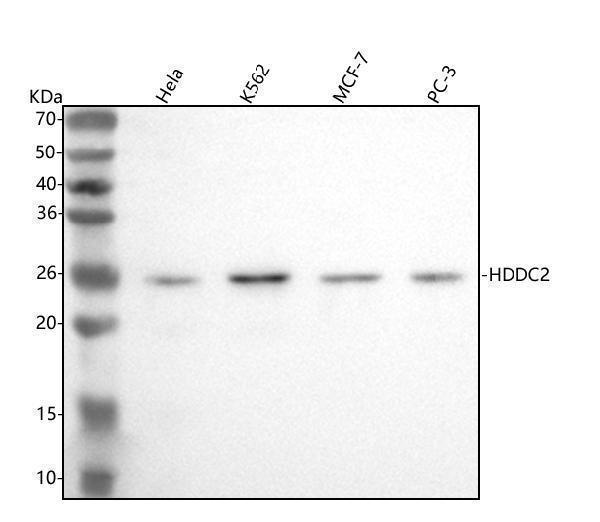
Figure 1. Western blot analysis of HDDC2 using anti-HDDC2 antibody (A08033-1).
Electrophoresis was performed on a 5-20% SDS-PAGE gel at 70V (Stacking gel) / 90V (Resolving gel) for 2-3 hours. The sample well of each lane was loaded with 30 ug of sample under reducing conditions.
Lane 1: human Hela whole cell lysates,
Lane 2: human K562 whole cell lysates,
Lane 3: human MCF-7 whole cell lysates,
Lane 4: human PC-3 whole cell lysates.
After electrophoresis, proteins were transferred to a nitrocellulose membrane at 150 mA for 50-90 minutes. Blocked the membrane with 5% non-fat milk/TBS for 1.5 hour at RT. The membrane was incubated with rabbit anti-HDDC2 antigen affinity purified polyclonal antibody (Catalog # A08033-1) at 0.5 μg/mL overnight at 4°C, then washed with TBS-0.1%Tween 3 times with 5 minutes each and probed with a goat anti-rabbit IgG-HRP secondary antibody at a dilution of 1:5000 for 1.5 hour at RT. The signal is developed using an Enhanced Chemiluminescent detection (ECL) kit (Catalog # EK1002) with Tanon 5200 system. A specific band was detected for HDDC2 at approximately 26 kDa. The expected band size for HDDC2 is at 23 kDa.

Click image to see more details
Figure 2. IF analysis of HDDC2 using anti-HDDC2 antibody (A08033-1).
HDDC2 was detected in an immunocytochemical section of U2OS cells. Enzyme antigen retrieval was performed using IHC enzyme antigen retrieval reagent (AR0022) for 15 mins. The cells were blocked with 10% goat serum. And then incubated with 5 μg/mL rabbit anti-HDDC2 Antibody (A08033-1) overnight at 4°C. Cy3 Conjugated Goat Anti-Rabbit IgG (BA1032) was used as secondary antibody at 1:500 dilution and incubated for 30 minutes at 37°C. The section was counterstained with DAPI. Visualize using a fluorescence microscope and filter sets appropriate for the label used.

Click image to see more details
Figure 3. Flow Cytometry analysis of SiHa cells using anti-HDDC2 antibody (A08033-1).
Overlay histogram showing SiHa cells stained with A08033-1 (Blue line). To facilitate intracellular staining, cells were fixed with 4% paraformaldehyde and permeabilized with permeabilization buffer. The cells were blocked with 10% normal goat serum. And then incubated with rabbit anti-HDDC2 Antibody (A08033-1, 1 μg/1x106 cells) for 30 min at 20°C. DyLight®488 conjugated goat anti-rabbit IgG (BA1127, 5-10 μg/1x106 cells) was used as secondary antibody for 30 minutes at 20°C. Isotype control antibody (Green line) was rabbit IgG (1 μg/1x106) used under the same conditions. Unlabelled sample (Red line) was also used as a control.
Protein Target Info & Infographic
Gene/Protein Information For HDDC2 (Source: Uniprot.org, NCBI)
Gene Name
HDDC2
Full Name
HD domain-containing protein 2
Weight
23.39kDa
Superfamily
HDDC2 family
Alternative Names
HD domain-containing protein 2 HDDC2 C6orf74, CGI-130, NS5ATP2, dJ167O5.2 HD domain containing 2 5-deoxynucleotidase HDDC2|HCV NS5A-transactivated protein 2|HD domain-containing protein 2|hepatitis C virus NS5A-transactivated protein 2|testicular tissue protein Li 83
*If product is indicated to react with multiple species, protein info is based on the gene entry specified above in "Species".For more info on HDDC2, check out the HDDC2 Infographic

We have 30,000+ of these available, one for each gene! Check them out.
In this infographic, you will see the following information for HDDC2: database IDs, superfamily, protein function, synonyms, molecular weight, chromosomal locations, tissues of expression, subcellular locations, post-translational modifications, and related diseases, research areas & pathways. If you want to see more information included, or would like to contribute to it and be acknowledged, please contact [email protected].
Specific Publications For Anti-HDDC2 Antibody Picoband™ (A08033-1)
Hello CJ!
No publications found for A08033-1
*Do you have publications using this product? Share with us and receive a reward. Ask us for more details.
Recommended Resources
Here are featured tools and databases that you might find useful.
- Boster's Pathways Library
- Protein Databases
- Bioscience Research Protocol Resources
- Data Processing & Analysis Software
- Photo Editing Software
- Scientific Literature Resources
- Research Paper Management Tools
- Molecular Biology Software
- Primer Design Tools
- Bioinformatics Tools
- Phylogenetic Tree Analysis
Customer Reviews
Have you used Anti-HDDC2 Antibody Picoband™?
Submit a review and receive an Amazon gift card.
- $30 for a review with an image
Be the first to review Anti-HDDC2 Antibody Picoband™
*The first user to submit a review for a product is eligible for Boster's Innovating Scientists Reward, which gives product credits. This is in addition to the gift card reward.
Customer Q&As
Have a question?
Find answers in Q&As, reviews.
Can't find your answer?
Submit your question
