
Click image to see more details
-

-
-
-
-
+1
Product Info Summary
| SKU: | PB9132 |
|---|---|
| Size: | 100 μg/vial |
| Reactive Species: | Human, Mouse, Rat |
| Host: | Rabbit |
| Application: | IHC, WB |
Customers Who Bought This Also Bought

Product info
Product Name
Anti-MCL1 Antibody Picoband™
SKU/Catalog Number
PB9132
Size
100 μg/vial
Form
Lyophilized
Description
Boster Bio Anti-MCL1 Antibody Picoband™ catalog # PB9132. Tested in IHC, WB applications. This antibody reacts with Human, Mouse, Rat.
Storage & Handling
Store at -20˚C for one year from date of receipt. After reconstitution, at 4˚C for one month. It can also be aliquotted and stored frozen at -20˚C for six months. Avoid repeated freeze-thaw cycles.
Cite This Product
Anti-MCL1 Antibody Picoband™ (Boster Biological Technology, Pleasanton CA, USA, Catalog # PB9132)
Host
Rabbit
Contents
Each vial contains 5mg BSA, 0.9mg NaCl, 0.2mg Na2HPO4, 0.05mg NaN3.
Clonality
Polyclonal
Isotype
Rabbit IgG
Immunogen
E.coli-derived human MCL1 recombinant protein (Position: M1-R350). Human MCL1 shares 82% and 78% amino acid (aa) sequences identity with mouse and rat MCL1, respectively.
*Blocking peptide can be purchased. Costs vary based on immunogen length. Contact us for pricing.
Cross-reactivity
No cross-reactivity with other proteins
Reactive Species
PB9132 is reactive to MCL1 in Human, Mouse, Rat
Applications
PB9132 is guaranteed for IHC, WB Boster Guarantee
Observed Molecular Weight
37 kDa
Calculated molecular weight
37.337kDa
Background of Mcl-1
MCL1, also known as myeloid cell leukemia 1, is a protein that in humans is encoded the MCL1 gene. It is mapped to 1q21.3. MCL1 is a potent multidomain antiapoptotic protein of the BCL2 family that heterodimerizes with other BCL2 family members to protect against apoptotic cell death. MCL1 as an attractive candidate for regulation of hematopoietic stem cell homeostasis that is highly expressed in hematopoietic stem cells and regulated by growth factor signals. MCL1 is a critical and specific regulator essential for ensuring the homeostasis of early hematopoietic progenitors. During mitotic arrest, MCL1 protein levels decline markedly, through a posttranslational mechanism, potentiating cell death. Phosphorylation of MCL1 directs its interaction with the tumor suppressor protein FBW7, which is the substrate-binding component of a ubiquitin ligase complex.
Antibody Validation
Boster validates all antibodies on WB, IHC, ICC, Immunofluorescence, and ELISA with known positive control and negative samples to ensure specificity and high affinity, including thorough antibody incubations.
Innovating Scientists Reward
If you are the first to review this product, or if you have results for a special sample, species or application this product is not validated in, share your results with us and receive product credits you can use towards any Boster products! Applicable to all scientists worldwide.
Submit A Review
Assay dilution & Images
Reconsitution
Add 0.2ml of distilled water will yield a concentration of 500ug/ml.
Assay Dilutions Recommendation
The recommendations below provide a starting point for assay optimization. The actual working concentration varies and should be decided by the user.
Immunohistochemistry (Paraffin-embedded Section), 0.5-1μg/ml, Human, Mouse, Rat, By Heat
Western blot, 0.1-0.5μg/ml, Human, Rat
Validation Images & Assay Conditions
Click image to see more details
Figure 1. Western blot analysis of MCL1 using anti-MCL1 antibody (PB9132).
Electrophoresis was performed on a 5-20% SDS-PAGE gel at 70V (Stacking gel) / 90V (Resolving gel) for 2-3 hours.
Lane 1: recombinant human MCL1 protein 0.5 ng.
After electrophoresis, proteins were transferred to a nitrocellulose membrane at 150 mA for 50-90 minutes. Blocked the membrane with 5% non-fat milk/TBS for 1.5 hour at RT. The membrane was incubated with rabbit anti-MCL1 antigen affinity purified polyclonal antibody (Catalog # PB9132) at 0.5 μg/mL overnight at 4°C, then washed with TBS-0.1%Tween 3 times with 5 minutes each and probed with a goat anti-rabbit IgG-HRP secondary antibody at a dilution of 1:5000 for 1.5 hour at RT. The signal is developed using an Enhanced Chemiluminescent detection (ECL) kit (Catalog # EK1002) with Tanon 5200 system. A specific band was detected for MCL1 at approximately 40 kDa. The expected band size for MCL1 is at 40 kDa.

Click image to see more details
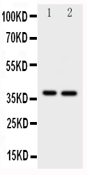
Figure 2. Western blot analysis of MCL1 using anti-MCL1 antibody (PB9132).
Electrophoresis was performed on a 5-20% SDS-PAGE gel at 70V (Stacking gel) / 90V (Resolving gel) for 2-3 hours. The sample well of each lane was loaded with 30 ug of sample under reducing conditions.
Lane 1: rat spleen tissue lysates,
Lane 2: human HepG2 whole cell lysates,
Lane 3: human MCF-7 whole cell lysates,
Lane 4: human COLO320 whole cell lysates.
After electrophoresis, proteins were transferred to a nitrocellulose membrane at 150 mA for 50-90 minutes. Blocked the membrane with 5% non-fat milk/TBS for 1.5 hour at RT. The membrane was incubated with rabbit anti-MCL1 antigen affinity purified polyclonal antibody (Catalog # PB9132) at 0.5 μg/mL overnight at 4°C, then washed with TBS-0.1%Tween 3 times with 5 minutes each and probed with a goat anti-rabbit IgG-HRP secondary antibody at a dilution of 1:5000 for 1.5 hour at RT. The signal is developed using an Enhanced Chemiluminescent detection (ECL) kit (Catalog # EK1002) with Tanon 5200 system. A specific band was detected for MCL1 at approximately 37 kDa. The expected band size for MCL1 is at 37 kDa.

Click image to see more details
Figure 3. IHC analysis of MCL1 using anti-MCL1 antibody (PB9132).
MCL1 was detected in a paraffin-embedded section of rat intestine tissue. Heat mediated antigen retrieval was performed in EDTA buffer (pH 8.0, epitope retrieval solution). The tissue section was blocked with 10% goat serum. The tissue section was then incubated with 1 μg/ml rabbit anti-MCL1 Antibody (PB9132) overnight at 4°C. Biotinylated goat anti-rabbit IgG was used as secondary antibody and incubated for 30 minutes at 37°C. The tissue section was developed using Strepavidin-Biotin-Complex (SABC) (Catalog # SA1022) with DAB as the chromogen.

Click image to see more details
Figure 4. IHC analysis of MCL1 using anti-MCL1 antibody (PB9132).
MCL1 was detected in a paraffin-embedded section of human intestinal cancer tissue. Heat mediated antigen retrieval was performed in EDTA buffer (pH 8.0, epitope retrieval solution). The tissue section was blocked with 10% goat serum. The tissue section was then incubated with 1 μg/ml rabbit anti-MCL1 Antibody (PB9132) overnight at 4°C. Biotinylated goat anti-rabbit IgG was used as secondary antibody and incubated for 30 minutes at 37°C. The tissue section was developed using Strepavidin-Biotin-Complex (SABC) (Catalog # SA1022) with DAB as the chromogen.

Click image to see more details
Figure 5. IHC analysis of MCL1 using anti-MCL1 antibody (PB9132).
MCL1 was detected in a paraffin-embedded section of mouse intestine tissue. Heat mediated antigen retrieval was performed in EDTA buffer (pH 8.0, epitope retrieval solution). The tissue section was blocked with 10% goat serum. The tissue section was then incubated with 1 μg/ml rabbit anti-MCL1 Antibody (PB9132) overnight at 4°C. Biotinylated goat anti-rabbit IgG was used as secondary antibody and incubated for 30 minutes at 37°C. The tissue section was developed using Strepavidin-Biotin-Complex (SABC) (Catalog # SA1022) with DAB as the chromogen.
Protein Target Info & Infographic
Gene/Protein Information For MCL1 (Source: Uniprot.org, NCBI)
Gene Name
MCL1
Full Name
Induced myeloid leukemia cell differentiation protein Mcl-1
Weight
37.337kDa
Superfamily
Bcl-2 family
Alternative Names
BCL2L3; bcl2-L-3; BCL2L3MGC104264; Bcl-2-like protein 3; Bcl-2-related protein EAT/mcl1; EAT; induced myeloid leukemia cell differentiation protein Mcl-1; Mcl1; Mcl-1; mcl1/EAT; MCL1-ES; MCL1L; MCL1S; MGC1839; myeloid cell leukemia ES; myeloid cell leukemia sequence 1 (BCL2-related); TM MCL1 BCL2L3, EAT-ES, MCL1L, MCL1S, Mcl-1, TM, bcl2-L-3, mcl1/EAT, MCL1 MCL1 apoptosis regulator, BCL2 family member induced myeloid leukemia cell differentiation protein Mcl-1|BCL2 family apoptosis regulator|MCL1, BCL2 family apoptosis regulator|bcl-2-like protein 3|bcl-2-related protein EAT/mcl1|myeloid cell leukemia 1|myeloid cell leukemia ES|myeloid cell leukemia sequence 1 (BCL2-related)
*If product is indicated to react with multiple species, protein info is based on the gene entry specified above in "Species".For more info on MCL1, check out the MCL1 Infographic

We have 30,000+ of these available, one for each gene! Check them out.
In this infographic, you will see the following information for MCL1: database IDs, superfamily, protein function, synonyms, molecular weight, chromosomal locations, tissues of expression, subcellular locations, post-translational modifications, and related diseases, research areas & pathways. If you want to see more information included, or would like to contribute to it and be acknowledged, please contact [email protected].
Specific Publications For Anti-MCL1 Antibody Picoband™ (PB9132)
Hello CJ!
PB9132 has been cited in 2 publications:
*The publications in this section are manually curated by our staff scientists. They may differ from Bioz's machine gathered results. Both are accurate. If you find a publication citing this product but is missing from this list, please let us know we will issue you a thank-you coupon.
Zhang L,Zhang J,Gong Y,Lv L.Systematic and experimental investigations of the anti-colorectal cancer mediated by genistein. Biofactors.2020 Nov;46(6):974-982.doi:10.1002/biof.1677.Epub 2020 Sep 20.PMID:32951326.
Species: Human,Mouse
PB9132 usage in article: APP:IF, SAMPLE:CRC TISSUE, DILUTION:1:100
Liu N, Chen T, Wang X, Yang D, Xue B, Zhu H. Febs Lett. 2015 Apr 2;589(8):897-903. Doi: 10.1016/J.Febslet.2015.02.026. Epub 2015 Mar 3. Msi1 Confers Resistance To Trail By Activating Erk In Liver Cancer Cells.
Recommended Resources
Here are featured tools and databases that you might find useful.
- Boster's Pathways Library
- Protein Databases
- Bioscience Research Protocol Resources
- Data Processing & Analysis Software
- Photo Editing Software
- Scientific Literature Resources
- Research Paper Management Tools
- Molecular Biology Software
- Primer Design Tools
- Bioinformatics Tools
- Phylogenetic Tree Analysis
Customer Reviews
Have you used Anti-MCL1 Antibody Picoband™?
Submit a review and receive an Amazon gift card.
- $30 for a review with an image
Be the first to review Anti-MCL1 Antibody Picoband™
*The first user to submit a review for a product is eligible for Boster's Innovating Scientists Reward, which gives product credits. This is in addition to the gift card reward.
Customer Q&As
Have a question?
Find answers in Q&As, reviews.
Can't find your answer?
Submit your question
16 Customer Q&As for Anti-MCL1 Antibody Picoband™
Question
Would anti-MCL1 antibody PB9132 work for IHC with epithelium of mammary gland?
Verified Customer
Verified customer
Asked: 2020-03-18
Answer
According to the expression profile of epithelium of mammary gland, MCL1 is highly expressed in epithelium of mammary gland. So, it is likely that anti-MCL1 antibody PB9132 will work for IHC with epithelium of mammary gland.
Boster Scientific Support
Answered: 2020-03-18
Question
I have attached the WB image, lot number and protocol we used for epithelium of mammary gland using anti-MCL1 antibody PB9132. Please let me know if you require anything else.
Verified Customer
Verified customer
Asked: 2020-02-19
Answer
Thank you very much for the data. Our lab team are working to resolve this as quickly as possible, and we appreciate your patience and understanding! You have provided everything we needed. Please let me know if there is anything you need in the meantime.
Boster Scientific Support
Answered: 2020-02-19
Question
We have been able to see staining in mouse myeloid leukemia cell neuroblastoma. Any tips? Is anti-MCL1 antibody supposed to stain myeloid leukemia cell neuroblastoma positively?
L. Wu
Verified customer
Asked: 2020-01-28
Answer
From literature myeloid leukemia cell neuroblastoma does express MCL1. From Uniprot.org, MCL1 is expressed in epithelium of mammary gland, myeloid leukemia cell, myeloid leukemia cell neuroblastoma, thalamus, mammary gland placenta, ewing sarcoma, cervix carcinoma, among other tissues. Regarding which tissues have MCL1 expression, here are a few articles citing expression in various tissues:
Cervix carcinoma, Pubmed ID: 18669648
Ewing sarcoma, Pubmed ID: 10634649
Mammary gland, and Placenta, Pubmed ID: 15489334
Myeloid leukemia cell, Pubmed ID: 7682708
Myeloid leukemia cell, and Neuroblastoma, Pubmed ID: 10766760
Thalamus, Pubmed ID: 14702039
Boster Scientific Support
Answered: 2020-01-28
Question
We were satisfied with the WB result of your anti-MCL1 antibody. However we have seen positive staining in cervix carcinoma membrane using this antibody. Is that expected? Could you tell me where is MCL1 supposed to be expressed?
Verified Customer
Verified customer
Asked: 2019-12-20
Answer
Based on literature, cervix carcinoma does express MCL1. Generally MCL1 expresses in membrane. Regarding which tissues have MCL1 expression, here are a few articles citing expression in various tissues:
Cervix carcinoma, Pubmed ID: 18669648
Ewing sarcoma, Pubmed ID: 10634649
Mammary gland, and Placenta, Pubmed ID: 15489334
Myeloid leukemia cell, Pubmed ID: 7682708
Myeloid leukemia cell, and Neuroblastoma, Pubmed ID: 10766760
Thalamus, Pubmed ID: 14702039
Boster Scientific Support
Answered: 2019-12-20
Question
I see that the anti-MCL1 antibody PB9132 works with IHC, what is the protocol used to produce the result images on the product page?
Verified Customer
Verified customer
Asked: 2019-12-20
Answer
You can find protocols for IHC on the "support/technical resources" section of our navigation menu. If you have any further questions, please send an email to [email protected]
Boster Scientific Support
Answered: 2019-12-20
Question
We are currently using anti-MCL1 antibody PB9132 for rat tissue, and we are happy with the IHC results. The species of reactivity given in the datasheet says human, mouse, rat. Is it likely that the antibody can work on feline tissues as well?
Verified Customer
Verified customer
Asked: 2019-10-18
Answer
The anti-MCL1 antibody (PB9132) has not been validated for cross reactivity specifically with feline tissues, though there is a good chance of cross reactivity. We have an innovator award program that if you test this antibody and show it works in feline you can get your next antibody for free. Please contact me if I can help you with anything.
Boster Scientific Support
Answered: 2019-10-18
Question
Will PB9132 anti-MCL1 antibody work on parafin embedded sections? If so, which fixation method do you recommend we use (PFA, paraformaldehyde, other)?
L. Zhang
Verified customer
Asked: 2019-09-23
Answer
It shows on the product datasheet, PB9132 anti-MCL1 antibody as been validated on IHC. It is best to use PFA for fixation because it has better tissue penetration ability. PFA needs to be prepared fresh before use. Long term stored PFA turns into formalin, as the PFA molecules congregate and become formalin.
Boster Scientific Support
Answered: 2019-09-23
Question
We appreciate helping with my inquiry over the phone. Here are the WB image, lot number and protocol we used for epithelium of mammary gland using anti-MCL1 antibody PB9132. Let me know if you need anything else.
Verified Customer
Verified customer
Asked: 2019-08-05
Answer
I appreciate the data. You have provided everything we needed. Our lab team are working to resolve your inquiry as quickly as possible, and we appreciate your patience and understanding! Please let me know if there is anything you need in the meantime.
Boster Scientific Support
Answered: 2019-08-05
Question
Is there a BSA free version of anti-MCL1 antibody PB9132 available?
Verified Customer
Verified customer
Asked: 2019-06-06
Answer
We appreciate your recent telephone inquiry. I can confirm that some lots of this anti-MCL1 antibody PB9132 are BSA free. For now, these lots are available and we can make a BSA free formula for you free of charge. It will take 3 extra days to prepare. If you require this antibody BSA free again in future, please do not hesitate to contact me and I will be pleased to check which lots we have in stock that are BSA free.
Boster Scientific Support
Answered: 2019-06-06
Question
Is this PB9132 anti-MCL1 antibody reactive to the isotypes of MCL1?
Verified Customer
Verified customer
Asked: 2019-03-22
Answer
The immunogen of PB9132 anti-MCL1 antibody is E.coli-derived human MCL1 recombinant protein (Position: M1-R350). Human MCL1 shares 82% and 78% amino acid (aa) sequences identity with mouse and rat MCL1, respectively. Could you tell me which isotype you are interested in so I can help see if the immunogen is part of this isotype?
Boster Scientific Support
Answered: 2019-03-22
Question
We purchased anti-MCL1 antibody for IHC on myeloid leukemia cell neuroblastoma a few months ago. I am using human, and I plan to use the antibody for WB next. We need examining myeloid leukemia cell neuroblastoma as well as ewing sarcoma in our next experiment. Could you please give me some suggestion on which antibody would work the best for WB?
D. Krishna
Verified customer
Asked: 2018-10-05
Answer
I looked at the website and datasheets of our anti-MCL1 antibody and it seems that PB9132 has been validated on human in both IHC and WB. Thus PB9132 should work for your application. Our Boster satisfaction guarantee will cover this product for WB in human even if the specific tissue type has not been validated. We do have a comprehensive range of products for WB detection and you can check out our website bosterbio.com to find out more information about them.
Boster Scientific Support
Answered: 2018-10-05
Question
I was wanting to use your anti-MCL1 antibody for IHC for mouse epithelium of mammary gland on frozen tissues, but I want to know if it has been tested for this particular application. Has this antibody been tested and is this antibody a good choice for mouse epithelium of mammary gland identification?
Verified Customer
Verified customer
Asked: 2018-01-24
Answer
You can see on the product datasheet, PB9132 anti-MCL1 antibody has been tested for IHC, WB on human, mouse, rat tissues. We have an innovator award program that if you test this antibody and show it works in mouse epithelium of mammary gland in IHC-frozen, you can get your next antibody for free.
Boster Scientific Support
Answered: 2018-01-24
Question
Can you help my question with product PB9132, anti-MCL1 antibody. I was wondering if it would be possible to conjugate this antibody with biotin. I would need it to be without BSA or sodium azide. I am planning on using a buffer exchange of sodium azide with PBS only. Would there be problems for me to conjugate the antibody and store it in -20 degrees in small aliquots?
L. Krishna
Verified customer
Asked: 2017-09-06
Answer
We do not recommend storing this antibody with PBS buffer only in -20 degrees. If you want to store it in -20 degrees it is best to add some cryoprotectant like glycerol. If you want carrier free PB9132 anti-MCL1 antibody, we can provide it to you in a special formula with trehalose and/or glycerol. These molecules will not interfere with conjugation chemistry and provide a good level of protection for the antibody from degradation. Please be sure to specify this in your purchase order.
Boster Scientific Support
Answered: 2017-09-06
Question
My question regards to test anti-MCL1 antibody PB9132 on mouse epithelium of mammary gland for research purposes, then I may be interested in using anti-MCL1 antibody PB9132 for diagnostic purposes as well. Is the antibody suitable for diagnostic purposes?
Verified Customer
Verified customer
Asked: 2017-08-25
Answer
The products we sell, including anti-MCL1 antibody PB9132, are only intended for research use. They would not be suitable for use in diagnostic work. If you have the means to develop a product into diagnostic use, and are interested in collaborating with us and develop our product into an IVD product, please contact us for more discussions.
Boster Scientific Support
Answered: 2017-08-25
Question
I am interested in using your anti-MCL1 antibody for positive regulation of oxidative stress-induced neuron intrinsic apoptotic signaling pathway studies. Has this antibody been tested with western blotting on rat spleen tissue? We would like to see some validation images before ordering.
G. Miller
Verified customer
Asked: 2016-06-08
Answer
Thank you for your inquiry. This PB9132 anti-MCL1 antibody is tested on rat spleen tissue, tissue lysate, hepg2 whole cell lysate, colo320 whole cell lysate, intestinal cancer tissue, mouse intestine tissue. It is guaranteed to work for IHC, WB in human, mouse, rat. Our Boster guarantee will cover your intended experiment even if the sample type has not been be directly tested.
Boster Scientific Support
Answered: 2016-06-08
Question
Is a blocking peptide available for product anti-MCL1 antibody (PB9132)?
G. Mitchell
Verified customer
Asked: 2015-07-16
Answer
We do provide the blocking peptide for product anti-MCL1 antibody (PB9132). If you would like to place an order for it please contact [email protected] and make a special request.
Boster Scientific Support
Answered: 2015-07-16
