Click image to see more details
-

-
-
-
-
+1
Product Info Summary
| SKU: | A01516-1 |
|---|---|
| Size: | 100 μg/vial |
| Reactive Species: | Human, Mouse, Rat |
| Host: | Rabbit |
| Application: | ELISA, IHC, WB |
Customers Who Bought This Also Bought


Product info
Product Name
Anti-NCR1 Antibody Picoband™
View all NKp46/NCR1 Antibodies
SKU/Catalog Number
A01516-1
Size
100 μg/vial
Form
Lyophilized
Description
Boster Bio Anti-NCR1 Antibody Picoband™ catalog # A01516-1. Tested in ELISA, IHC, WB applications. This antibody reacts with Human, Mouse, Rat.
Storage & Handling
Store at -20˚C for one year from date of receipt. After reconstitution, at 4˚C for one month. It can also be aliquotted and stored frozen at -20˚C for six months. Avoid repeated freeze-thaw cycles.
Cite This Product
Anti-NCR1 Antibody Picoband™ (Boster Biological Technology, Pleasanton CA, USA, Catalog # A01516-1)
Host
Rabbit
Contents
Each vial contains 4 mg Trehalose, 0.9 mg NaCl and 0.2 mg Na2HPO4.
Clonality
Polyclonal
Isotype
Rabbit IgG
Immunogen
E. coli-derived human NCR1 recombinant protein (Position: Q22-R258).
*Blocking peptide can be purchased. Costs vary based on immunogen length. Contact us for pricing.
Cross-reactivity
No cross-reactivity with other proteins.
Reactive Species
A01516-1 is reactive to NCR1 in Human, Mouse, Rat
Applications
A01516-1 is guaranteed for ELISA, IHC, WB Boster Guarantee
Observed Molecular Weight
36 kDa
Calculated molecular weight
34.481kDa
Background of NKp46/NCR1
Natural cytotoxicity triggering receptor 1, also known as NKp46, is a protein that in humans is encoded by the NCR1 gene. This gene is mapped to chromosome 19, where genes encoding other NK inhibitory and activator structures are also located. NKP46 participates in NK-cell-mediated lysis of cells infected with an intracellular bacterium and that reduced functional capacity of NK cells is associated with severe manifestations of infectious disease.
Antibody Validation
Boster validates all antibodies on WB, IHC, ICC, Immunofluorescence, and ELISA with known positive control and negative samples to ensure specificity and high affinity, including thorough antibody incubations.
Innovating Scientists Reward
If you are the first to review this product, or if you have results for a special sample, species or application this product is not validated in, share your results with us and receive product credits you can use towards any Boster products! Applicable to all scientists worldwide.
Submit A Review
Assay dilution & Images
Reconsitution
Add 0.2ml of distilled water will yield a concentration of 500ug/ml.
Assay Dilutions Recommendation
The recommendations below provide a starting point for assay optimization. The actual working concentration varies and should be decided by the user.
Western blot, 0.1-0.5μg/ml
Immunohistochemistry (Paraffin-embedded Section), 0.5-1μg/ml
Direct ELISA, 0.1-0.5μg/ml
Validation Images & Assay Conditions
Click image to see more details
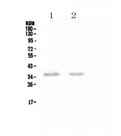
Figure 1. Western blot analysis of NCR1 using anti-NCR1 antibody (A01516-1).
Electrophoresis was performed on a 5-20% SDS-PAGE gel at 70V (Stacking gel) / 90V (Resolving gel) for 2-3 hours. The sample well of each lane was loaded with 50ug of sample under reducing conditions.
Lane 1: rat spleen tissue lysates,
Lane 2: mouse spleen tissue lysates.
After Electrophoresis, proteins were transferred to a Nitrocellulose membrane at 150mA for 50-90 minutes. Blocked the membrane with 5% Non-fat Milk/ TBS for 1.5 hour at RT. The membrane was incubated with rabbit anti-NCR1 antigen affinity purified polyclonal antibody (Catalog # A01516-1) at 0.5 μg/mL overnight at 4°C, then washed with TBS-0.1%Tween 3 times with 5 minutes each and probed with a goat anti-rabbit IgG-HRP secondary antibody at a dilution of 1:10000 for 1.5 hour at RT. The signal is developed using an Enhanced Chemiluminescent detection (ECL) kit (Catalog # EK1002) with Tanon 5200 system. A specific band was detected for NCR1 at approximately 36KD. The expected band size for NCR1 is at 34KD.

Click image to see more details
Figure 2. IHC analysis of NCR1 using anti-NCR1 antibody (A01516-1).
NCR1 was detected in paraffin-embedded section of rat spleen tissue . Heat mediated antigen retrieval was performed in citrate buffer (pH6, epitope retrieval solution) for 20 mins. The tissue section was blocked with 10% goat serum. The tissue section was then incubated with 1μg/ml rabbit anti-NCR1 Antibody (A01516-1) overnight at 4°C. Biotinylated goat anti-rabbit IgG was used as secondary antibody and incubated for 30 minutes at 37°C. The tissue section was developed using Strepavidin-Biotin-Complex (SABC)(Catalog # SA1022) with DAB as the chromogen.

Click image to see more details
Figure 3. IHC analysis of NCR1 using anti-NCR1 antibody (A01516-1).
NCR1 was detected in paraffin-embedded section of mouse spleen tissue . Heat mediated antigen retrieval was performed in citrate buffer (pH6, epitope retrieval solution) for 20 mins. The tissue section was blocked with 10% goat serum. The tissue section was then incubated with 1μg/ml rabbit anti-NCR1 Antibody (A01516-1) overnight at 4°C. Biotinylated goat anti-rabbit IgG was used as secondary antibody and incubated for 30 minutes at 37°C. The tissue section was developed using Strepavidin-Biotin-Complex (SABC)(Catalog # SA1022) with DAB as the chromogen.

Click image to see more details
Figure 4. IHC analysis of NCR1 using anti-NCR1 antibody (A01516-1).
NCR1 was detected in paraffin-embedded section of mouse lung tissues. Heat mediated antigen retrieval was performed in citrate buffer (pH6, epitope retrieval solution) for 20 mins. The tissue section was blocked with 10% goat serum. The tissue section was then incubated with 1μg/ml rabbit anti-NCR1 Antibody (A01516-1) overnight at 4°C. Biotinylated goat anti-rabbit IgG was used as secondary antibody and incubated for 30 minutes at 37°C. The tissue section was developed using Strepavidin-Biotin-Complex (SABC)(Catalog # SA1022) with DAB as the chromogen.

Click image to see more details
Figure 5. IHC analysis of NCR1 using anti-NCR1 antibody (A01516-1).
NCR1 was detected in paraffin-embedded section of human tonsil tissue. Heat mediated antigen retrieval was performed in EDTA buffer (pH8.0, epitope retrieval solution). The tissue section was blocked with 10% goat serum. The tissue section was then incubated with 1μg/ml rabbit anti-NCR1 Antibody (A01516-1) overnight at 4°C. Biotinylated goat anti-rabbit IgG was used as secondary antibody and incubated for 30 minutes at 37°C. The tissue section was developed using Strepavidin-Biotin-Complex (SABC) (Catalog # SA1022) with DAB as the chromogen.
Protein Target Info & Infographic
Gene/Protein Information For NCR1 (Source: Uniprot.org, NCBI)
Gene Name
NCR1
Full Name
Natural cytotoxicity triggering receptor 1
Weight
34.481kDa
Superfamily
natural cytotoxicity receptor (NCR) family
Alternative Names
CD335 antigen; CD335; hNKp46; Ly94; LY94lymphocyte antigen 94 homolog (activating NK-receptor; NK-p46); Lymphocyte antigen 94 homolog; MAR-1; natural cytotoxicity triggering receptor 1; Natural killer cell p46-related protein; NCR1; NK cell-activating receptor; NKp46; NKP46FLJ99094; NK-p46lymphocyte antigen 94 (mouse) homolog (activating NK-receptor; NK-p46) NCR1 CD335, LY94, NK-p46, NKP46 natural cytotoxicity triggering receptor 1 natural cytotoxicity triggering receptor 1|NK cell-activating receptor|lymphocyte antigen 94 homolog (activating NK-receptor; NK-p46)|natural killer cell p46-related protein
*If product is indicated to react with multiple species, protein info is based on the gene entry specified above in "Species".For more info on NCR1, check out the NCR1 Infographic

We have 30,000+ of these available, one for each gene! Check them out.
In this infographic, you will see the following information for NCR1: database IDs, superfamily, protein function, synonyms, molecular weight, chromosomal locations, tissues of expression, subcellular locations, post-translational modifications, and related diseases, research areas & pathways. If you want to see more information included, or would like to contribute to it and be acknowledged, please contact [email protected].
Specific Publications For Anti-NCR1 Antibody Picoband™ (A01516-1)
Hello CJ!
No publications found for A01516-1
*Do you have publications using this product? Share with us and receive a reward. Ask us for more details.
Recommended Resources
Here are featured tools and databases that you might find useful.
- Boster's Pathways Library
- Protein Databases
- Bioscience Research Protocol Resources
- Data Processing & Analysis Software
- Photo Editing Software
- Scientific Literature Resources
- Research Paper Management Tools
- Molecular Biology Software
- Primer Design Tools
- Bioinformatics Tools
- Phylogenetic Tree Analysis
Customer Reviews
Have you used Anti-NCR1 Antibody Picoband™?
Submit a review and receive an Amazon gift card.
- $30 for a review with an image
Be the first to review Anti-NCR1 Antibody Picoband™
*The first user to submit a review for a product is eligible for Boster's Innovating Scientists Reward, which gives product credits. This is in addition to the gift card reward.
Customer Q&As
Have a question?
Find answers in Q&As, reviews.
Can't find your answer?
Submit your question
6 Customer Q&As for Anti-NCR1 Antibody Picoband™
Question
We are currently using anti-NCR1 antibody A01516-1 for mouse tissue, and we are content with the IHC results. The species of reactivity given in the datasheet says human, mouse, rat. Is it possible that the antibody can work on monkey tissues as well?
Verified Customer
Verified customer
Asked: 2020-04-02
Answer
The anti-NCR1 antibody (A01516-1) has not been validated for cross reactivity specifically with monkey tissues, but there is a good chance of cross reactivity. We have an innovator award program that if you test this antibody and show it works in monkey you can get your next antibody for free. Please contact me if I can help you with anything.
Boster Scientific Support
Answered: 2020-04-02
Question
Would A01516-1 anti-NCR1 antibody work on parafin embedded sections? If so, which fixation method do you recommend we use (PFA, paraformaldehyde, other)?
Verified Customer
Verified customer
Asked: 2020-02-28
Answer
You can see on the product datasheet, A01516-1 anti-NCR1 antibody as been tested on WB. It is best to use PFA for fixation because it has better tissue penetration ability. PFA needs to be prepared fresh before use. Long term stored PFA turns into formalin, as the PFA molecules congregate and become formalin.
Boster Scientific Support
Answered: 2020-02-28
Question
Would anti-NCR1 antibody A01516-1 work for WB with blood?
Verified Customer
Verified customer
Asked: 2020-01-03
Answer
According to the expression profile of blood, NCR1 is highly expressed in blood. So, it is likely that anti-NCR1 antibody A01516-1 will work for WB with blood.
Boster Scientific Support
Answered: 2020-01-03
Question
Please see the WB image, lot number and protocol we used for blood using anti-NCR1 antibody A01516-1. Please let me know if you require anything else.
Verified Customer
Verified customer
Asked: 2019-09-13
Answer
Thank you very much for the data. Our lab team are working to resolve this as quickly as possible, and we appreciate your patience and understanding! You have provided everything we needed. Please let me know if there is anything you need in the meantime.
Boster Scientific Support
Answered: 2019-09-13
Question
Do you have a BSA free version of anti-NCR1 antibody A01516-1 available?
Verified Customer
Verified customer
Asked: 2019-08-13
Answer
Thank you for your recent telephone inquiry. I can confirm that some lots of this anti-NCR1 antibody A01516-1 are BSA free. For now, these lots are available and we can make a BSA free formula for you free of charge. It will take 3 extra days to prepare. If you require this antibody BSA free again in future, please do not hesitate to contact me and I will be pleased to check which lots we have in stock that are BSA free.
Boster Scientific Support
Answered: 2019-08-13
Question
I am looking for to test anti-NCR1 antibody A01516-1 on mouse blood for research purposes, then I may be interested in using anti-NCR1 antibody A01516-1 for diagnostic purposes as well. Is the antibody suitable for diagnostic purposes?
Verified Customer
Verified customer
Asked: 2019-04-12
Answer
The products we sell, including anti-NCR1 antibody A01516-1, are only intended for research use. They would not be suitable for use in diagnostic work. If you have the means to develop a product into diagnostic use, and are interested in collaborating with us and develop our product into an IVD product, please contact us for more discussions.
Boster Scientific Support
Answered: 2019-04-12
