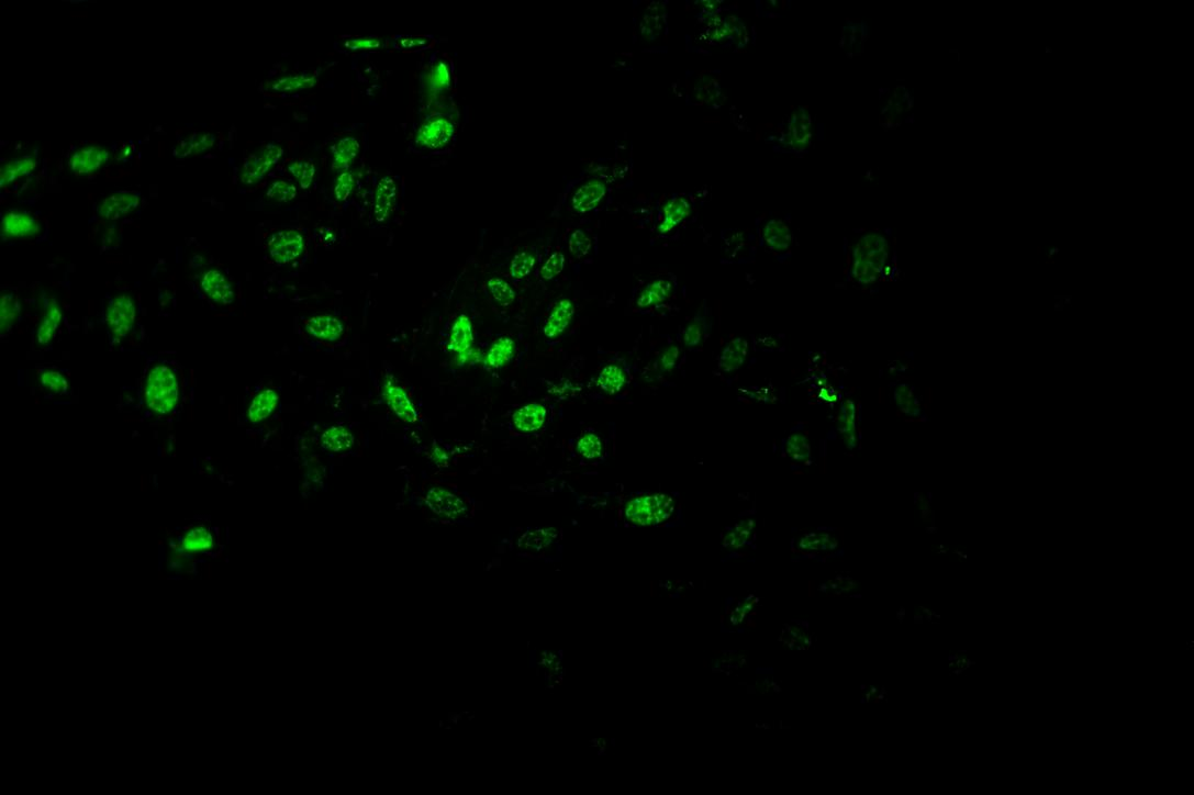
Pcna antibody (M00125-6) performs well in IHC, ICC, WB and shows reactivity with Human, Mouse, Rat.
  • Validated for inter-lot consistency, specificity, and reproducibility
  • Committed to quality since 1993
placeholder
Designing a Western blot for PCNA? The guide covers the expected band size, antibodies backed by real blot images, the controls to run alongside, and protocols taken from published papers.
Open the PCNA Western Blot Guide

Anti-PCNA Antibody (Monoclonal, 36P00)

Pcna antibody

Reactive SpeciesHostApplication
Human, Mouse, RatMouseIHC, ICC, WB

SKU: M00125-6

Prefer to Label Your Own Antibody?

Explore our Bundle & Save labeling kits, select your options below to add to cart, and earn 10% back in Boster Credit.

Select a kit to view available options.

Size: 100 μg

Bundle to save: I do not want free secondary ab
Price:
$370/100 μg

1 in stock in USA, Ships Next Business Day.

Quantity: 1

1
Get A Quote
Guarantee / Product Promise

Recommend Dilution

Application
Dilution
Species
Immunohistochemistry (Paraffin-embedded Section)
0.4-1μg/ml
Human, mouse, rat
Immunocytochemistry
1μg/ml
Human, mouse, rat, -
Immunohistochemistry (Frozen Section)
0.4-1μg/ml
Human, mouse, rat, -
Western blot
2μg/ml
Human, mouse, rat

* It is recommended to optimize the amount of this reagent in each experimental system to achieve the best results.

Product Information

Product Name
Anti-PCNA Antibody (Monoclonal, 36P00)
View all PCNA Antibodies
Form
Lyophilized
Uniprot
Gene ID
Storage & Handling
Store at -20˚C for one year from date of receipt. After reconstitution, at 4˚C for one month. It can also be aliquotted and stored frozen at -20˚C for six months. Avoid repeated freeze-thaw cycles.
Host
Mouse
Contents
Mouse IgG in stabilizing components, 1.2% sodium acetate and 0.01mg NaN3.
Clonality
Monoclonal
Clone Number
36P00
Isotype
Mouse IgG2a
Immunogen
Protein A fusion protein.
Cross-Reactivity
No cross-reactivity with other proteins
Reconstitution
Add 1ml of PBS buffer will yield a concentration of 100ug/ml.
Calculated Molecular Weight

28.7 kDa

Background of Pcna
Proliferating cell nuclear antigen (PCNA) was originally identified by immunofluorescence as a nuclear protein whose appearance correlated with the proliferative state of the cell. PCNA/cyclin has been localized by in situ hybridization to the short arm of human chromosome 20 with a peak of grains over band 20p13. PCNA gene is present in single copy and has 6 exons. It spans 4,961 bp. Synthesis of the nuclear protein cyclin and DNA in quiescent mouse fibroblasts is coordinately induced by serum and purified growth factors. PCNA controls establishment of sister chromatid cohesion during S phase.
Cite This Product
Anti-PCNA Antibody (Monoclonal, 36P00) (Boster Biological Technology, Pleasanton CA, USA, Catalog # M00125-6)

Review

Have you used Anti-PCNA Antibody (Monoclonal, 36P00)?

Submit a review and receive $370 in product credits, a $185 Amazon gift card, or our carefully curated gift bundles.
2 Reviews For Anti-PCNA Antibody (Monoclonal, 36P00)

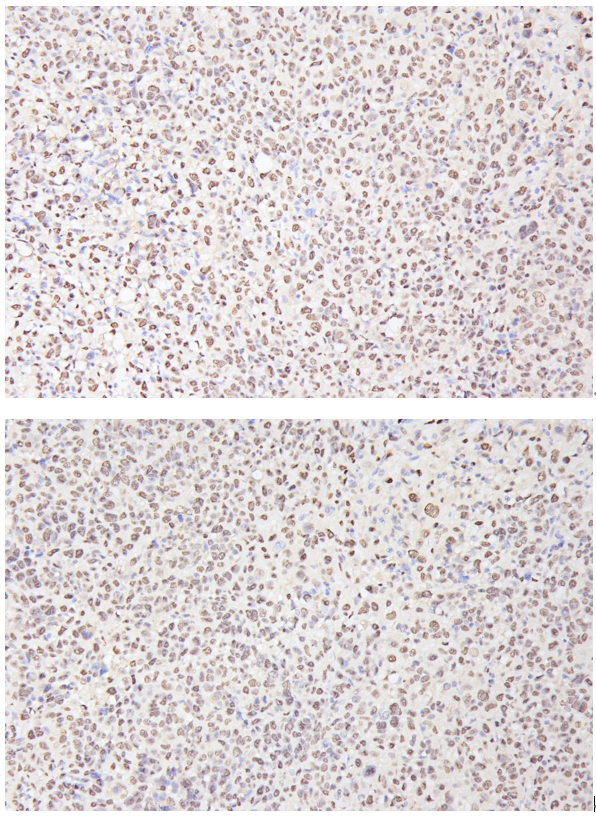
The PCNA antibody (Cat# M00125-6) showed clear and distinct nuclear positivity in HepG2-derived subcutaneous tumors, indicating reliable detection.

MA1083 Immunohistochemistry
SKU M00125-6
Application Immunohistochemistry
Sample HepG2 subcutaneous xenograft in nude mice
Sample Processing Description HepG2 cells were expanded and then implanted subcutaneously into nude mice. After 2 weeks, tumors formed, which were excised, fixed in formalin for 48 hours, and processed for paraffin embedding and sectioning.
Other Reagents Goat serum, DAB chromogen solution
Primary Antibody PCNA Antibody (Monoclonal, PC 10)
Primary Incubation 1:500, overnight at 4 ℃
Secondary Antibody Two-step IHC detection kit
Secondary Incubation 30 min in 37℃
Detection Image system: Leica DM2500
Results Summary PCNA is a key marker for cell proliferation studies; however, besides being a proliferation marker, it is a core component of the DNA replication and repair complex. Its long half-life and involvement in DNA damage repair mean that PCNA positivity does not solely indicate cell proliferation but may also reflect cells responding to DNA damage. Combined with Ki67 staining, it can accurately indicate proliferative status. In our experiment, the staining was clear, with distinct nuclear positivity.

Verified customer

Submitted

Highly prone to positive signals, with precise localization of expression

MA1083 Immunofluorenscence
SKU MA1083
Application Immunofluorenscence
Sample Mouse MC-8 cells
Sample Processing Description 4% paraformaldehyde for 15 minutes
Primary Antibody Anti-PCNA Antibody (Monoclonal, PC 10)
Primary Incubation 1:1000, overnight at 4 ℃
Blocking Agent Goat serum
Secondary Antibody DyLight 550-conjugated goat anti-rabbit antibody.
Secondary Incubation Incubate at room temperature for 1 hour
Detection Laser confocal microscopy
Results Summary I will purchase Boster products again and recommend them to my classmates and colleagues.

Verified customer

Submitted

Product Promise

We promise Anti-PCNA Antibody (Monoclonal, 36P00) performs excellently in relevant applications for IHC, ICC, WB. If it doesn't perform as promised, we will refund the product. Learn more


In stock
Order Product
M00125-6
Buy one primary antibody get one 0.5ml HRP or Biotin secondary antibody for free.
*Sample sizes are prepared on demand and will take extra lead time. (cannot be conjugated)
$370.00

Troubleshooting

troubleshooting-box-image

Download troubleshooting handbooks for IHC, Western blot and ELISA for FREE.

Download Free PDFs Now

Boster Guarantee

Guaranteed product quality

Guaranteed product quality

We promise all of our products perform as described in datasheets.

This product is for Research Use Only. Not for use in diagnostic procedures.

Update date: Oct 16, 2026