Click image to see more details
Product Info Summary
| SKU: | A01992-2 |
|---|---|
| Size: | 100 μg/vial |
| Reactive Species: | Human, Mouse, Rat |
| Host: | Rabbit |
| Application: | ELISA, Flow Cytometry, WB |
Customers Who Bought This Also Bought

Product info
Product Name
Anti-PERK/EIF2AK3 Antibody Picoband™
SKU/Catalog Number
A01992-2
Size
100 μg/vial
Form
Lyophilized
Description
Boster Bio Anti-PERK/EIF2AK3 Antibody Picoband™ catalog # A01992-2. Tested in ELISA, Flow Cytometry, WB applications. This antibody reacts with Human, Mouse, Rat.
Storage & Handling
Store at -20˚C for one year from date of receipt. After reconstitution, at 4˚C for one month. It can also be aliquotted and stored frozen at -20˚C for six months. Avoid repeated freeze-thaw cycles.
Cite This Product
Anti-PERK/EIF2AK3 Antibody Picoband™ (Boster Biological Technology, Pleasanton CA, USA, Catalog # A01992-2)
Host
Rabbit
Contents
Each vial contains 4mg Trehalose, 0.9mg NaCl, 0.2mg Na2HPO4, 0.05mg NaN3.
Clonality
Polyclonal
Isotype
Rabbit IgG
Immunogen
E. coli-derived human PERK recombinant protein (Position: R222-Q334).
*Blocking peptide can be purchased. Costs vary based on immunogen length. Contact us for pricing.
Cross-reactivity
No cross-reactivity with other proteins.
Reactive Species
A01992-2 is reactive to EIF2AK3 in Human, Mouse, Rat
Applications
A01992-2 is guaranteed for ELISA, Flow Cytometry, WB Boster Guarantee
Observed Molecular Weight
140 kDa
Calculated molecular weight
125.216kDa
Background of PERK
Eukaryotic translation initiation factor 2-alpha kinase 3, also known as protein kinase R (PKR)-like endoplasmic reticulum kinase (PERK), is an enzyme that in humans is encoded by the EIF2AK3 gene. The protein encoded by this gene phosphorylates the alpha subunit of eukaryotic translation-initiation factor 2, leading to its inactivation, and thus to a rapid reduction of translational initiation and repression of global protein synthesis. This protein is thought to modulate mitochondrial function. It is a type I membrane protein located in the endoplasmic reticulum (ER), where it is induced by ER stress caused by malfolded proteins. Mutations in this gene are associated with Wolcott-Rallison syndrome.
Antibody Validation
Boster validates all antibodies on WB, IHC, ICC, Immunofluorescence, and ELISA with known positive control and negative samples to ensure specificity and high affinity, including thorough antibody incubations.
Innovating Scientists Reward
If you are the first to review this product, or if you have results for a special sample, species or application this product is not validated in, share your results with us and receive product credits you can use towards any Boster products! Applicable to all scientists worldwide.
Submit A Review
Assay dilution & Images
Reconsitution
Add 0.2ml of distilled water will yield a concentration of 500ug/ml.
Assay Dilutions Recommendation
The recommendations below provide a starting point for assay optimization. The actual working concentration varies and should be decided by the user.
Western blot, 0.1-0.5μg/ml
Flow Cytometry, 1-3μg/1x106 cells
Direct ELISA, 0.1-0.5μg/ml
Validation Images & Assay Conditions
Click image to see more details
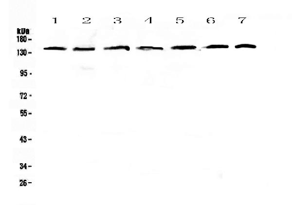
Figure 1. Western blot analysis of PERK using anti-PERK antibody (A01992-2).
Electrophoresis was performed on a 5-20% SDS-PAGE gel at 70V (Stacking gel) / 90V (Resolving gel) for 2-3 hours. The sample well of each lane was loaded with 50ug of sample under reducing conditions.
Lane 1: human Hela whole cell lysates,
Lane 2: human COLO-320 whole cell lysates,
Lane 3: human A549 whole cell lysates,
Lane 4: human SK-OV-3 whole cell lysates,
Lane 5: Human A431 whole cell lysates,
Lane 6: rat brain tissue lysates,
Lane 7: mouse brain tissue lysates.
After Electrophoresis, proteins were transferred to a Nitrocellulose membrane at 150mA for 50-90 minutes. Blocked the membrane with 5% Non-fat Milk/ TBS for 1.5 hour at RT. The membrane was incubated with rabbit anti-PERK antigen affinity purified polyclonal antibody (Catalog # A01992-2) at 0.5 μg/mL overnight at 4°C, then washed with TBS-0.1%Tween 3 times with 5 minutes each and probed with a goat anti-rabbit IgG-HRP secondary antibody at a dilution of 1:10000 for 1.5 hour at RT. The signal is developed using an Enhanced Chemiluminescent detection (ECL) kit (Catalog # EK1002) with Tanon 5200 system. A specific band was detected for PERK at approximately 140KD. The expected band size for PERK is at 125KD.

Click image to see more details
Figure 2. Flow Cytometry analysis of HepG2 cells using anti-PERK antibody (A01992-2).
Overlay histogram showing HepG2 cells stained with A01992-2 (Blue line).The cells were blocked with 10% normal goat serum. And then incubated with rabbit anti-PERK Antibody (A01992-2,1μg/1x106 cells) for 30 min at 20°C. DyLight®488 conjugated goat anti-rabbit IgG (BA1127, 5-10μg/1x106 cells) was used as secondary antibody for 30 minutes at 20°C. Isotype control antibody (Green line) was rabbit IgG (1μg/1x106) used under the same conditions. Unlabelled sample (Red line) was also used as a control.
Protein Target Info & Infographic
Gene/Protein Information For EIF2AK3 (Source: Uniprot.org, NCBI)
Gene Name
EIF2AK3
Full Name
Eukaryotic translation initiation factor 2-alpha kinase 3
Weight
125.216kDa
Superfamily
protein kinase superfamily
Alternative Names
DKFZp781H1925; EC 2.7.11.1; EIF2AK3; eukaryotic translation initiation factor 2 alpha kinase 3; eukaryotic translation initiation factor 2-alpha kinase 3; hsPEK; PEK; PEKPERKPancreatic eIF2-alpha kinase; PERK; PRKR-like endoplasmic reticulum kinase; WRS EIF2AK3 PEK, PERK, WRS eukaryotic translation initiation factor 2 alpha kinase 3 eukaryotic translation initiation factor 2-alpha kinase 3|PRKR-like endoplasmic reticulum kinase|pancreatic EIF2-alpha kinase|truncated eukaryotic translation initiation factor 2 alpha kinase 3
*If product is indicated to react with multiple species, protein info is based on the gene entry specified above in "Species".For more info on EIF2AK3, check out the EIF2AK3 Infographic

We have 30,000+ of these available, one for each gene! Check them out.
In this infographic, you will see the following information for EIF2AK3: database IDs, superfamily, protein function, synonyms, molecular weight, chromosomal locations, tissues of expression, subcellular locations, post-translational modifications, and related diseases, research areas & pathways. If you want to see more information included, or would like to contribute to it and be acknowledged, please contact [email protected].
Specific Publications For Anti-PERK/EIF2AK3 Antibody Picoband™ (A01992-2)
Hello CJ!
A01992-2 has been cited in 7 publications:
*The publications in this section are manually curated by our staff scientists. They may differ from Bioz's machine gathered results. Both are accurate. If you find a publication citing this product but is missing from this list, please let us know we will issue you a thank-you coupon.
Recombinant Newcastle disease virus (rL-RVG) triggers autophagy and apoptosis in gastric carcinoma cells by inducing ER stress
Endoplasmic reticulum stress in diethylnitrosamine‑induced rat liver cancer
Total Panax notoginseng saponin inhibits balloon injury-induced neointimal hyperplasia in rat carotid artery models by suppressing pERK/p38 MAPK pathways
ASK1 Enhances Angiotensin II-Induced Liver Fibrosis In Vitro by Mediating Endoplasmic Reticulum Stress-Dependent Exosomes
Chi L,Jiao D,Nan G,Yuan H,Shen J,Gao Y.miR-9-5p attenuates ischemic stroke through targeting ERMP1-mediated endoplasmic reticulum stress.Acta Histochem.2019 Nov;121(8):151438.doi:10.1016/j.acthis.2019.08.005.Epub 2019 Sep 7.PMID:31500865.
Species: Human,Rat
A01992-2 usage in article: APP:WB, SAMPLE:ISCHEMIC PENUMBRA PART AND SH-SY5Y CELL, DILUTION:NA
Pei-pei Fang,Chen-wei Pan,Wei Lin,Jie Li,Shan-shan Huang,Guang-yao Zhou,Wen-jun Du,Qiang Li, "ASK1 Enhances Angiotensin II-Induced Liver Fibrosis In Vitro by Mediating Endoplasmic Reticulum Stress-Dependent Exosomes",Mediators of Inflammation,vol.2020,Art
Species: Human
A01992-2 usage in article: APP:WB, SAMPLE:LX-2 CELLS, DILUTION:1:1500
Recombinant Newcastle disease virus (rL-RVG) triggers autophagy and apoptosis in gastric carcinoma cells by inducing ER stress
Recommended Resources
Here are featured tools and databases that you might find useful.
- Boster's Pathways Library
- Protein Databases
- Bioscience Research Protocol Resources
- Data Processing & Analysis Software
- Photo Editing Software
- Scientific Literature Resources
- Research Paper Management Tools
- Molecular Biology Software
- Primer Design Tools
- Bioinformatics Tools
- Phylogenetic Tree Analysis
Customer Reviews
Have you used Anti-PERK/EIF2AK3 Antibody Picoband™?
Submit a review and receive an Amazon gift card.
- $30 for a review with an image
Be the first to review Anti-PERK/EIF2AK3 Antibody Picoband™
*The first user to submit a review for a product is eligible for Boster's Innovating Scientists Reward, which gives product credits. This is in addition to the gift card reward.
Customer Q&As
Have a question?
Find answers in Q&As, reviews.
Can't find your answer?
Submit your question
5 Customer Q&As for Anti-PERK/EIF2AK3 Antibody Picoband™
Question
We are currently using anti-PERK/EIF2AK3 antibody A01992-2 for mouse tissue, and we are satisfied with the Flow Cytometry results. The species of reactivity given in the datasheet says human, mouse, rat. Is it likely that the antibody can work on canine tissues as well?
Verified Customer
Verified customer
Asked: 2019-08-27
Answer
The anti-PERK/EIF2AK3 antibody (A01992-2) has not been tested for cross reactivity specifically with canine tissues, though there is a good chance of cross reactivity. We have an innovator award program that if you test this antibody and show it works in canine you can get your next antibody for free. Please contact me if I can help you with anything.
Boster Scientific Support
Answered: 2019-08-27
Question
We have observed staining in mouse nasal cavity mucosa. Any tips? Is anti-PERK/EIF2AK3 antibody supposed to stain nasal cavity mucosa positively?
K. Li
Verified customer
Asked: 2019-08-21
Answer
From literature nasal cavity mucosa does express EIF2AK3. From Uniprot.org, EIF2AK3 is expressed in nasal cavity mucosa, liver, pancreas testis, brain pancreas, brain, cervix carcinoma, among other tissues. Regarding which tissues have EIF2AK3 expression, here are a few articles citing expression in various tissues:
Brain, Pubmed ID: 14702039
Brain, and Pancreas, Pubmed ID: 10677345
Cervix carcinoma, Pubmed ID: 20068231
Liver, Pancreas, and Testis, Pubmed ID: 10026192
Boster Scientific Support
Answered: 2019-08-21
Question
I was wanting to use using your anti-PERK/EIF2AK3 antibody for bone mineralization studies. Has this antibody been tested with western blotting on hepg2 cells? We would like to see some validation images before ordering.
Verified Customer
Verified customer
Asked: 2019-06-03
Answer
Thank you for your inquiry. This A01992-2 anti-PERK/EIF2AK3 antibody is validated on human hela, hela whole cell lysates, a549 whole cell lysates, a431 whole cell lysates, rat brain tissue, mouse brain, hepg2 cells. It is guaranteed to work for ELISA, Flow Cytometry, WB in human, mouse, rat. Our Boster guarantee will cover your intended experiment even if the sample type has not been be directly tested.
Boster Scientific Support
Answered: 2019-06-03
Question
We bought anti-PERK/EIF2AK3 antibody for ELISA on nasal cavity mucosa in the past. I am using rat, and I plan to use the antibody for Flow Cytometry next. I am interested in examining nasal cavity mucosa as well as liver in our next experiment. Could give a recommendation on which antibody would work the best for Flow Cytometry?
Verified Customer
Verified customer
Asked: 2019-05-27
Answer
I looked at the website and datasheets of our anti-PERK/EIF2AK3 antibody and it seems that A01992-2 has been validated on rat in both ELISA and Flow Cytometry. Thus A01992-2 should work for your application. Our Boster satisfaction guarantee will cover this product for Flow Cytometry in rat even if the specific tissue type has not been validated. We do have a comprehensive range of products for Flow Cytometry detection and you can check out our website bosterbio.com to find out more information about them.
Boster Scientific Support
Answered: 2019-05-27
Question
Our team were well pleased with the WB result of your anti-PERK/EIF2AK3 antibody. However we have been able to see positive staining in nasal cavity mucosa endoplasmic reticulum membrane using this antibody. Is that expected? Could you tell me where is EIF2AK3 supposed to be expressed?
T. Wu
Verified customer
Asked: 2019-01-29
Answer
From literature, nasal cavity mucosa does express EIF2AK3. Generally EIF2AK3 expresses in endoplasmic reticulum membrane. Regarding which tissues have EIF2AK3 expression, here are a few articles citing expression in various tissues:
Brain, Pubmed ID: 14702039
Brain, and Pancreas, Pubmed ID: 10677345
Cervix carcinoma, Pubmed ID: 20068231
Liver, Pancreas, and Testis, Pubmed ID: 10026192
Boster Scientific Support
Answered: 2019-01-29
