Click image to see more details
-

-
-
-
-
+3
Product Info Summary
| SKU: | M00236 |
|---|---|
| Size: | 100 μl |
| Reactive Species: | Human, Mouse, Rat |
| Host: | Rabbit |
| Application: | WB |
Customers Who Bought This Also Bought

Product info
Product Name
Anti-PGC1 alpha PPARGC1A Rabbit Monoclonal Antibody
SKU/Catalog Number
M00236
BM4898 is an alternative SKU for this antibody, used in previous lots.
Size
100 μl
Form
Liquid
Description
Boster Bio Anti-PGC1 alpha PPARGC1A Rabbit Monoclonal Antibody catalog # M00236. Tested in WB application. This antibody reacts with Human, Mouse, Rat.
Storage & Handling
Store at -20°C for one year. For short term storage and frequent use, store at 4°C for up to one month. Avoid repeated freeze-thaw cycles.
Cite This Product
Anti-PGC1 alpha PPARGC1A Rabbit Monoclonal Antibody (Boster Biological Technology, Pleasanton CA, USA, Catalog # M00236)
Host
Rabbit
Contents
Rabbit IgG in stabilizing components, phosphate buffered saline, pH 7.4, 150mM NaCl, 0.02% sodium azide and 50% glycerol.
*This antibody is supplied in a stabilized formulation.
Compatibility with conjugation reactions depends on the chemistry of the conjugation method used.
For conjugation methods that are not compatible with the stabilizing components present in this formulation, a carrier-free antibody format is required.
Clonality
Monoclonal
Clone Number
AOBC-16
Isotype
Rabbit IgG
Immunogen
A synthesized peptide derived from human PGC1 alpha
Reactive Species
M00236 is reactive to PPARGC1A in Human, Mouse, Rat
Observed Molecular Weight
113 kDa
Calculated molecular weight
91.0 kDa
Antibody Validation
Boster validates all antibodies on WB, IHC, ICC, Immunofluorescence, and ELISA with known positive control and negative samples to ensure specificity and high affinity, including thorough antibody incubations.
Application & Images
Applications
M00236 is guaranteed for WB Boster Guarantee
Recommend Dilution
WB 1:500-2000
ICC/IF 1:50-200
Validation Images & Assay Conditions
Click image to see more details
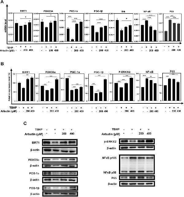
Arbutin exerted protective effects via the SIRT1/FOXO3A/PGC-1α/β and NF-κB/p65 signaling pathway. (A) qRT-PCR was used to measure transcript levels of the SIRT1/FOXO3a/PGC-1α/β pathway and NF-κB/p65 genes. TBHP decreased the expression of SIRT1, FOXO3a, and PGC-1α/β and increased the expression of NF-κB/p65, whereas mRNA levels in the groups that were pretreated with Arbutin showed reversed trend. (B) western blots were conducted to detect the proteins level of SIRT1, FOXO3a, PGC-1α/β, p-ERK, and NFKB1/P65. (C) imageJ was used to analyze the relative expression level of the proteins mentioned above (* p < 0.05, ** p < 0.01, *** p < 0.001, n = 3, bars represent SD).
Index in PubMed under a CC BY license. PMID: 36051689

Click image to see more details
Sirtinol diminished the capability of Arbutin to assist ARPE-19 cells to defend against oxidative stress. (A) (B) flow cytometric analysis showed that cells treated with only sirtinol, TBHP, cotreated with sirtinol, and TBHP displayed decreased cellular viability. However, sirtinol conduction diminished the protective capacity of Arbutin. (C) ARPE-19 cells were seeded in a 24-well plate and applied wounds at the confluence of 80%. The cells were pretreated with or without Arbutin and then subjected to TBHP (350 µM); meanwhile, cells in certain groups were incubated with sirtinol. Photos were taken at different time points post distinct treatments. (D) fluorescence images observed that ARPE-19 treated with Arbutin while subjected to sirtinol and then exposed to TBHP was unable to recuperate ΔΨm. (E) with sirtinol administration, the protein levels of the SIRT1/FOXO3a/PGC-1α/β pathway decreased in the presence of Arbutin (* p < 0.05, ** p < 0.01, *** p < 0.001, n = 3, bars represent SD).
Index in PubMed under a CC BY license. PMID: 36051689

Click image to see more details
All lanes use the Antibody at 1:1K dilution for 1 hour at room temperature.

Click image to see more details
Immunofluorescent analysis using the Antibody at 1:150 dilution.

Click image to see more details
Western blot analysis of PGC1 Alpha/PPARGC1A using anti-PGC1 Alpha/PPARGC1A antibody (M00236).
Electrophoresis was performed on a 5-20% SDS-PAGE gel at 70V (Stacking gel) / 90V (Resolving gel) for 2-3 hours. The sample well of each lane was loaded with 30 ug of sample under reducing conditions.
Lane 1: rat heart tissue lysates,
Lane 2: rat liver tissue lysates,
Lane 3: mouse heart tissue lysates,
Lane 4: mouse liver tissue lysates.
After electrophoresis, proteins were transferred to a nitrocellulose membrane at 150 mA for 50-90 minutes. Blocked the membrane with 5% non-fat milk/TBS for 1.5 hour at RT. The membrane was incubated with rabbit anti-PGC1 Alpha/PPARGC1A antigen affinity purified monoclonal antibody (M00236) at 1:500 overnight at 4°C, then washed with TBS-0.1%Tween 3 times with 5 minutes each and probed with a goat anti-rabbit IgG-HRP secondary antibody at a dilution of 1:5000 for 1.5 hour at RT. The signal is developed using an Enhanced Chemiluminescent detection (ECL) kit (Catalog # EK1002) with Tanon 5200 system. A specific band was detected for PGC1 Alpha/PPARGC1A at approximately 113 kDa. The expected band size for PGC1 Alpha/PPARGC1A is at 91 kDa.

Click image to see more details
Western blot analysis of PGC1 alpha/beta expression in (1) HeLa cell lysate; (2) NIH 3T3 cell lysate; (3) C6 cell lysate.

Click image to see more details
Western blot analysis of PGC1 Alpha/PPARGC1A using anti-PGC1 Alpha/PPARGC1A antibody (M00236).
Electrophoresis was performed on a 5-20% SDS-PAGE gel at 70V (Stacking gel) / 90V (Resolving gel) for 2-3 hours. The sample well of each lane was loaded with 30 ug of sample under reducing conditions.
Lane 1: rat liver tissue lysates,
Lane 2: rat C6 whole cell lysates,
Lane 3: mouse liver tissue lysates,
Lane 4: mouse NIH/3T3 whole cell lysates.
After electrophoresis, proteins were transferred to a nitrocellulose membrane at 150 mA for 50-90 minutes. Blocked the membrane with 5% non-fat milk/TBS for 1.5 hour at RT. The membrane was incubated with rabbit anti-PGC1 Alpha/PPARGC1A antigen affinity purified monoclonal antibody (M00236) at 1:500 overnight at 4°C, then washed with TBS-0.1%Tween 3 times with 5 minutes each and probed with a goat anti-rabbit IgG-HRP secondary antibody at a dilution of 1:5000 for 1.5 hour at RT. The signal is developed using an Enhanced Chemiluminescent detection (ECL) kit (Catalog # EK1002) with Tanon 5200 system. A specific band was detected for PGC1 Alpha/PPARGC1A at approximately 113 kDa. The expected band size for PGC1 Alpha/PPARGC1A is at 91 kDa.
Specific Publications For Anti-PGC1 alpha PPARGC1A Rabbit Monoclonal Antibody (M00236)
Loading publications
Recommended Resources
Here are featured tools and databases that you might find useful.
- Boster's Pathways Library
- Protein Databases
- Bioscience Research Protocol Resources
- Data Processing & Analysis Software
- Photo Editing Software
- Scientific Literature Resources
- Research Paper Management Tools
- Molecular Biology Software
- Primer Design Tools
- Bioinformatics Tools
- Phylogenetic Tree Analysis
Customer Reviews
Have you used Anti-PGC1 alpha PPARGC1A Rabbit Monoclonal Antibody?
Share your experimental results or join a short interview to earn up to $1,000 in product credits or other rewards.
0 Reviews For Anti-PGC1 alpha PPARGC1A Rabbit Monoclonal Antibody
Customer Q&As
Have a question?
Find answers in Q&As, reviews.
Can't find your answer?
Submit your question
4 Customer Q&As for Anti-PGC1 alpha PPARGC1A Rabbit Monoclonal Antibody
Question
We are currently using anti-PGC1 alpha Rabbit Monoclonal antibody M00236 for mouse tissue, and we are well pleased with the WB results. The species of reactivity given in the datasheet says human, mouse, rat. Is it true that the antibody can work on monkey tissues as well?
Verified Customer
Verified customer
Asked: 2019-07-19
Answer
The anti-PGC1 alpha Rabbit Monoclonal antibody (M00236) has not been validated for cross reactivity specifically with monkey tissues, but there is a good chance of cross reactivity. We have an innovator award program that if you test this antibody and show it works in monkey you can get your next antibody for free. Please contact me if I can help you with anything.
Boster Scientific Support
Answered: 2019-07-19
Question
What is the clone name of M00236?
Verified customer
Asked: 2019-04-19
Answer
The clone name of the Anti-PGC1 Alpha PPARGC1A Rabbit Monoclonal Antibody (M00236) is AOBC-16.
Boster Scientific Support
Answered: 2019-04-22
Question
Is this M00236 anti-PGC1 alpha Rabbit Monoclonal antibody reactive to the isotypes of PPARGC1A?
Verified Customer
Verified customer
Asked: 2017-06-09
Answer
The immunogen of M00236 anti-PGC1 alpha Rabbit Monoclonal antibody is A synthesized peptide derived from human PGC1 alpha. Could you tell me which isotype you are interested in so I can help see if the immunogen is part of this isotype?
Boster Scientific Support
Answered: 2017-06-09
Question
Do you have a BSA free version of anti-PGC1 alpha Rabbit Monoclonal antibody M00236 available?
J. Krishna
Verified customer
Asked: 2015-10-23
Answer
Thank you for your recent telephone inquiry. I can confirm that some lots of this anti-PGC1 alpha Rabbit Monoclonal antibody M00236 are BSA free. For now, these lots are available and we can make a BSA free formula for you free of charge. It will take 3 extra days to prepare. If you require this antibody BSA free again in future, please do not hesitate to contact me and I will be pleased to check which lots we have in stock that are BSA free.
Boster Scientific Support
Answered: 2015-10-23
