Click image to see more details
Product Info Summary
| SKU: | A01183-1 |
|---|---|
| Size: | 100 μg/vial |
| Reactive Species: | Human, Mouse |
| Host: | Rabbit |
| Application: | ELISA, Flow Cytometry, WB |
Customers Who Bought This Also Bought


Product info
Product Name
Anti-PHO1/APOBEC3A Antibody Picoband™
SKU/Catalog Number
A01183-1
Size
100 μg/vial
Form
Lyophilized
Description
Boster Bio Anti-PHO1/APOBEC3A Antibody Picoband™ catalog # A01183-1. Tested in ELISA, Flow Cytometry, WB applications. This antibody reacts with Human, Mouse.
Storage & Handling
Store at -20˚C for one year from date of receipt. After reconstitution, at 4˚C for one month. It can also be aliquotted and stored frozen at -20˚C for six months. Avoid repeated freeze-thaw cycles.
Cite This Product
Anti-PHO1/APOBEC3A Antibody Picoband™ (Boster Biological Technology, Pleasanton CA, USA, Catalog # A01183-1)
Host
Rabbit
Contents
Each vial contains 4mg Trehalose, 0.9mg NaCl, 0.2mg Na2HPO4, 0.05mg NaN3.
Clonality
Polyclonal
Isotype
Rabbit IgG
Immunogen
E. coli-derived human PHO1 recombinant protein (Position: M1-L63).
*Blocking peptide can be purchased. Costs vary based on immunogen length. Contact us for pricing.
Cross-reactivity
No cross-reactivity with other proteins.
Reactive Species
A01183-1 is reactive to APOBEC3A in Human, Mouse
Applications
A01183-1 is guaranteed for ELISA, Flow Cytometry, WB Boster Guarantee
Observed Molecular Weight
28 kDa
Calculated molecular weight
23.012kDa
Background of APOBEC3A
Apolipoprotein B mRNA editing enzyme, catalytic polypeptide-like 3A, also known as APOBEC3A, is a gene of the APOBEC3 family found in humans, non-human primates, and some other mammals.This gene is a member of the cytidine deaminase gene family. It is one of seven related genes or pseudogenes found in a cluster, thought to result from gene duplication, on chromosome 22.The protein plays a role in immunity, by restricting transmission of foreign DNA such as viruses. One mechanism of foreign DNA restriction is deamination of foreign double-stranded DNA cytidines to uridines, which leads to DNA degradation. However, other mechanisms are also thought to be involved, as anti-viral effect is not dependent on deaminase activity. Two transcript variants encoding different isoforms have been found for this gene.
Antibody Validation
Boster validates all antibodies on WB, IHC, ICC, Immunofluorescence, and ELISA with known positive control and negative samples to ensure specificity and high affinity, including thorough antibody incubations.
Innovating Scientists Reward
If you are the first to review this product, or if you have results for a special sample, species or application this product is not validated in, share your results with us and receive product credits you can use towards any Boster products! Applicable to all scientists worldwide.
Submit A Review
Assay dilution & Images
Reconsitution
Add 0.2ml of distilled water will yield a concentration of 500ug/ml.
Assay Dilutions Recommendation
The recommendations below provide a starting point for assay optimization. The actual working concentration varies and should be decided by the user.
Western blot, 0.1-0.5μg/ml
Flow Cytometry, 1-3μg/1x106 cells
Direct ELISA, 0.1-0.5μg/ml
Validation Images & Assay Conditions
Click image to see more details
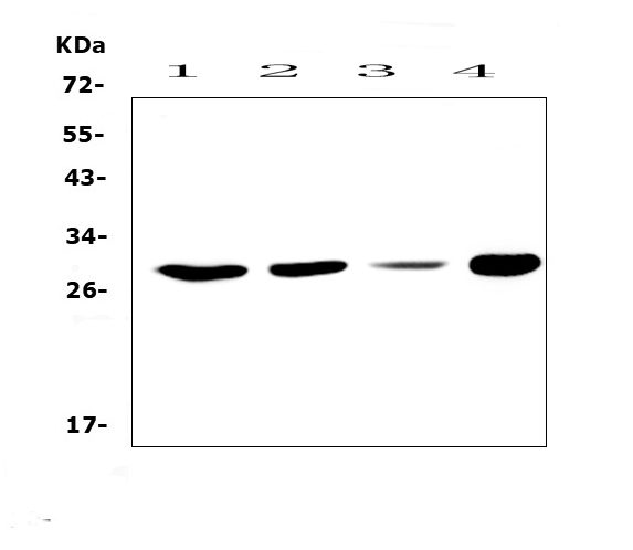
Figure 1. Western blot analysis of PHO1 using anti-PHO1 antibody (A01183-1).
Electrophoresis was performed on a 5-20% SDS-PAGE gel at 70V (Stacking gel) / 90V (Resolving gel) for 2-3 hours. The sample well of each lane was loaded with 50ug of sample under reducing conditions.
Lane 1: human Hela whole cell lysates,
Lane 2: human MCF-7 whole cell lysates,
Lane 3: human HepG2 whole cell lysates,
Lane 4: mouse SP20 whole cell lysates.
After Electrophoresis, proteins were transferred to a Nitrocellulose membrane at 150mA for 50-90 minutes. Blocked the membrane with 5% Non-fat Milk/ TBS for 1.5 hour at RT. The membrane was incubated with rabbit anti-PHO1 antigen affinity purified polyclonal antibody (Catalog # A01183-1) at 0.5 μg/mL overnight at 4°C, then washed with TBS-0.1%Tween 3 times with 5 minutes each and probed with a goat anti-rabbit IgG-HRP secondary antibody at a dilution of 1:10000 for 1.5 hour at RT. The signal is developed using an Enhanced Chemiluminescent detection (ECL) kit (Catalog # EK1002) with Tanon 5200 system. A specific band was detected for PHO1 at approximately 28KD. The expected band size for PHO1 is at 23KD.

Click image to see more details
Figure 2. Flow Cytometry analysis of A549 cells using anti-PHO1 antibody (A01183-1).
Overlay histogram showing A549 cells stained with A01183-1 (Blue line).The cells were blocked with 10% normal goat serum. And then incubated with rabbit anti-PHO1 Antibody (A01183-1,1μg/1x106 cells) for 30 min at 20°C. DyLight®488 conjugated goat anti-rabbit IgG (BA1127, 5-10μg/1x106 cells) was used as secondary antibody for 30 minutes at 20°C. Isotype control antibody (Green line) was rabbit IgG (1μg/1x106) used under the same conditions. Unlabelled sample (Red line) was also used as a control.
Protein Target Info & Infographic
Gene/Protein Information For APOBEC3A (Source: Uniprot.org, NCBI)
Gene Name
APOBEC3A
Full Name
DNA dC->dU-editing enzyme APOBEC-3A
Weight
23.012kDa
Superfamily
cytidine and deoxycytidylate deaminase family
Alternative Names
apolipoprotein B mRNA editing enzyme, catalytic polypeptide-like 3A; ARP3; bK150C2.1; EC 3.5.4; EC 3.5.4.-; phorbolin 1; phorbolin-1; PHRBNprobable DNA dC->dU-editing enzyme APOBEC-3A APOBEC3A A3A, ARP3, PHRBN, bK150C2.1 apolipoprotein B mRNA editing enzyme catalytic subunit 3A DNA dC->dU-editing enzyme APOBEC-3A|apolipoprotein B mRNA editing enzyme, catalytic polypeptide-like 3A|phorbolin-1|probable DNA dC->dU-editing enzyme APOBEC-3A
*If product is indicated to react with multiple species, protein info is based on the gene entry specified above in "Species".For more info on APOBEC3A, check out the APOBEC3A Infographic

We have 30,000+ of these available, one for each gene! Check them out.
In this infographic, you will see the following information for APOBEC3A: database IDs, superfamily, protein function, synonyms, molecular weight, chromosomal locations, tissues of expression, subcellular locations, post-translational modifications, and related diseases, research areas & pathways. If you want to see more information included, or would like to contribute to it and be acknowledged, please contact [email protected].
Specific Publications For Anti-PHO1/APOBEC3A Antibody Picoband™ (A01183-1)
Hello CJ!
No publications found for A01183-1
*Do you have publications using this product? Share with us and receive a reward. Ask us for more details.
Recommended Resources
Here are featured tools and databases that you might find useful.
- Boster's Pathways Library
- Protein Databases
- Bioscience Research Protocol Resources
- Data Processing & Analysis Software
- Photo Editing Software
- Scientific Literature Resources
- Research Paper Management Tools
- Molecular Biology Software
- Primer Design Tools
- Bioinformatics Tools
- Phylogenetic Tree Analysis
Customer Reviews
Have you used Anti-PHO1/APOBEC3A Antibody Picoband™?
Submit a review and receive an Amazon gift card.
- $30 for a review with an image
Be the first to review Anti-PHO1/APOBEC3A Antibody Picoband™
*The first user to submit a review for a product is eligible for Boster's Innovating Scientists Reward, which gives product credits. This is in addition to the gift card reward.
Customer Q&As
Have a question?
Find answers in Q&As, reviews.
Can't find your answer?
Submit your question
7 Customer Q&As for Anti-PHO1/APOBEC3A Antibody Picoband™
Question
Does anti-PHO1/APOBEC3A antibody A01183-1 work on primate ELISA with brain?
Verified Customer
Verified customer
Asked: 2020-02-25
Answer
Our lab technicians have not tested anti-PHO1/APOBEC3A antibody A01183-1 on primate. You can run a BLAST between primate and the immunogen sequence of anti-PHO1/APOBEC3A antibody A01183-1 to see if they may cross-react. If the sequence homology is close, then you can perform a pilot test. Keep in mind that since we have not validated primate samples, this use of the antibody is not covered by our guarantee. However we have an innovator award program that if you test this antibody and show it works in primate brain in ELISA, you can get your next antibody for free.
Boster Scientific Support
Answered: 2020-02-25
Question
Is this A01183-1 anti-PHO1/APOBEC3A antibody reactive to the isotypes of APOBEC3A?
B. Jackson
Verified customer
Asked: 2018-07-27
Answer
The immunogen of A01183-1 anti-PHO1/APOBEC3A antibody is E. coli-derived human PHO1 recombinant protein (Position: M1-L63). Could you tell me which isotype you are interested in so I can help see if the immunogen is part of this isotype?
Boster Scientific Support
Answered: 2018-07-27
Question
I was wanting to use your anti-PHO1/APOBEC3A antibody for ELISA for mouse keratinocyte on frozen tissues, but I want to know if it has been tested for this particular application. Has this antibody been tested and is this antibody a good choice for mouse keratinocyte identification?
Verified Customer
Verified customer
Asked: 2018-04-10
Answer
It shows on the product datasheet, A01183-1 anti-PHO1/APOBEC3A antibody has been tested for ELISA, Flow Cytometry, WB on human, mouse tissues. We have an innovator award program that if you test this antibody and show it works in mouse keratinocyte in IHC-frozen, you can get your next antibody for free.
Boster Scientific Support
Answered: 2018-04-10
Question
Does anti-PHO1/APOBEC3A antibody A01183-1 work for ELISA with keratinocyte?
Verified Customer
Verified customer
Asked: 2018-03-06
Answer
According to the expression profile of keratinocyte, APOBEC3A is highly expressed in keratinocyte. So, it is likely that anti-PHO1/APOBEC3A antibody A01183-1 will work for ELISA with keratinocyte.
Boster Scientific Support
Answered: 2018-03-06
Question
Thanks for helping with my inquiry over the phone. Here are the WB image, lot number and protocol we used for keratinocyte using anti-PHO1/APOBEC3A antibody A01183-1. Let me know if you need anything else.
Verified Customer
Verified customer
Asked: 2017-09-25
Answer
Thank you for the data. You have provided everything we needed. Our lab team are working to resolve your inquiry as quickly as possible, and we appreciate your patience and understanding! Please let me know if there is anything you need in the meantime.
Boster Scientific Support
Answered: 2017-09-25
Question
We are currently using anti-PHO1/APOBEC3A antibody A01183-1 for human tissue, and we are satisfied with the ELISA results. The species of reactivity given in the datasheet says human, mouse. Is it possible that the antibody can work on zebrafish tissues as well?
G. Huang
Verified customer
Asked: 2017-07-04
Answer
The anti-PHO1/APOBEC3A antibody (A01183-1) has not been validated for cross reactivity specifically with zebrafish tissues, though there is a good chance of cross reactivity. We have an innovator award program that if you test this antibody and show it works in zebrafish you can get your next antibody for free. Please contact me if I can help you with anything.
Boster Scientific Support
Answered: 2017-07-04
Question
We want to test anti-PHO1/APOBEC3A antibody A01183-1 on mouse keratinocyte for research purposes, then I may be interested in using anti-PHO1/APOBEC3A antibody A01183-1 for diagnostic purposes as well. Is the antibody suitable for diagnostic purposes?
A. Brown
Verified customer
Asked: 2013-10-28
Answer
The products we sell, including anti-PHO1/APOBEC3A antibody A01183-1, are only intended for research use. They would not be suitable for use in diagnostic work. If you have the means to develop a product into diagnostic use, and are interested in collaborating with us and develop our product into an IVD product, please contact us for more discussions.
Boster Scientific Support
Answered: 2013-10-28
