
Click image to see more details
-

-
-
-
-
+4
Product Info Summary
| SKU: | PB9676 |
|---|---|
| Size: | 100 μg/vial |
| Reactive Species: | Human, Mouse, Rat |
| Host: | Rabbit |
| Application: | Flow Cytometry, IF, IHC, ICC, WB |
Customers Who Bought This Also Bought

Product info
Product Name
Anti-S100 alpha 6/S100A6 Antibody Picoband™
SKU/Catalog Number
PB9676
Size
100 μg/vial
Form
Lyophilized
Description
Boster Bio Anti-S100 alpha 6/S100A6 Antibody Picoband™ catalog # PB9676. Tested in Flow Cytometry, IF, IHC, ICC, WB applications. This antibody reacts with Human, Mouse, Rat.
Storage & Handling
Store at -20˚C for one year from date of receipt. After reconstitution, at 4˚C for one month. It can also be aliquotted and stored frozen at -20˚C for six months. Avoid repeated freeze-thaw cycles.
Cite This Product
Anti-S100 alpha 6/S100A6 Antibody Picoband™ (Boster Biological Technology, Pleasanton CA, USA, Catalog # PB9676)
Host
Rabbit
Contents
Each vial contains 5mg BSA, 0.9mg NaCl, 0.2mg Na2HPO4, 0.05mg NaN3.
Clonality
Polyclonal
Isotype
Rabbit IgG
Immunogen
E.coli-derived human S100 alpha 6 recombinant protein (Position: M1-G90). Human S100 alpha 6 shares 96.6% and 95.5% amino acid (aa) sequence identity with mouse and rat S100 alpha 6, respectively.
*Blocking peptide can be purchased. Costs vary based on immunogen length. Contact us for pricing.
Cross-reactivity
No cross-reactivity with other proteins
Reactive Species
PB9676 is reactive to S100A6 in Human, Mouse, Rat
Applications
PB9676 is guaranteed for Flow Cytometry, IF, IHC, ICC, WB Boster Guarantee
Observed Molecular Weight
10 kDa
Calculated molecular weight
10.18kDa
Background of S100A6
S100 calcium-binding protein A6 (S100A6) is a protein that in humans is encoded by the S100A6 gene. The protein encoded by this gene is a member of the S100 family of proteins containing 2 EF-hand calcium-binding motifs. S100 proteins are localized in the cytoplasm and/or nucleus of a wide range of cells, and involved in the regulation of a number of cellular processes such as cell cycle progression and differentiation. And S100 genes include at least 13 members which are located as a cluster on chromosome 1q21. This protein may function in stimulation of Ca2+-dependent insulin release, stimulation of prolactin secretion, and exocytosis. Chromosomal rearrangements and altered expression of this gene have been implicated in melanoma.
Antibody Validation
Boster validates all antibodies on WB, IHC, ICC, Immunofluorescence, and ELISA with known positive control and negative samples to ensure specificity and high affinity, including thorough antibody incubations.
Innovating Scientists Reward
If you are the first to review this product, or if you have results for a special sample, species or application this product is not validated in, share your results with us and receive product credits you can use towards any Boster products! Applicable to all scientists worldwide.
Submit A Review
Assay dilution & Images
Reconsitution
Add 0.2ml of distilled water will yield a concentration of 500ug/ml.
Assay Dilutions Recommendation
The recommendations below provide a starting point for assay optimization. The actual working concentration varies and should be decided by the user.
Western blot, 0.1-0.5μg/ml, Human, Mouse
Immunohistochemistry (Paraffin-embedded Section), 0.5-1μg/ml, Human, Mouse, Rat, By Heat
Immunocytochemistry/Immunofluorescence, 5μg/ml, Human
Flow Cytometry, 1-3μg/1x106 cells, Human
Validation Images & Assay Conditions
Click image to see more details
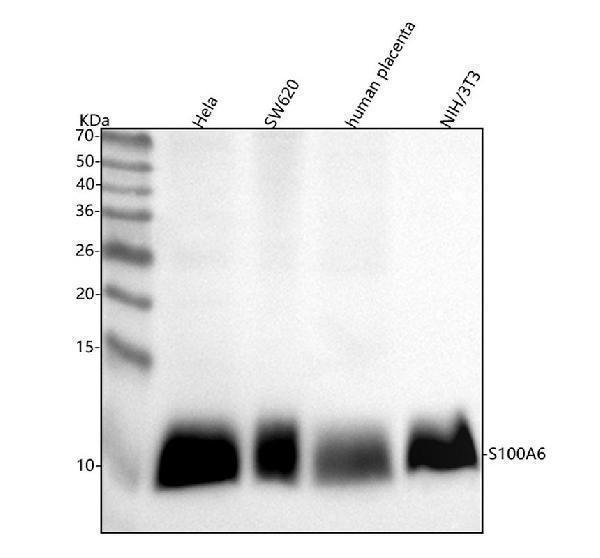
Figure 1. Western blot analysis of S100A6 using anti-S100A6 antibody (PB9676).
Electrophoresis was performed on a 5-20% SDS-PAGE gel at 70V (Stacking gel) / 90V (Resolving gel) for 2-3 hours. The sample well of each lane was loaded with 30 ug of sample under reducing conditions.
Lane 1: human Hela whole cell lysates,
Lane 2: human SW620 whole cell lysates,
Lane 3: human placenta tissue lysates,
Lane 4: mouse NIH/3T3 whole cell lysates.
After electrophoresis, proteins were transferred to a nitrocellulose membrane at 150 mA for 50-90 minutes. Blocked the membrane with 5% non-fat milk/TBS for 1.5 hour at RT. The membrane was incubated with rabbit anti-S100A6 antigen affinity purified polyclonal antibody (Catalog # PB9676) at 0.5 μg/mL overnight at 4°C, then washed with TBS-0.1%Tween 3 times with 5 minutes each and probed with a goat anti-rabbit IgG-HRP secondary antibody at a dilution of 1:5000 for 1.5 hour at RT. The signal is developed using an Enhanced Chemiluminescent detection (ECL) kit (Catalog # EK1002) with Tanon 5200 system. A specific band was detected for S100A6 at approximately 10 kDa. The expected band size for S100A6 is at 10 kDa.

Click image to see more details
Figure 2. IHC analysis of S100A6 using anti-S100A6 antibody (PB9676).
S100A6 was detected in paraffin-embedded section of Mouse Testis tissues. Heat mediated antigen retrieval was performed in citrate buffer (pH6, epitope retrieval solution) for 20 mins. The tissue section was blocked with 10% goat serum. The tissue section was then incubated with 1μg/ml rabbit anti-S100A6 Antibody (PB9676) overnight at 4°C. Biotinylated goat anti-rabbit IgG was used as secondary antibody and incubated for 30 minutes at 37°C. The tissue section was developed using Strepavidin-Biotin-Complex (SABC)(Catalog # SA1022) with DAB as the chromogen.

Click image to see more details
Figure 3. IHC analysis of S100A6 using anti-S100A6 antibody (PB9676).
S100A6 was detected in paraffin-embedded section of Rat Lung tissues. Heat mediated antigen retrieval was performed in citrate buffer (pH6, epitope retrieval solution) for 20 mins. The tissue section was blocked with 10% goat serum. The tissue section was then incubated with 1μg/ml rabbit anti-S100A6 Antibody (PB9676) overnight at 4°C. Biotinylated goat anti-rabbit IgG was used as secondary antibody and incubated for 30 minutes at 37°C. The tissue section was developed using Strepavidin-Biotin-Complex (SABC)(Catalog # SA1022) with DAB as the chromogen.

Click image to see more details
Figure 4. IHC analysis of S100A6 using anti-S100A6 antibody (PB9676).
S100A6 was detected in paraffin-embedded section of Human Mammary Cancer tissues. Heat mediated antigen retrieval was performed in citrate buffer (pH6, epitope retrieval solution) for 20 mins. The tissue section was blocked with 10% goat serum. The tissue section was then incubated with 1μg/ml rabbit anti-S100A6 Antibody (PB9676) overnight at 4°C. Biotinylated goat anti-rabbit IgG was used as secondary antibody and incubated for 30 minutes at 37°C. The tissue section was developed using Strepavidin-Biotin-Complex (SABC)(Catalog # SA1022) with DAB as the chromogen.

Click image to see more details
Figure 5. IHC analysis of S100A6 using anti-S100A6 antibody (PB9676).
S100A6 was detected in paraffin-embedded section of Human Placenta tissues. Heat mediated antigen retrieval was performed in citrate buffer (pH6, epitope retrieval solution) for 20 mins. The tissue section was blocked with 10% goat serum. The tissue section was then incubated with 1μg/ml rabbit anti-S100A6 Antibody (PB9676) overnight at 4°C. Biotinylated goat anti-rabbit IgG was used as secondary antibody and incubated for 30 minutes at 37°C. The tissue section was developed using Strepavidin-Biotin-Complex (SABC)(Catalog # SA1022) with DAB as the chromogen.

Click image to see more details
Figure 6. IF analysis of S100A6 using anti-S100A6 antibody (PB9677).
S100A6 was detected in an immunocytochemical section of A431 cells. Enzyme antigen retrieval was performed using IHC enzyme antigen retrieval reagent (AR0022) for 15 mins. The cells were blocked with 10% goat serum. And then incubated with 5 μg/mL rabbit anti-S100A6 Antibody (PB9677) overnight at 4°C. DyLight®488 Conjugated Goat Anti-Rabbit IgG (BA1127) was used as secondary antibody at 1:100 dilution and incubated for 30 minutes at 37°C. The tissue section was developed using Phalloidin-iFluor 594 Conjugated. Visualize using a fluorescence microscope and filter sets appropriate for the label used.

Click image to see more details
Figure 7. Flow Cytometry analysis of SiHa cells using anti-S100A6 antibody (PB9676).
Overlay histogram showing SiHa cells stained with PB9676 (Blue line).The cells were blocked with 10% normal goat serum. And then incubated with rabbit anti-S100A6 Antibody (PB9676,1μg/1x106 cells) for 30 min at 20°C. DyLight®488 conjugated goat anti-rabbit IgG (BA1127, 5-10μg/1x106 cells) was used as secondary antibody for 30 minutes at 20°C. Isotype control antibody (Green line) was rabbit IgG (1μg/1x106) used under the same conditions. Unlabelled sample (Red line) was also used as a control.

Click image to see more details
Figure 8. Flow Cytometry analysis of A431 cells using anti-S100A6 antibody (PB9676).
Overlay histogram showing A431 cells stained with PB9676 (Blue line).The cells were blocked with 10% normal goat serum. And then incubated with rabbit anti-S100A6 Antibody (PB9676,1μg/1x106 cells) for 30 min at 20°C. DyLight®488 conjugated goat anti-rabbit IgG (BA1127, 5-10μg/1x106 cells) was used as secondary antibody for 30 minutes at 20°C. Isotype control antibody (Green line) was rabbit IgG (1μg/1x106) used under the same conditions. Unlabelled sample (Red line) was also used as a control.
Protein Target Info & Infographic
Gene/Protein Information For S100A6 (Source: Uniprot.org, NCBI)
Gene Name
S100A6
Full Name
Protein S100-A6
Weight
10.18kDa
Superfamily
S-100 family
Alternative Names
2A9; CABP; CACY; CACY5B10; calcyclin; Growth factor-inducible protein 2A9; MLN 4; PRA; PRAS100 calcium binding protein A6 (calcyclin); Prolactin receptor-associated protein; protein S100-A6; S100 calcium binding protein A6; S100 calcium-binding protein A6 (calcyclin); S100 calcium-binding protein A6; S100A6 S100A6 2A9, 5B10, CABP, CACY, PRA, S10A6 S100 calcium binding protein A6 protein S100-A6|MLN 4|calcyclin|growth factor-inducible protein 2A9|prolactin receptor-associated protein
*If product is indicated to react with multiple species, protein info is based on the gene entry specified above in "Species".For more info on S100A6, check out the S100A6 Infographic

We have 30,000+ of these available, one for each gene! Check them out.
In this infographic, you will see the following information for S100A6: database IDs, superfamily, protein function, synonyms, molecular weight, chromosomal locations, tissues of expression, subcellular locations, post-translational modifications, and related diseases, research areas & pathways. If you want to see more information included, or would like to contribute to it and be acknowledged, please contact [email protected].
Specific Publications For Anti-S100 alpha 6/S100A6 Antibody Picoband™ (PB9676)
Hello CJ!
PB9676 has been cited in 1 publications:
*The publications in this section are manually curated by our staff scientists. They may differ from Bioz's machine gathered results. Both are accurate. If you find a publication citing this product but is missing from this list, please let us know we will issue you a thank-you coupon.
Song WH,Baik J,Yang S,Choi EK,Park SM,Jeong CW.Animal model evaluation of a novel renal denervation system for future laparoscopic treatment of resistant hypertension.Investig Clin Urol.2020 Jan;61(1):107-113.doi:10.4111/icu.2020.61.1.107.Epub 2019 Dec 23
Species: Pig
PB9676 usage in article: APP:IHC, SAMPLE:RENAL TISSUE, DILUTION:NA
Recommended Resources
Here are featured tools and databases that you might find useful.
- Boster's Pathways Library
- Protein Databases
- Bioscience Research Protocol Resources
- Data Processing & Analysis Software
- Photo Editing Software
- Scientific Literature Resources
- Research Paper Management Tools
- Molecular Biology Software
- Primer Design Tools
- Bioinformatics Tools
- Phylogenetic Tree Analysis
Customer Reviews
Have you used Anti-S100 alpha 6/S100A6 Antibody Picoband™?
Submit a review and receive an Amazon gift card.
- $30 for a review with an image
Be the first to review Anti-S100 alpha 6/S100A6 Antibody Picoband™
*The first user to submit a review for a product is eligible for Boster's Innovating Scientists Reward, which gives product credits. This is in addition to the gift card reward.
Customer Q&As
Have a question?
Find answers in Q&As, reviews.
Can't find your answer?
Submit your question
1 Customer Q&As for Anti-S100 alpha 6/S100A6 Antibody Picoband™
Question
We are currently using anti-S100 alpha 6/S100A6 antibody PB9676 for rat tissue, and we are happy with the IHC-P results. The species of reactivity given in the datasheet says human, mouse, rat. Is it possible that the antibody can work on bovine tissues as well?
E. Johnson
Verified customer
Asked: 2020-02-18
Answer
The anti-S100 alpha 6/S100A6 antibody (PB9676) has not been tested for cross reactivity specifically with bovine tissues, though there is a good chance of cross reactivity. We have an innovator award program that if you test this antibody and show it works in bovine you can get your next antibody for free. Please contact me if I can help you with anything.
Boster Scientific Support
Answered: 2020-02-18
