Click image to see more details
-

-
-
-
-
+9
Product Info Summary
| SKU: | M01194 |
|---|---|
| Size: | 100 μl |
| Reactive Species: | Human, Mouse, Rat |
| Host: | Rabbit |
| Application: | Flow Cytometry, IP, IF, IHC, ICC, WB |
Customers Who Bought This Also Bought


Product info
Product Name
Anti-Stathmin 1 STMN1 Rabbit Monoclonal Antibody
View all Stathmin 1 Antibodies
SKU/Catalog Number
M01194
Size
100 μl
Form
Liquid
Description
Boster Bio Anti-Stathmin 1 STMN1 Rabbit Monoclonal Antibody catalog # M01194. Tested in WB, IHC, ICC/IF, IP, Flow Cytometry applications. This antibody reacts with Human, Mouse, Rat.
Storage & Handling
Store at -20°C for one year. For short term storage and frequent use, store at 4°C for up to one month. Avoid repeated freeze-thaw cycles.
Cite This Product
Anti-Stathmin 1 STMN1 Rabbit Monoclonal Antibody (Boster Biological Technology, Pleasanton CA, USA, Catalog # M01194)
Host
Rabbit
Contents
Rabbit IgG in phosphate buffered saline, pH 7.4, 150mM NaCl, 0.02% sodium azide and 50% glycerol, 0.4-0.5mg/ml BSA.
Clonality
Monoclonal
Clone Number
DDA-19
Isotype
Rabbit IgG
Immunogen
A synthesized peptide derived from human Stathmin 1
*Blocking peptide can be purchased. Costs vary based on immunogen length. Contact us for pricing.
Reactive Species
M01194 is reactive to STMN1 in Human, Mouse, Rat
Reconstitution
Restore with deionized water (or equivalent) for reconstitution volume of 1.0 mL
Observed Molecular Weight
17 kDa
Calculated molecular weight
17303 MW
Antibody Validation
Boster validates all antibodies on WB, IHC, ICC, Immunofluorescence, and ELISA with known positive control and negative samples to ensure specificity and high affinity, including thorough antibody incubations.
Application & Images
Applications
M01194 is guaranteed for Flow Cytometry, IP, IF, IHC, ICC, WB Boster Guarantee
Assay Dilutions Recommendation
The recommendations below provide a starting point for assay optimization. The actual working concentration varies and should be decided by the user.
WB 1:500-1:2000
IHC 1:50-1:100
ICC/IF 1:50-1:100
IP 1:
50
FC 1:50
Positive Control
WB: human MDA-MB-453 whole cell, human SH-SY5Y whole cell, human Raji whole cell, rat brain tissue, rat testis tissue, rat PC-12 whole cell, mouse brain tissue, mouse testis tissue
IHC: human colon tissue, human colon tissue, human colon cancer tissue, human colon cancer tissue, human ovarian cancer tissue, human ovarian cancer tissue, neuroplexus of human colon tissue, neuroplexus of human colon tissue, neuroplexus of rat brain tissue, neuroplexus of rat brain tissue, neuroplexus of rat brain tissue, neuroplexus of rat brain tissue
Validation Images & Assay Conditions
Click image to see more details
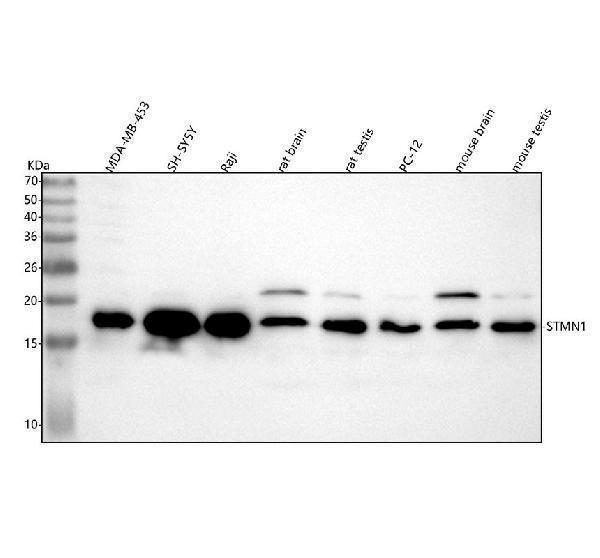
Figure 1. Western blot analysis of Stathmin 1 using anti-Stathmin 1 antibody (M00994).
Electrophoresis was performed on a 5-20% SDS-PAGE gel at 70V (Stacking gel) / 90V (Resolving gel) for 2-3 hours. The sample well of each lane was loaded with 30 ug of sample under reducing conditions.
Lane 1: human MDA-MB-453 whole cell lysates,
Lane 2: human SH-SY5Y whole cell lysates,
Lane 3: human Raji whole cell lysates,
Lane 4: rat brain tissue lysates,
Lane 5: rat testis tissue lysates,
Lane 6: rat PC-12 whole cell lysates,
Lane 7: mouse brain tissue lysates,
Lane 8: mouse testis tissue lysates.
After electrophoresis, proteins were transferred to a nitrocellulose membrane at 150 mA for 50-90 minutes. Blocked the membrane with 5% non-fat milk/TBS for 1.5 hour at RT. The membrane was incubated with rabbit anti-Stathmin 1 antigen affinity purified monoclonal antibody (Catalog # M00994) at 1:500 overnight at 4°C, then washed with TBS-0.1%Tween 3 times with 5 minutes each and probed with a goat anti-rabbit IgG-HRP secondary antibody at a dilution of 1:500 for 1.5 hour at RT. The signal is developed using an Enhanced Chemiluminescent detection (ECL) kit (Catalog # EK1002) with Tanon 5200 system. A specific band was detected for Stathmin 1 at approximately 17 kDa. The expected band size for Stathmin 1 is at 17 kDa.

Click image to see more details
Figure 2. IHC analysis of STMN1 using anti-STMN1 antibody (M01194).
STMN1 was detected in a paraffin-embedded section of human colon tissue. Heat mediated antigen retrieval was performed in EDTA buffer (pH 8.0, epitope retrieval solution). The tissue section was blocked with 10% goat serum. The tissue section was then incubated with 1:50 rabbit anti-STMN1 Antibody (M01194) overnight at 4°C. Peroxidase Conjugated Goat Anti-rabbit IgG was used as secondary antibody and incubated for 30 minutes at 37°C. The tissue section was developed using HRP Conjugated Rabbit IgG Super Vision Assay Kit (Catalog # SV0002) with DAB as the chromogen.

Click image to see more details
Figure 3. IHC analysis of STMN1 using anti-STMN1 antibody (M01194).
STMN1 was detected in a paraffin-embedded section of human colon tissue. Heat mediated antigen retrieval was performed in EDTA buffer (pH 8.0, epitope retrieval solution). The tissue section was blocked with 10% goat serum. The tissue section was then incubated with 1:50 rabbit anti-STMN1 Antibody (M01194) overnight at 4°C. Peroxidase Conjugated Goat Anti-rabbit IgG was used as secondary antibody and incubated for 30 minutes at 37°C. The tissue section was developed using HRP Conjugated Rabbit IgG Super Vision Assay Kit (Catalog # SV0002) with DAB as the chromogen.

Click image to see more details
Figure 4. IHC analysis of STMN1 using anti-STMN1 antibody (M01194).
STMN1 was detected in a paraffin-embedded section of human colon cancer tissue. Heat mediated antigen retrieval was performed in EDTA buffer (pH 8.0, epitope retrieval solution). The tissue section was blocked with 10% goat serum. The tissue section was then incubated with 1:50 rabbit anti-STMN1 Antibody (M01194) overnight at 4°C. Peroxidase Conjugated Goat Anti-rabbit IgG was used as secondary antibody and incubated for 30 minutes at 37°C. The tissue section was developed using HRP Conjugated Rabbit IgG Super Vision Assay Kit (Catalog # SV0002) with DAB as the chromogen.

Click image to see more details
Figure 5. IHC analysis of STMN1 using anti-STMN1 antibody (M01194).
STMN1 was detected in a paraffin-embedded section of human colon cancer tissue. Heat mediated antigen retrieval was performed in EDTA buffer (pH 8.0, epitope retrieval solution). The tissue section was blocked with 10% goat serum. The tissue section was then incubated with 1:50 rabbit anti-STMN1 Antibody (M01194) overnight at 4°C. Peroxidase Conjugated Goat Anti-rabbit IgG was used as secondary antibody and incubated for 30 minutes at 37°C. The tissue section was developed using HRP Conjugated Rabbit IgG Super Vision Assay Kit (Catalog # SV0002) with DAB as the chromogen.

Click image to see more details
Figure 6. IHC analysis of STMN1 using anti-STMN1 antibody (M01194).
STMN1 was detected in a paraffin-embedded section of human ovarian cancer tissue. Heat mediated antigen retrieval was performed in EDTA buffer (pH 8.0, epitope retrieval solution). The tissue section was blocked with 10% goat serum. The tissue section was then incubated with 1:50 rabbit anti-STMN1 Antibody (M01194) overnight at 4°C. Peroxidase Conjugated Goat Anti-rabbit IgG was used as secondary antibody and incubated for 30 minutes at 37°C. The tissue section was developed using HRP Conjugated Rabbit IgG Super Vision Assay Kit (Catalog # SV0002) with DAB as the chromogen.

Click image to see more details
Figure 7. IHC analysis of STMN1 using anti-STMN1 antibody (M01194).
STMN1 was detected in a paraffin-embedded section of human ovarian cancer tissue. Heat mediated antigen retrieval was performed in EDTA buffer (pH 8.0, epitope retrieval solution). The tissue section was blocked with 10% goat serum. The tissue section was then incubated with 1:50 rabbit anti-STMN1 Antibody (M01194) overnight at 4°C. Peroxidase Conjugated Goat Anti-rabbit IgG was used as secondary antibody and incubated for 30 minutes at 37°C. The tissue section was developed using HRP Conjugated Rabbit IgG Super Vision Assay Kit (Catalog # SV0002) with DAB as the chromogen.

Click image to see more details
Figure 8. IHC analysis of STMN1 using anti-STMN1 antibody (M01194).
STMN1 was detected in a paraffin-embedded section of neuroplexus of human colon tissue. Heat mediated antigen retrieval was performed in EDTA buffer (pH 8.0, epitope retrieval solution). The tissue section was blocked with 10% goat serum. The tissue section was then incubated with 1:50 rabbit anti-STMN1 Antibody (M01194) overnight at 4°C. Peroxidase Conjugated Goat Anti-rabbit IgG was used as secondary antibody and incubated for 30 minutes at 37°C. The tissue section was developed using HRP Conjugated Rabbit IgG Super Vision Assay Kit (Catalog # SV0002) with DAB as the chromogen.

Click image to see more details
Figure 9. IHC analysis of STMN1 using anti-STMN1 antibody (M01194).
STMN1 was detected in a paraffin-embedded section of neuroplexus of human colon tissue. Heat mediated antigen retrieval was performed in EDTA buffer (pH 8.0, epitope retrieval solution). The tissue section was blocked with 10% goat serum. The tissue section was then incubated with 1:50 rabbit anti-STMN1 Antibody (M01194) overnight at 4°C. Peroxidase Conjugated Goat Anti-rabbit IgG was used as secondary antibody and incubated for 30 minutes at 37°C. The tissue section was developed using HRP Conjugated Rabbit IgG Super Vision Assay Kit (Catalog # SV0002) with DAB as the chromogen.

Click image to see more details
Figure 10. IHC analysis of STMN1 using anti-STMN1 antibody (M01194).
STMN1 was detected in a paraffin-embedded section of neuroplexus of rat brain tissue. Heat mediated antigen retrieval was performed in EDTA buffer (pH 8.0, epitope retrieval solution). The tissue section was blocked with 10% goat serum. The tissue section was then incubated with 1:50 rabbit anti-STMN1 Antibody (M01194) overnight at 4°C. Peroxidase Conjugated Goat Anti-rabbit IgG was used as secondary antibody and incubated for 30 minutes at 37°C. The tissue section was developed using HRP Conjugated Rabbit IgG Super Vision Assay Kit (Catalog # SV0002) with DAB as the chromogen.

Click image to see more details
Figure 11. IHC analysis of STMN1 using anti-STMN1 antibody (M01194).
STMN1 was detected in a paraffin-embedded section of neuroplexus of rat brain tissue. Heat mediated antigen retrieval was performed in EDTA buffer (pH 8.0, epitope retrieval solution). The tissue section was blocked with 10% goat serum. The tissue section was then incubated with 1:50 rabbit anti-STMN1 Antibody (M01194) overnight at 4°C. Peroxidase Conjugated Goat Anti-rabbit IgG was used as secondary antibody and incubated for 30 minutes at 37°C. The tissue section was developed using HRP Conjugated Rabbit IgG Super Vision Assay Kit (Catalog # SV0002) with DAB as the chromogen.

Click image to see more details
Figure 12. IHC analysis of STMN1 using anti-STMN1 antibody (M01194).
STMN1 was detected in a paraffin-embedded section of neuroplexus of rat brain tissue. Heat mediated antigen retrieval was performed in EDTA buffer (pH 8.0, epitope retrieval solution). The tissue section was blocked with 10% goat serum. The tissue section was then incubated with 1:50 rabbit anti-STMN1 Antibody (M01194) overnight at 4°C. Peroxidase Conjugated Goat Anti-rabbit IgG was used as secondary antibody and incubated for 30 minutes at 37°C. The tissue section was developed using HRP Conjugated Rabbit IgG Super Vision Assay Kit (Catalog # SV0002) with DAB as the chromogen.

Click image to see more details
Figure 13. IHC analysis of STMN1 using anti-STMN1 antibody (M01194).
STMN1 was detected in a paraffin-embedded section of neuroplexus of rat brain tissue. Heat mediated antigen retrieval was performed in EDTA buffer (pH 8.0, epitope retrieval solution). The tissue section was blocked with 10% goat serum. The tissue section was then incubated with 1:50 rabbit anti-STMN1 Antibody (M01194) overnight at 4°C. Peroxidase Conjugated Goat Anti-rabbit IgG was used as secondary antibody and incubated for 30 minutes at 37°C. The tissue section was developed using HRP Conjugated Rabbit IgG Super Vision Assay Kit (Catalog # SV0002) with DAB as the chromogen.
Protein Target Info & Infographic
Gene/Protein Information For STMN1 (Source: Uniprot.org, NCBI)
Gene Name
STMN1
Full Name
Stathmin
Weight
17303 MW
Superfamily
stathmin family
Alternative Names
Stathmin;Leukemia-associated phosphoprotein p18;Metablastin;Oncoprotein 18;Op18;Phosphoprotein p19;pp19;Prosolin;Protein Pr22;pp17;STMN1;C1orf215, LAP18, OP18; STMN1 C1orf215, LAP18, Lag, OP18, PP17, PP19, PR22, SMN stathmin 1 stathmin|leukemia-associated phosphoprotein p18|metablastin|oncoprotein 18|phosphoprotein 19|phosphoprotein p19|prosolin|stathmin 1/oncoprotein 18|testicular tissue protein Li 189|transmembrane protein C1orf215
*If product is indicated to react with multiple species, protein info is based on the gene entry specified above in "Species".For more info on STMN1, check out the STMN1 Infographic

We have 30,000+ of these available, one for each gene! Check them out.
In this infographic, you will see the following information for STMN1: database IDs, superfamily, protein function, synonyms, molecular weight, chromosomal locations, tissues of expression, subcellular locations, post-translational modifications, and related diseases, research areas & pathways. If you want to see more information included, or would like to contribute to it and be acknowledged, please contact [email protected].
Specific Publications For Anti-Stathmin 1 STMN1 Rabbit Monoclonal Antibody (M01194)
Hello CJ!
No publications found for M01194
*Do you have publications using this product? Share with us and receive a reward. Ask us for more details.
Recommended Resources
Here are featured tools and databases that you might find useful.
- Boster's Pathways Library
- Protein Databases
- Bioscience Research Protocol Resources
- Data Processing & Analysis Software
- Photo Editing Software
- Scientific Literature Resources
- Research Paper Management Tools
- Molecular Biology Software
- Primer Design Tools
- Bioinformatics Tools
- Phylogenetic Tree Analysis
Customer Reviews
Have you used Anti-Stathmin 1 STMN1 Rabbit Monoclonal Antibody?
Submit a review and receive an Amazon gift card.
- $30 for a review with an image
0 Reviews For Anti-Stathmin 1 STMN1 Rabbit Monoclonal Antibody
Customer Q&As
Have a question?
Find answers in Q&As, reviews.
Can't find your answer?
Submit your question
7 Customer Q&As for Anti-Stathmin 1 STMN1 Rabbit Monoclonal Antibody
Question
Is there a BSA free version of anti-Stathmin 1 Rabbit Monoclonal antibody M01194 available?
J. Collins
Verified customer
Asked: 2019-10-25
Answer
Thanks for your recent telephone inquiry. I can confirm that some lots of this anti-Stathmin 1 Rabbit Monoclonal antibody M01194 are BSA free. For now, these lots are available and we can make a BSA free formula for you free of charge. It will take 3 extra days to prepare. If you require this antibody BSA free again in future, please do not hesitate to contact me and I will be pleased to check which lots we have in stock that are BSA free.
Boster Scientific Support
Answered: 2019-10-25
Question
Is this M01194 anti-Stathmin 1 Rabbit Monoclonal antibody reactive to the isotypes of STMN1?
Verified Customer
Verified customer
Asked: 2019-09-13
Answer
The immunogen of M01194 anti-Stathmin 1 Rabbit Monoclonal antibody is A synthesized peptide derived from human Stathmin 1. Could you tell me which isotype you are interested in so I can help see if the immunogen is part of this isotype?
Boster Scientific Support
Answered: 2019-09-13
Question
Does M01194 anti-Stathmin 1 Rabbit Monoclonal antibody work on parafin embedded sections? If so, which fixation method do you recommend we use (PFA, paraformaldehyde, other)?
Verified Customer
Verified customer
Asked: 2019-08-07
Answer
It shows on the product datasheet, M01194 anti-Stathmin 1 Rabbit Monoclonal antibody as been tested on IHC. It is best to use PFA for fixation because it has better tissue penetration ability. PFA needs to be prepared fresh before use. Long term stored PFA turns into formalin, as the PFA molecules congregate and become formalin.
Boster Scientific Support
Answered: 2019-08-07
Question
My question regarding product M01194, anti-Stathmin 1 Rabbit Monoclonal antibody. I was wondering if it would be possible to conjugate this antibody with biotin. I would need it to be without BSA or sodium azide. I am planning on using a buffer exchange of sodium azide with PBS only. Would there be problems for me to conjugate the antibody and store it in -20 degrees in small aliquots?
S. Walker
Verified customer
Asked: 2019-01-25
Answer
We do not advise storing this antibody with PBS buffer only in -20 degrees. If you want to store it in -20 degrees it is best to add some cryoprotectant like glycerol. If you want carrier free M01194 anti-Stathmin 1 Rabbit Monoclonal antibody, we can provide it to you in a special formula with trehalose and/or glycerol. These molecules will not interfere with conjugation chemistry and provide a good level of protection for the antibody from degradation. Please be sure to specify this in your purchase order.
Boster Scientific Support
Answered: 2019-01-25
Question
Please see the WB image, lot number and protocol we used for cajal-retzius cell fetal brain cortex using anti-Stathmin 1 Rabbit Monoclonal antibody M01194. Please let me know if you require anything else.
Verified Customer
Verified customer
Asked: 2018-08-27
Answer
Thank you very much for the data. Our lab team are working to resolve this as quickly as possible, and we appreciate your patience and understanding! You have provided everything we needed. Please let me know if there is anything you need in the meantime.
Boster Scientific Support
Answered: 2018-08-27
Question
Is a blocking peptide available for product anti-Stathmin 1 Rabbit Monoclonal antibody (M01194)?
V. Baker
Verified customer
Asked: 2014-12-31
Answer
We do provide the blocking peptide for product anti-Stathmin 1 Rabbit Monoclonal antibody (M01194). If you would like to place an order for it please contact [email protected] and make a special request.
Boster Scientific Support
Answered: 2014-12-31
Question
I appreciate helping with my inquiry over the phone. Here are the WB image, lot number and protocol we used for cajal-retzius cell fetal brain cortex using anti-Stathmin 1 Rabbit Monoclonal antibody M01194. Let me know if you need anything else.
E. Li
Verified customer
Asked: 2013-03-01
Answer
Thank you for the data. You have provided everything we needed. Our lab team are working to resolve your inquiry as quickly as possible, and we appreciate your patience and understanding! Please let me know if there is anything you need in the meantime.
Boster Scientific Support
Answered: 2013-03-01
