Click image to see more details
-

-
-
-
-
+3
Product Info Summary
| SKU: | A01282-2 |
|---|---|
| Size: | 100 μg/vial |
| Reactive Species: | Human, Mouse, Rat |
| Host: | Rabbit |
| Application: | ELISA, Flow Cytometry, IF, IHC, ICC, WB |
Customers Who Bought This Also Bought

Product info
Product Name
Anti-SUMO2/3 Antibody Picoband™
SKU/Catalog Number
A01282-2
Size
100 μg/vial
Form
Lyophilized
Description
Boster Bio Anti-SUMO2/3 Antibody Picoband™ catalog # A01282-2. Tested in ELISA, Flow Cytometry, IF, IHC, ICC, WB applications. This antibody reacts with Human, Mouse, Rat.
Storage & Handling
Store at -20˚C for one year from date of receipt. After reconstitution, at 4˚C for one month. It can also be aliquotted and stored frozen at -20˚C for six months. Avoid repeated freeze-thaw cycles.
Cite This Product
Anti-SUMO2/3 Antibody Picoband™ (Boster Biological Technology, Pleasanton CA, USA, Catalog # A01282-2)
Host
Rabbit
Contents
Each vial contains 4mg Trehalose, 0.9mg NaCl, 0.2mg Na2HPO4.
Clonality
Polyclonal
Isotype
Rabbit IgG
Immunogen
E.coli-derived human SUMO2/3 recombinant protein (Position: Q24-T72).
*Blocking peptide can be purchased. Costs vary based on immunogen length. Contact us for pricing.
Cross-reactivity
No cross-reactivity with other proteins.
Reactive Species
A01282-2 is reactive to in Human, Mouse, Rat
Applications
A01282-2 is guaranteed for ELISA, Flow Cytometry, IF, IHC, ICC, WB Boster Guarantee
Observed Molecular Weight
20 kDa
Calculated molecular weight
Background of
Small ubiquitin-related modifier 2,3 is a protein that in humans is encoded by the SUMO2,3 gene. This gene encodes a protein that is a member of the SUMO (small ubiquitin-like modifier) protein family. It functions in a manner similar to ubiquitin in that it is bound to target proteins as part of a post-translational modification system. However, unlike ubiquitin which targets proteins for degradation, this protein is involved in a variety of cellular processes, such as nuclear transport, transcriptional regulation, apoptosis, and protein stability. It is not active until the last two amino acids of the carboxy-terminus have been cleaved off. Numerous pseudogenes have been reported for this gene. Alternate transcriptional splice variants, encoding different isoforms, have been characterized.
Antibody Validation
Boster validates all antibodies on WB, IHC, ICC, Immunofluorescence, and ELISA with known positive control and negative samples to ensure specificity and high affinity, including thorough antibody incubations.
Innovating Scientists Reward
If you are the first to review this product, or if you have results for a special sample, species or application this product is not validated in, share your results with us and receive product credits you can use towards any Boster products! Applicable to all scientists worldwide.
Submit A Review
Assay dilution & Images
Reconsitution
Add 0.2ml of distilled water will yield a concentration of 500ug/ml.
Assay Dilutions Recommendation
The recommendations below provide a starting point for assay optimization. The actual working concentration varies and should be decided by the user.
Western blot, 0.25-0.5μg/ml, Human, Mouse, Rat
Immunohistochemistry (Paraffin-embedded Section), 2-5μg/ml, Human, Rat
Immunocytochemistry/Immunofluorescence, 5μg/ml, Human
Flow Cytometry, 1-3μg/1x106 cells, Human
Direct ELISA, 0.1-0.5μg/ml, Human
Validation Images & Assay Conditions
Click image to see more details
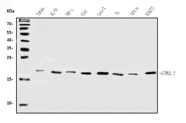
Figure 1. Western blot analysis of SUMO2/3 using anti-SUMO2/3 antibody (A01282-2).
Electrophoresis was performed on a 5-20% SDS-PAGE gel at 70V (Stacking gel) / 90V (Resolving gel) for 2-3 hours. The sample well of each lane was loaded with 30 ug of sample under reducing conditions.
Lane 1: human Jurkat whole cell lysates,
Lane 2: human HL-60 whole cell lysates,
Lane 3: human THP-1 whole cell lysates,
Lane 4: human K562 whole cell lysates,
Lane 5: human Caco-2 whole cell lysates,
Lane 6: rat C6 whole cell lysates,
Lane 8: mouse SP2/0 whole cell lysates,
Lane 7: mouse NIH/3T3 whole cell lysates.
After electrophoresis, proteins were transferred to a nitrocellulose membrane at 150 mA for 50-90 minutes. Blocked the membrane with 5% non-fat milk/TBS for 1.5 hour at RT. The membrane was incubated with rabbit anti-A01282-2 antigen affinity purified polyclonal antibody (Catalog # A01282-2) at 0.5 μg/mL overnight at 4°C, then washed with TBS-0.1%Tween 3 times with 5 minutes each and probed with a goat anti-rabbit IgG-HRP secondary antibody at a dilution of 1:5000 for 1.5 hour at RT. The signal is developed using an Enhanced Chemiluminescent detection (ECL) kit (Catalog # EK1002) with Tanon 5200 system. A specific band was detected for SUMO2/3 at approximately 20 kDa. The expected band size for SUMO2/3 is at 20 kDa.

Click image to see more details
Figure 2. IHC analysis of SUMO2/3 using anti-SUMO2/3 antibody (A01282-2).
SUMO2/3 was detected in a paraffin-embedded section of human bladder cancer tissue. Heat mediated antigen retrieval was performed in EDTA buffer (pH 8.0, epitope retrieval solution). The tissue section was blocked with 10% goat serum. The tissue section was then incubated with 2 μg/ml rabbit anti-SUMO2/3 Antibody (A01282-2) overnight at 4°C. Biotinylated goat anti-rabbit IgG was used as secondary antibody and incubated for 30 minutes at 37°C. The tissue section was developed using Strepavidin-Biotin-Complex (SABC) (Catalog # SA1022) with DAB as the chromogen.

Click image to see more details
Figure 3. IHC analysis of SUMO2/3 using anti-SUMO2/3 antibody (A01282-2).
SUMO2/3 was detected in a paraffin-embedded section of human gallbladder adenocarcinoma tissue. Heat mediated antigen retrieval was performed in EDTA buffer (pH 8.0, epitope retrieval solution). The tissue section was blocked with 10% goat serum. The tissue section was then incubated with 2 μg/ml rabbit anti-SUMO2/3 Antibody (A01282-2) overnight at 4°C. Biotinylated goat anti-rabbit IgG was used as secondary antibody and incubated for 30 minutes at 37°C. The tissue section was developed using Strepavidin-Biotin-Complex (SABC) (Catalog # SA1022) with DAB as the chromogen.

Click image to see more details
Figure 4. IHC analysis of SUMO2/3 using anti-SUMO2/3 antibody (A01282-2).
SUMO2/3 was detected in a paraffin-embedded section of human breast cancer tissue. Heat mediated antigen retrieval was performed in EDTA buffer (pH 8.0, epitope retrieval solution). The tissue section was blocked with 10% goat serum. The tissue section was then incubated with 2 μg/ml rabbit anti-SUMO2/3 Antibody (A01282-2) overnight at 4°C. Biotinylated goat anti-rabbit IgG was used as secondary antibody and incubated for 30 minutes at 37°C. The tissue section was developed using Strepavidin-Biotin-Complex (SABC) (Catalog # SA1022) with DAB as the chromogen.

Click image to see more details
Figure 5. IF analysis of SUMO2/3 using anti-SUMO2/3 antibody (A01282-2).
SUMO2/3 was detected in an immunocytochemical section of A431 cells. Enzyme antigen retrieval was performed using IHC enzyme antigen retrieval reagent (AR0022) for 15 mins. The cells were blocked with 10% goat serum. And then incubated with 5 μg/mL rabbit anti-SUMO2/3 Antibody (A01282-2) overnight at 4°C. DyLight®488 Conjugated Goat Anti-Rabbit IgG (BA1127) was used as secondary antibody at 1:100 dilution and incubated for 30 minutes at 37°C. The section was counterstained with DAPI. Visualize using a fluorescence microscope and filter sets appropriate for the label used.

Click image to see more details
Figure 6. Flow Cytometry analysis of A431 cells using anti-SUMO2/3 antibody (A01282-2).
Overlay histogram showing A431 cells stained with A01282-2 (Blue line). The cells were blocked with 10% normal goat serum. And then incubated with rabbit anti-SUMO2/3 Antibody (A01282-2, 1 μg/1x106 cells) for 30 min at 20°C. DyLight®488 conjugated goat anti-rabbit IgG (BA1127, 5-10 μg/1x106 cells) was used as secondary antibody for 30 minutes at 20°C. Isotype control antibody (Green line) was rabbit IgG (1 μg/1x106) used under the same conditions. Unlabelled sample (Red line) was also used as a control.

Click image to see more details
Figure 7. IHC analysis of SUMO2/3 using anti-SUMO2/3 antibody (A01282-2).
SUMO2/3 was detected in a paraffin-embedded section of rat intestine tissue. Heat mediated antigen retrieval was performed in EDTA buffer (pH 8.0, epitope retrieval solution). The tissue section was blocked with 10% goat serum. The tissue section was then incubated with 2 μg/ml rabbit anti-SUMO2/3 Antibody (A01282-2) overnight at 4°C. Biotinylated goat anti-rabbit IgG was used as secondary antibody and incubated for 30 minutes at 37°C. The tissue section was developed using Strepavidin-Biotin-Complex (SABC) (Catalog # SA1022) with DAB as the chromogen.
Specific Publications For Anti-SUMO2/3 Antibody Picoband™ (A01282-2)
Hello CJ!
No publications found for A01282-2
*Do you have publications using this product? Share with us and receive a reward. Ask us for more details.
Recommended Resources
Here are featured tools and databases that you might find useful.
- Boster's Pathways Library
- Protein Databases
- Bioscience Research Protocol Resources
- Data Processing & Analysis Software
- Photo Editing Software
- Scientific Literature Resources
- Research Paper Management Tools
- Molecular Biology Software
- Primer Design Tools
- Bioinformatics Tools
- Phylogenetic Tree Analysis
Customer Reviews
Have you used Anti-SUMO2/3 Antibody Picoband™?
Submit a review and receive an Amazon gift card.
- $30 for a review with an image
Be the first to review Anti-SUMO2/3 Antibody Picoband™
*The first user to submit a review for a product is eligible for Boster's Innovating Scientists Reward, which gives product credits. This is in addition to the gift card reward.
Customer Q&As
Have a question?
Find answers in Q&As, reviews.
Can't find your answer?
Submit your question
