Click image to see more details
-

-
-
-
-
+7
Product Info Summary
| SKU: | M03989-3 |
|---|---|
| Size: | 100 μg/vial |
| Reactive Species: | Human, Mouse, Rat |
| Host: | Mouse |
| Application: | Flow Cytometry, IF, IHC, ICC, WB |
Customers Who Bought This Also Bought

Product info
Product Name
Anti-Tubulin alpha Picoband™ Antibody (monoclonal, 7B12)
View all alpha Tubulin Antibodies
SKU/Catalog Number
M03989-3
Size
100 μg/vial
Form
Lyophilized
Description
Boster Bio Anti-Tubulin alpha Picoband™ Antibody (monoclonal, 7B12) catalog # M03989-3. Tested in Flow Cytometry, IF, IHC, ICC, WB applications. This antibody reacts with Human, Mouse, Rat.
Storage & Handling
Store at -20˚C for one year from date of receipt. After reconstitution, at 4˚C for one month. It can also be aliquotted and stored frozen at -20˚C for six months. Avoid repeated freeze-thaw cycles.
Cite This Product
Anti-Tubulin alpha Picoband™ Antibody (monoclonal, 7B12) (Boster Biological Technology, Pleasanton CA, USA, Catalog # M03989-3)
Host
Mouse
Contents
Each vial contains 4mg Trehalose, 0.9mg NaCl and 0.2mg Na2HPO4.
Clonality
Monoclonal
Clone Number
7B12
Isotype
Mouse IgG2b
Immunogen
E.coli-derived human Tubulin alpha recombinant protein (Position: N18-A403).
*Blocking peptide can be purchased. Costs vary based on immunogen length. Contact us for pricing.
Cross-reactivity
No cross-reactivity with other proteins.
Reactive Species
M03989-3 is reactive to TUBA1A in Human, Mouse, Rat
Applications
M03989-3 is guaranteed for Flow Cytometry, IF, IHC, ICC, WB Boster Guarantee
Observed Molecular Weight
56 kDa
Calculated molecular weight
50.136kDa
Background of alpha Tubulin
Tubulin is the major constituent of microtubules. It binds two moles of GTP, one at an exchangeable site on the beta chain and one at a non-exchangeable site on the alpha chain. Microtubules of the eukaryotic cytoskeleton perform essential and diverse functions and are composed of a heterodimer of alpha and beta tubulins. The genes encoding these microtubule constituents belong to the tubulin superfamily, which is composed of six distinct families. Genes from the alpha, beta and gamma tubulin families are found in all eukaryotes. The alpha and beta tubulins represent the major components of microtubules, while gamma tubulin plays a critical role in the nucleation of microtubule assembly. There are multiple alpha and beta tubulin genes, which are highly conserved among species. This gene encodes alpha tubulin and is highly similar to the mouse and rat Tuba1 genes. Northern blot studies have shown that the gene expression is predominantly found in morphologically differentiated neurologic cells. This gene is one of three alpha-tubulin genes in a cluster on chromosome 12q. Mutations in this gene cause lissencephaly type 3 (LIS3) - a neurological condition characterized by microcephaly, intellectual disability, and early-onset epilepsy caused by defective neuronal migration. Alternative splicing results in multiple transcript variants encoding distinct isoforms.
Antibody Validation
Boster validates all antibodies on WB, IHC, ICC, Immunofluorescence, and ELISA with known positive control and negative samples to ensure specificity and high affinity, including thorough antibody incubations.
Assay dilution & Images
Reconsitution
Add 0.2ml of distilled water will yield a concentration of 500ug/ml.
Assay Dilutions Recommendation
The recommendations below provide a starting point for assay optimization. The actual working concentration varies and should be decided by the user.
Western blot, 0.1-0.25μg/ml, Human, Mouse, Rat
Immunohistochemistry (Paraffin-embedded Section), 2-5μg/ml, Human, Mouse, Rat
Immunocytochemistry/Immunofluorescence, 5μg/ml, Human
Flow Cytometry, 1-3μg/1x106 cells, Human
Validation Images & Assay Conditions
Click image to see more details
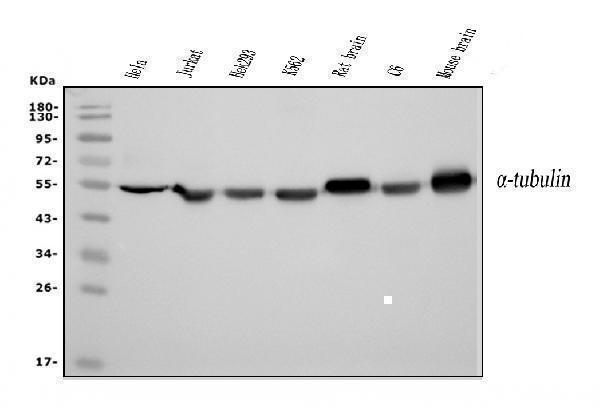
Figure 1. Western blot analysis of Tubulin alpha using anti-Tubulin alpha antibody (M03989-3).
Electrophoresis was performed on a 5-20% SDS-PAGE gel at 70V (Stacking gel) / 90V (Resolving gel) for 2-3 hours. The sample well of each lane was loaded with 50ug of sample under reducing conditions.
Lane 1: human HELA whole cell lysates,
Lane 2: human Jurkat whole cell lysates,
Lane 3: human HEK293 whole cell lysates,
Lane 4: human K562 whole cell lysates,
Lane 5: rat brain tissue lysates,
Lane 6: rat C6 whole cell lysates,
Lane 7: mouse brain tissue lysates.
After Electrophoresis, proteins were transferred to a Nitrocellulose membrane at 150mA for 50-90 minutes. Blocked the membrane with 5% Non-fat Milk/ TBS for 1.5 hour at RT. The membrane was incubated with mouse anti-Tubulin alpha antigen affinity purified monoclonal antibody (Catalog # M03989-3) at 0.25 μg/mL overnight at 4°C, then washed with TBS-0.1%Tween 3 times with 5 minutes each and probed with a goat anti-mouse IgG-HRP secondary antibody at a dilution of 1:10000 for 1.5 hour at RT. The signal is developed using an Enhanced Chemiluminescent detection (ECL) kit (Catalog # EK1001) with Tanon 5200 system. A specific band was detected for Tubulin alpha at approximately 56KD. The expected band size for Tubulin alpha is at 56KD.

Click image to see more details
Figure 10. Flow Cytometry analysis of A431 cells using anti-Tubulin alpha antibody (M03989-3).
Overlay histogram showing A431 cells stained with M03989-3 (Blue line).The cells were blocked with 10% normal goat serum. And then incubated with mouse anti-Tubulin alpha Antibody (M03989-3, 1μg/1x106 cells) for 30 min at 20°C. DyLight®488 conjugated goat anti-mouse IgG (BA1126, 5-10μg/1x106 cells) was used as secondary antibody for 30 minutes at 20°C. Isotype control antibody (Green line) was mouse IgG (1μg/1x106) used under the same conditions. Unlabelled sample (Red line) was also used as a control.

Click image to see more details
Figure 2. IHC analysis of Tubulin alpha using anti-Tubulin alpha antibody (M03989-3).
Tubulin alpha was detected in paraffin-embedded section of mouse brain tissue. Heat mediated antigen retrieval was performed in EDTA buffer (pH8.0, epitope retrieval solution). The tissue section was blocked with 10% goat serum. The tissue section was then incubated with 2μg/ml mouse anti-Tubulin alpha Antibody (M03989-3) overnight at 4°C. Biotinylated goat anti-mouse IgG was used as secondary antibody and incubated for 30 minutes at 37°C. The tissue section was developed using Strepavidin-Biotin-Complex (SABC) (Catalog # SA1021) with DAB as the chromogen.

Click image to see more details
Figure 3. IHC analysis of Tubulin alpha using anti-Tubulin alpha antibody (M03989-3).
Tubulin alpha was detected in paraffin-embedded section of rat brain tissue. Heat mediated antigen retrieval was performed in EDTA buffer (pH8.0, epitope retrieval solution). The tissue section was blocked with 10% goat serum. The tissue section was then incubated with 2μg/ml mouse anti-Tubulin alpha Antibody (M03989-3) overnight at 4°C. Biotinylated goat anti-mouse IgG was used as secondary antibody and incubated for 30 minutes at 37°C. The tissue section was developed using Strepavidin-Biotin-Complex (SABC) (Catalog # SA1021) with DAB as the chromogen.

Click image to see more details
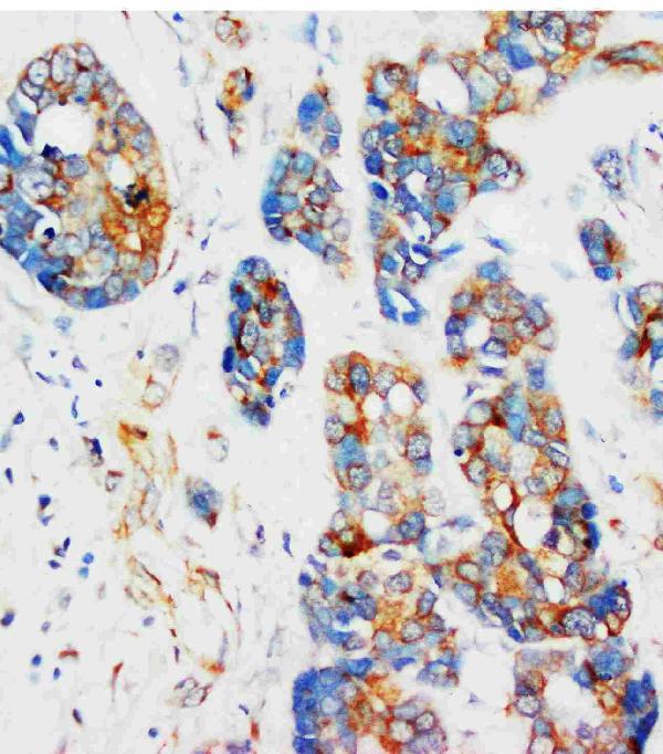
Figure 4. IHC analysis of Tubulin alpha using anti-Tubulin alpha antibody (M03989-3).
Tubulin alpha was detected in paraffin-embedded section of human ovarian serous adenocarcinoma tissue. Heat mediated antigen retrieval was performed in EDTA buffer (pH8.0, epitope retrieval solution). The tissue section was blocked with 10% goat serum. The tissue section was then incubated with 2μg/ml mouse anti-Tubulin alpha Antibody (M03989-3) overnight at 4°C. Biotinylated goat anti-mouse IgG was used as secondary antibody and incubated for 30 minutes at 37°C. The tissue section was developed using Strepavidin-Biotin-Complex (SABC) (Catalog # SA1021) with DAB as the chromogen.

Click image to see more details
Figure 5. IHC analysis of Tubulin alpha using anti-Tubulin alpha antibody (M03989-3).
Tubulin alpha was detected in paraffin-embedded section of human lung cancer tissue. Heat mediated antigen retrieval was performed in EDTA buffer (pH8.0, epitope retrieval solution). The tissue section was blocked with 10% goat serum. The tissue section was then incubated with 2μg/ml mouse anti-Tubulin alpha Antibody (M03989-3) overnight at 4°C. Biotinylated goat anti-mouse IgG was used as secondary antibody and incubated for 30 minutes at 37°C. The tissue section was developed using Strepavidin-Biotin-Complex (SABC) (Catalog # SA1021) with DAB as the chromogen.

Click image to see more details
Figure 6. IHC analysis of Tubulin alpha using anti-Tubulin alpha antibody (M03989-3).
Tubulin alpha was detected in paraffin-embedded section of human breast cancer tissue. Heat mediated antigen retrieval was performed in EDTA buffer (pH8.0, epitope retrieval solution). The tissue section was blocked with 10% goat serum. The tissue section was then incubated with 2μg/ml mouse anti-Tubulin alpha Antibody (M03989-3) overnight at 4°C. Biotinylated goat anti-mouse IgG was used as secondary antibody and incubated for 30 minutes at 37°C. The tissue section was developed using Strepavidin-Biotin-Complex (SABC) (Catalog # SA1021) with DAB as the chromogen.

Click image to see more details
Figure 7. IHC analysis of Tubulin alpha using anti-Tubulin alpha antibody (M03989-3).
Tubulin alpha was detected in paraffin-embedded section of human liver cancer tissue. Heat mediated antigen retrieval was performed in EDTA buffer (pH8.0, epitope retrieval solution). The tissue section was blocked with 10% goat serum. The tissue section was then incubated with 2μg/ml mouse anti-Tubulin alpha Antibody (M03989-3) overnight at 4°C. Biotinylated goat anti-mouse IgG was used as secondary antibody and incubated for 30 minutes at 37°C. The tissue section was developed using Strepavidin-Biotin-Complex (SABC) (Catalog # SA1021) with DAB as the chromogen.

Click image to see more details
Figure 8. IHC analysis of Tubulin alpha using anti-Tubulin alpha antibody (M03989-3).
Tubulin alpha was detected in paraffin-embedded section of human pancreatic cancer tissue. Heat mediated antigen retrieval was performed in EDTA buffer (pH8.0, epitope retrieval solution). The tissue section was blocked with 10% goat serum. The tissue section was then incubated with 2μg/ml mouse anti-Tubulin alpha Antibody (M03989-3) overnight at 4°C. Biotinylated goat anti-mouse IgG was used as secondary antibody and incubated for 30 minutes at 37°C. The tissue section was developed using Strepavidin-Biotin-Complex (SABC) (Catalog # SA1021) with DAB as the chromogen.

Click image to see more details
Figure 9. IF analysis of Tubulin alpha using anti-Tubulin alpha antibody (M03989-3).
Tubulin alpha was detected in immunocytochemical section of A431 cells. Enzyme antigen retrieval was performed using IHC enzyme antigen retrieval reagent (AR0022) for 15 mins. The cells were blocked with 10% goat serum. And then incubated with 5μg/mL mouse anti-Tubulin alpha Antibody (M03989-3) overnight at 4°C. DyLight®488 Conjugated Goat Anti-Mouse IgG (BA1126) was used as secondary antibody at 1:100 dilution and incubated for 30 minutes at 37°C. The section was counterstained with DAPI. Visualize using a fluorescence microscope and filter sets appropriate for the label used.

Click image to see more details
Figure 11. IF analysis of Tubulin alpha using anti-Tubulin alpha antibody (M03989-3).
Tubulin alpha was detected in immunocytochemical section of CACO-2 cells. Enzyme antigen retrieval was performed using IHC enzyme antigen retrieval reagent (AR0022) for 15 mins. The cells were blocked with 10% goat serum. And then incubated with 5μg/mL mouse anti-Tubulin alpha Antibody (M03989-3) overnight at 4°C. DyLight®594 Conjugated Goat Anti-Mouse IgG (BA1141) was used as secondary antibody at 1:100 dilution and incubated for 30 minutes at 37°C. The section was counterstained with DAPI. Visualize using a fluorescence microscope and filter sets appropriate for the label used.
Protein Target Info & Infographic
Gene/Protein Information For TUBA1A (Source: Uniprot.org, NCBI)
Gene Name
TUBA1A
Full Name
Tubulin alpha-1A chain
Weight
50.136kDa
Superfamily
tubulin family
Alternative Names
alpha tubulin loading control; alpha tubulin monoclonal; alpha Tubulin; Alpha-Tubulin 3; B-ALPHA-1; DM1A tubulin; dm1a monoclonal; DM1a tubulin; DM1A; FLJ25113; hum-a-tub1; hum-a-tub2; LIS3; TUBA1 monoclonal; TUBA1A; TUBA3; tubulin alpha-1A chain; Tubulin alpha-3 chain; Tubulin B-Alpha-1; tubulin monoclonal; tubulin, alpha 1a; tubulin, alpha 3; tubulin, alpha, brain-specific TUBA1A B-ALPHA-1, LIS3, TUBA3 tubulin alpha 1a tubulin alpha-1A chain|hum-a-tub1|hum-a-tub2|tubulin B-alpha-1|tubulin alpha-3 chain|tubulin, alpha, brain-specific
*If product is indicated to react with multiple species, protein info is based on the gene entry specified above in "Species".For more info on TUBA1A, check out the TUBA1A Infographic

We have 30,000+ of these available, one for each gene! Check them out.
In this infographic, you will see the following information for TUBA1A: database IDs, superfamily, protein function, synonyms, molecular weight, chromosomal locations, tissues of expression, subcellular locations, post-translational modifications, and related diseases, research areas & pathways. If you want to see more information included, or would like to contribute to it and be acknowledged, please contact [email protected].
Specific Publications For Anti-Tubulin alpha Picoband™ Antibody (monoclonal, 7B12) (M03989-3)
Hello CJ!
M03989-3 has been cited in 9 publications:
*The publications in this section are manually curated by our staff scientists. They may differ from Bioz's machine gathered results. Both are accurate. If you find a publication citing this product but is missing from this list, please let us know we will issue you a thank-you coupon.
Protein Nanoparticle-Related Osmotic Pressure Modifies Nonselective Permeability of the Blood–Brain Barrier by Increasing Membrane Fluidity
A proteomic view of Caenorhabditis elegans caused by short-term hypoxic stress
Yu-Yuan Chen,Yin-Peng Bai,Bin Li,Xiao-Bo Zhao,Cheng-Jie Yang,Ying-Qian Liu,Jian-Mei Gao,Jun Guo,Chun Li,Jing-Wen Peng,Zhong-Min Zhao,Zhi-Jun Zhang,Chuan-Rui Xu,Design and Synthesis of Novel 20(S)-α-aminophosphonate Derivatives of Camptothecin as Potent Antitumor Agents,Bioorganic Chemistry,2021,105065,ISSN 0045-2068, https://doi.org/10.1016/j.bioorg.2021.105065.
Species: Human,Mouse
M03989-3 usage in article: APP:WB, SAMPLE:MCF-7 CELL, DILUTION:1:1000
Danyang Chong,Zhong Chen,Shan Guan,Tongyu Zhang,Na Xu,Yue Zhao,Chaojun Li,Geranylgeranyl pyrophosphate-mediated protein geranylgeranylation regulates endothelial cell proliferation and apoptosis during vasculogenesis in mouse embryo,Journal of Genetics and Genomics,2021,,ISSN 1673-8527,https://doi.org/10.1016/j.jgg. 2021.03.009.
Species: Mouse
M03989-3 usage in article: APP:WB, SAMPLE:HUVECS, DILUTION:1:1000
Feng LH,Sun HC,Zhu XD,Zhang SZ,Li XL,Li KS,Liu XF,Lei M,Li Y, Tang ZY. Irbesartan inhibits metastasis by interrupting the adherence of tumor cell to endothelial cell induced by angiotensin II in hepatocellular carcinoma. Ann Transl Med.2021 Feb;9(3):207.doi:10.21037/atm-20-5293.PMID:33708834;PMCID:PMC7940954.
Species: Human,Mouse
M03989-3 usage in article: APP:WB, SAMPLE:HCC AND HEPATOCYES, DILUTION:1:1000
Li C,Chen L,Wang Y,Wang T,Di D,Zhang H,Zhao H,Shen X,Guo J. Protein Nanoparticle-Related Osmotic Pressure Modifies Nonselective Permeability of the Blood-Brain Barrier by Increasing Membrane Fluidity. Int J Nanomedicine. 2021 Mar 1;16:1663-1680.doi:10.2147/IJN.S291286.PMID:33688184;PMCID:PMC7935347.
Species: Human,Rat
Zhu J,Lv Y,Hao J,Shi T,Wang S,Wang K,Fan X,Guo Y,Zhang J,Li J.N-myc downstream-regulated gene 2 promotes the protein stability of estrogen receptor beta via inhibition of ubiquitin-protein ligase E3A to suppress colorectal cancer.J Gastrointest Oncol.2020
Species: Human,Mouse
M03989-3 usage in article: APP:WB, SAMPLE:COLON TISSUE,HCT116 CELL AND SW620 CELL, DILUTION:1:1000
Yuan Y,Li B,Kuang Y,Ni S,Zhuge A,Yang J,Lv L,Gu S,Yan R,Li Y,Wang K,Yang L,Zhu X,Wu J,Bian X,Li L. The fiber metabolite butyrate reduces gp130 by targeting TRAF5 in colorectal cancer cells. Cancer Cell Int.2020 Jun 3;20:212.doi:10. 1186/s12935-020-01305-9
Species: Human,Mouse
M03989-3 usage in article: APP:WB, SAMPLE:HCT-116 CELL AND HT-29 CELL, DILUTION:1:1000
Ni S,Kuang Y,Yuan Y,Yu B. Mitochondrion-mediated iron accumulation promotes carcinogenesis and Warburg effect through reactive oxygen species in osteosarcoma. Cancer Cell Int.2020 Aug 18;20:399. doi:10.1186/s12935-020-01494-3.PMID:32831652;PMCID:PMC743701
Species: Human,Mouse
M03989-3 usage in article: APP:WB, SAMPLE:SAOS-2 CELL AND U2OS CELL, DILUTION:1:1000
Recommended Resources
Here are featured tools and databases that you might find useful.
- Boster's Pathways Library
- Protein Databases
- Bioscience Research Protocol Resources
- Data Processing & Analysis Software
- Photo Editing Software
- Scientific Literature Resources
- Research Paper Management Tools
- Molecular Biology Software
- Primer Design Tools
- Bioinformatics Tools
- Phylogenetic Tree Analysis
Customer Reviews
Have you used Anti-Tubulin alpha Picoband™ Antibody (monoclonal, 7B12)?
Submit a review and receive an Amazon gift card.
- $30 for a review with an image
0 Reviews For Anti-Tubulin alpha Picoband™ Antibody (monoclonal, 7B12)
Customer Q&As
Have a question?
Find answers in Q&As, reviews.
Can't find your answer?
Submit your question
