This website uses cookies to ensure you get the best experience on our website.
- Table of Contents

Nitrocellulose membranes are one of the top matrices used in protein blotting. They have high protein-binding affinity, compatibility with a variety of detection methods, and the ability to immobilize proteins, glycoproteins, or nucleic acids. Examples of compatible detection methods include chemiluminescence, chromogenic, and fluorescence. It is proven to produce excellent signal-to-noise results when used for amino acid analysis and western, northern, and Southern blotting.
Western blotting (also called Protein Immunoblotting) is an analytical technique used to detect specific proteins in the given sample. It uses SDS-polyacrylamide gel electrophoresis (SDS-PAGE) to separate various proteins contained in the sample. The separated proteins are then transferred or blotted onto a matrix, where they are stained with antibodies specific to the target protein. Expression details of the target proteins in the given cells or tissue homogenate can then be obtained through analyzing the location and intensity of the specific reaction. Western blotting analysis can detect target protein as low as 1 ng due to high resolution of the gel electrophoresis and strong specificity and high sensitivity of the immunoassay. This method is used in the fields of molecular biology, biochemistry, immunogenetics and other molecular biology disciplines for various experiments.
It is believed that protein immobilization is the result of hydrophobic interactions. Nitrocellulose membranes have a non-hydrophobic nature, allowing the proteins to bind to them through electrostatic interactions. This is enhanced with high salt and low methanol concentrations, which assist with protein immobilization during the electrophoretic transfer, especially for proteins with higher molecular weights. However, nitrocellulose membranes are not optimal for the electrophoretic transfer of nucleic acids, as the high salt concentrations required for efficient binding will eliminate some or all of the charged nucleic acid fragments. Nitrocellulose membranes will also readily dissolve in organic solvents such as acetone and methanol, requiring low concentrations of the latter to prevent disruption. Nevertheless, nitrocellulose membranes can still provide substantial results when used with nucleic acid.
The three main types of transfer membranes are nitrocellulose, polyvinylidene fluoride (PVDF), and nylon (polyamide). The pros and cons and differences are described in the following table:
| PVDF | Nitrocellulose | Nylon | |
|---|---|---|---|
| Protein size | Better with high molecular weight proteins | Better with mid to low molecular weight proteins (< 20 kDa) | Better with nucleic acids |
| Binding capacity | 100 – 200 μg/cm2 | 80 – 100 μg/cm2 | > 400 μg/cm2 |
| Background noise | Slightly low | Very low | High |
| Strip and re-probe | Performs well | Possible but may lose sensitivity | Performs well |
| Mechanical strength | High | Brittle when dry | High |
| Solvent resistance | Strong | Weak | weak |
| Pre-treatment | Wet with methanol | Wet with buffer | Wet with buffer |
| Pore size | 0.2 μm and 0.45 μm options | 0.2 μm and 0.45 μm options | 0.45 μm |
One of the greatest benefits of using the nitrocellulose membrane over the other membranes is the very low background noise. The background is the darkening of the blot as the protein travels through the membrane. A high background noise equates to a highly darkened blot, which means it is difficult to distinguish specific bands. The very low background noise seen with the nitrocellulose membranes helps to ensure the experiment can be run with minimal difficulty interpreting the results.

Boster Bio specializes in manufacturing primary antibodies for WB, IHC, and ICC/IF as well as producing high quality ELISA and reagent kits. Our lab runs hundreds of WB every week for our internal production quality control, and we use the reagents and kits we manufacture ourselves for our ELISA kits and IHC- and WB-validated antibodies, so we ensure all our extraction kits, buffers, controls, solutions, etc. will help you obtain clean and specific results for accurate localization and identification of antigens.
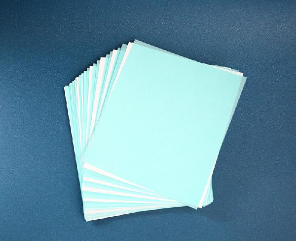
We provide nitrocellulose membrane for western blot transfer with both pore 0.2 μm (Catalog# AR0135-02) and pore 0.45 μm (Catalog# AR0135-04) options. Boster’s nitrocellulose membranes can be used for a wide range of protein molecular weights and nucleic acids > 500 base pairs in size. Each ordered pack contains 20 sheets measuring 9 cm x 10 cm in size. They can remain stable for one year when stored at room temperature. The products are high quality, pure 100% nitrocellulose membranes with a high surface area and excellent uniformity. The variety optimizes for peptide applications with the 0.2 μm pore size and protein applications with the 0.45 μm pore size.
References
Accelerate your research and gain invaluable insights from your samples with the help of our exceptional service. Reach out to us now to discuss your project requirements and experience the difference our Western blotting service can bring to your work.
Unleash innovation in your research with our Recombinant Protein Expression Service. Elevate your experiments today!