This website uses cookies to ensure you get the best experience on our website.


- Table of Contents
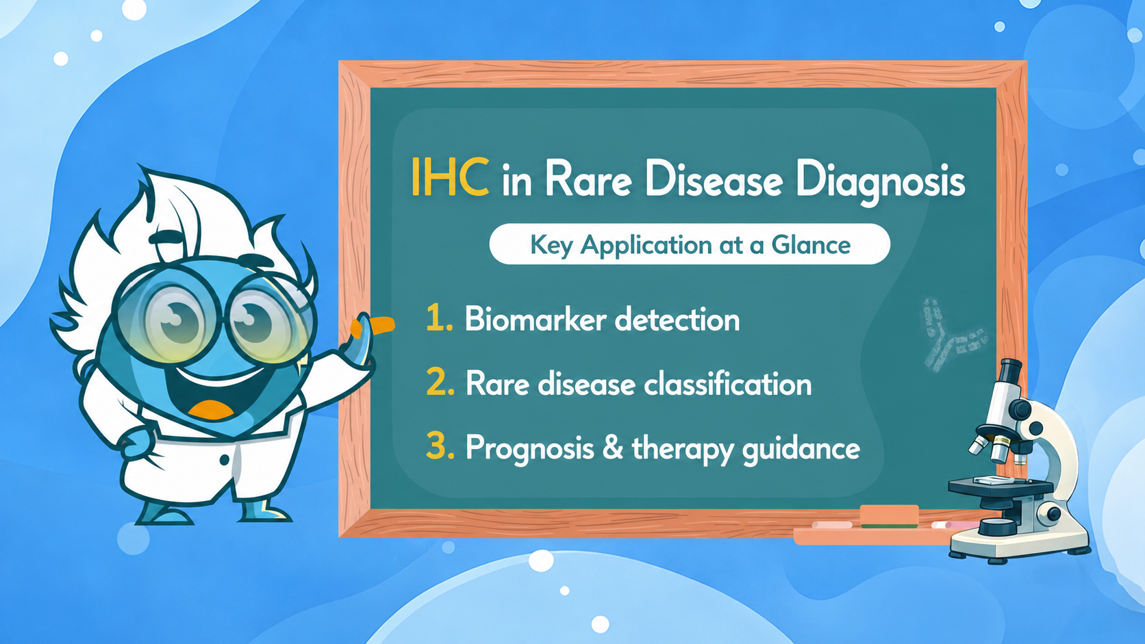
How antibody-based tissue analysis drives diagnosis, biomarker discovery, and targeted therapy decisions in rare and complex diseases — from basic principles to clinical practice.
Immunohistochemistry, or IHC, is a laboratory technique that uses antibodies to detect specific proteins, also called antigens, within preserved tissue sections. In pathology and rare disease diagnosis, IHC staining helps reveal molecular details that routine stains such as hematoxylin and eosin (H&E) may not show. This makes it especially useful when diseases look similar under the microscope but differ in the proteins they express.
The field developed from traditional, nonspecific tissue stains into a more targeted molecular method. A major turning point came in 1941, when researchers showed that antibodies could be linked to fluorescent dyes to identify antigens in tissue. Since then, IHC has become a core diagnostic tool for identifying tissue origin, confirming rare disease subtypes, evaluating tumor markers, and supporting treatment decisions.
In practice, IHC turns a biopsy sample into a visible map of protein expression through a defined workflow:
A positive IHC result indicates that the target protein was detected in a meaningful pattern, while a negative result can help rule out certain diagnoses. For rare diseases, this protein-level evidence is often critical for narrowing the diagnosis when morphology alone is not enough.
What is IHC used for in a clinical rare disease context? The answer spans the full diagnostic-to-therapeutic continuum. IHC is employed for initial diagnosis, confirmation of rare entities, prognostic stratification, and selection of targeted therapies — often in a single tissue block when lesional material is limited.
What is immunohistochemistry used for in pathology specifically? Three broad categories define its clinical utility:
IHC panels enable accurate classification of tumors by detecting tumor markers and tumor antigens, supporting precise tumor diagnosis even in complex or rare cases. In rare tumors, morphology alone is frequently insufficient. Small blue round cell tumors, spindle-cell sarcomas, and undifferentiated carcinomas can appear histologically indistinguishable. An immunohistochemistry application panel — selecting markers that differentially mark rhabdomyosarcoma versus Ewing sarcoma versus neuroblastoma, for example — narrows the differential to a single entity with high confidence.
In cancers of unknown primary (CUP) workups, IHC panels can help narrow the likely tissue of origin and, in selected settings, support site-specific classification. However, diagnostic accuracy varies substantially by tumor type, differentiation status, sample quality, and marker panel design.
Immunohistochemistry in pathology extends beyond neoplasia. In tissue-invasive infections caused by organisms that do not grow readily in culture — Whipple’s disease (Tropheryma whipplei), disseminated histoplasmosis, or CNS toxoplasmosis — IHC can identify organism-specific antigens directly in paraffin sections. The same principle applies to viral pathogens: IHC was instrumental in demonstrating SARS-CoV-2 tropism in the CNS and myocardium, providing pathological explanation for neurological sequelae.
For rare autoimmune and inflammatory conditions, IHC localizes autoantibody deposits, complement components, or immune cell infiltrates in a spatially resolved manner that serological testing cannot provide.
IHC has become indispensable for mapping protein aggregation in rare neurological diseases. By detecting tau, amyloid-beta, alpha-synuclein, TDP-43, FUS, and prion protein (PrPSc) in specific brain regions, neuropathologists can precisely subtype rare dementias, movement disorders, and prion diseases. Braak staging of tau pathology — the basis for understanding Alzheimer's disease progression — is entirely dependent on serial IHC-based mapping of post-mortem tissue.
Why is immunohistochemistry considered crucial in pathology for rare disease diagnosis? Because rare diseases are defined partly by their infrequency — which means large morphology-based reference datasets rarely exist. IHC compensates by providing objective protein-expression evidence that is reproducible, archivable, and cross-referenceable across institutions. Rare sarcomas, orphan lymphomas, and unusual infectious presentations all benefit from a standardized IHC language that allows global comparison even across small case series.
In clinical practice, a comprehensive IHC profile serves as the principal evidentiary standard for many rare tumor diagnoses. It is the reason why rare disease experts worldwide can confer on equivocal cases using slide scans and IHC data, arriving at a reproducible diagnostic consensus without requiring fresh tissue.
Researchers and clinicians working with rare or complex tumors frequently face the question of which diagnostic modality to deploy when tissue is limited and time is critical. Genetic testing methods — FISH, next-generation sequencing (NGS), polymerase chain reaction (PCR) — offer different and often complementary information to IHC.
A key practical principle in rare tumor diagnostics is the use of IHC markers as surrogates for molecular alterations. This strategy allows laboratories without access to molecular platforms to make clinically equivalent diagnostic and therapeutic decisions. Expert-endorsed examples include:
Practical guidance: When deciding between IHC and molecular testing for a rare tumor, apply an "IHC first" algorithm where validated surrogates exist. Reserve NGS for cases where IHC is equivocal, multiple alterations need profiling simultaneously, or a rare diagnosis requires molecular confirmation before initiating targeted therapy.
Immunohistochemical markers are not simply laboratory reagents — they are clinically validated diagnostic tools with defined performance characteristics in specific disease contexts. The following marker overview covers categories most relevant to rare-disease pathology, organized by disease domain.
| Disease Category | Key IHC Markers | Clinical Significance |
|---|---|---|
| Rare Sarcomas | SMARCB1/INI1, SMARCA4/BRG1, MDM2, CDK4, STAT6, TLE1 | SWI/SNF complex members (SMARCB1, SMARCA4) define rhabdoid and dedifferentiated tumor subtypes; STAT6 nuclear expression is pathognomonic for solitary fibrous tumor. |
| Rare Lymphomas | CD20, CD3, CD30, CD15, ALK, MYC | Lineage markers (CD20, CD3) establish B- vs. T-cell origin; CD30/CD15 co-expression defines classical Hodgkin lymphoma; ALK marks anaplastic large cell lymphoma. |
| Neurodegeneration | Tau (AT8), Amyloid-β, TDP-43, α-synuclein, PrPSc | IHC-based mapping of protein aggregates underpins Braak staging (tau), ABC scoring (amyloid), and subtyping of rare frontotemporal dementias by TDP-43 versus FUS versus tau. |
| Rare Carcinomas | CK5/6, p40, Napsin A, PAX8, INSM1 | Cytokeratin and transcription factor panels establish lineage in undifferentiated and poorly differentiated rare carcinomas; INSM1 is sensitive for neuroendocrine differentiation. |
| MMR & Lynch Syndrome | MLH1, MSH2, MSH6, PMS2 | Loss of one or more MMR proteins by IHC triggers MSI testing and germline counseling; dMMR status also qualifies patients for pembrolizumab regardless of tumor type (FDA tumor-agnostic approval). |
| SDH-Deficient Tumors | SDHB, SDHA | SDHB loss detected by IHC identifies SDH-deficient GISTs, paragangliomas, and pheochromocytomas that require germline SDH gene testing; SDHA loss is specific for SDHA mutations. |
| Infectious & Inflammatory | CMV, EBV (LMP1), HPV (p16), SARS-CoV-2 N | Pathogen-specific IHC confirms in-situ infection in tissue, providing spatial localization of pathogen-host interaction that serological tests cannot offer. |
| Proliferation & Prognosis | Ki-67, p53, BCL-2, PD-L1 | Ki-67 quantifies proliferative index for tumor grading; p53 mutation pattern (aberrant IHC) identifies TP53-mutant tumors without sequencing; PD-L1 companion diagnostics guide checkpoint immunotherapy selection. |
A note on antibody validation for rare diseases: The clinical weight of an IHC result depends entirely on antibody validation. For rare entities where large validation cohorts do not exist, cross-platform validation (IHC + molecular confirmation in a reference cohort) and inter-laboratory concordance studies are essential before adopting a marker as a diagnostic standard.
Immunohistochemistry results do not end at diagnosis. They are the primary data inputs for prognostic scoring and therapy selection in a wide range of rare diseases. The transition from descriptive pathology to actionable precision medicine is mediated, in most rare tumor settings, by IHC profile interpretation.
| IHC Marker / Pattern | Disease Context | Prognostic Implication | Therapy Direction |
|---|---|---|---|
| HER2 3+ / FISH-amplified | Breast, gastroesophageal rare variants | Poor prognosis if untreated | Trastuzumab, pertuzumab, T-DM1, T-DXd |
| PD-L1 CPS positive | Selected tumor types / rare carcinomas | Response predictor for ICIs | ICIs such as pembrolizumab in PD-L1–guided indications |
| dMMR (MLH1/MSH2 loss) | CRC, endometrial, rare Lynch-associated | Better prognosis in early disease; ICI-sensitive | Pembrolizumab; avoid 5-FU monotherapy |
| Ki-67 >20% | Neuroendocrine tumors (grade 3) | Aggressive behavior, high recurrence | Platinum-based chemotherapy over somatostatin |
| ALK strong/diffuse positive | NSCLC, IMT, ALCL | Targetable; favorable response to ALK inhibitors | Crizotinib, alectinib, brigatinib, lorlatinib |
| SMARCB1/INI1 loss | Rhabdoid tumors, epithelioid sarcoma | Aggressive; EZH2-dependent mechanism | Tazemetostat (EZH2 inhibitor) |
| ER/PR positive (≥1%) | Breast, rare gynecologic | Hormone-sensitive; favorable with endocrine Rx | Tamoxifen, aromatase inhibitors, CDK4/6 inhibitors |
| p53 aberrant (null/diffuse) | High-grade serous, TP53-mutant rare sarcomas | Genomic instability indicator; poor prognosis | Guides exclusion from low-grade pathways |
Beyond single-timepoint diagnosis, IHC supports longitudinal analysis of disease progression through serial biopsy studies and post-mortem cohort analysis. Tracking changes in Ki-67 index, receptor expression, or immune checkpoint marker density across treatment cycles provides dynamic biomarker data that informs therapy escalation or de-escalation decisions.
In rare neurodegenerative disorders, longitudinal IHC mapping from staged autopsy cohorts continues to define the spatiotemporal spread of protein pathology — enabling staging systems that directly inform clinical trial design (e.g., enriching for participants at specific Braak tau stages) and biomarker development (e.g., correlating ante-mortem CSF tau levels with IHC-confirmed stage).
Beyond clinical diagnostics, immunohistochemistry application in research-grade settings enables the discovery of novel drug targets, elucidation of disease mechanisms, and translation of pre-clinical findings into actionable clinical hypotheses. Many FDA-approved targeted therapies and biologics were developed against targets first characterized and spatially validated through IHC.
Candidate drug targets identified in genomic screens must be confirmed at the protein level, in the relevant cell type, and at the correct subcellular location. IHC provides this spatial protein validation in human disease tissue before committing to expensive drug development programs. For rare diseases — where model organism fidelity is often limited — tissue microarrays (TMAs) constructed from rare-disease cohorts enable IHC-based screening of tens to hundreds of candidate markers across hundreds of cases simultaneously.
Multiplex IHC and multispectral imaging platforms now enable simultaneous quantification of multiple immune cell populations (CD8+ T cells, FOXP3+ Tregs, CD68+ macrophages, PD-L1-expressing tumor cells) in the same tissue section. This spatial immune profiling of the tumor immune microenvironment is transforming our understanding of how rare tumors evade immune surveillance and how they respond to immunotherapy. For biotech researchers working on immune microenvironment profiling, validated multiplex IHC panels represent the gold standard for spatial biomarker co-localization.
Research-grade IHC is instrumental in biomarker tracking studies that follow protein expression trajectories from early precursor lesions through invasive disease to metastasis or therapeutic resistance. In rare disease contexts — where longitudinal biopsies are often the only source of mechanistic insight — IHC-based biomarker tracking provides the temporal resolution that single-timepoint molecular snapshots cannot offer.
Key considerations for research-grade IHC longitudinal studies include standardized antibody lot management, digital image analysis with validated scoring algorithms, and co-registration of IHC data with matched genomic and clinical datasets. These are the methodological underpinnings of reproducible biomarker research that can withstand regulatory-grade scrutiny.
The integration of digital pathology — whole-slide image scanning and AI-driven analysis — is dramatically expanding IHC's research utility. Deep learning algorithms trained on large IHC datasets can now quantify marker expression with sub-cellular spatial resolution across entire tissue sections, eliminating the sampling bias inherent in manual scoring. For rare disease research, where cohort sizes are small, AI-augmented IHC maximizes statistical power by extracting more granular data from each available tissue specimen.
Emerging "pathomics" approaches integrate IHC-derived spatial protein data with bulk and single-cell transcriptomics, proteomics, and clinical outcomes, building multidimensional predictive models of disease behavior that were unthinkable a decade ago.
What role does immunohistochemistry play in identifying rare diseases?
Immunohistochemistry helps identify rare diseases by detecting specific protein markers within tissue, enabling precise classification when morphology alone is insufficient.
How do positive and negative immunohistochemical test results impact rare disease diagnosis?
Positive results confirm the presence of disease-specific markers, supporting diagnosis, while negative results help exclude conditions and narrow differential diagnoses.
How are antibodies used in immunohistochemistry to detect specific disease markers?
Antibodies bind selectively to target proteins in tissue sections, allowing visualization of specific markers through chromogenic or fluorescent detection systems.
Why is immunohistochemistry considered crucial in pathology for rare disease diagnosis?
It provides protein-level, spatial information that links molecular findings to tissue structure, improving diagnostic accuracy in complex or rare conditions.
What are the steps involved in performing an immunohistochemistry test?
The process includes tissue fixation, sectioning, antigen retrieval, antibody incubation, signal detection, and visualization under a microscope.
Can IHC be used for longitudinal disease monitoring and biomarker tracking?
Yes, IHC can track changes in biomarker expression over time using serial tissue samples, supporting disease progression analysis and treatment evaluation.




