This website uses cookies to ensure you get the best experience on our website.
- Table of Contents
Nitrocellulose membranes are one of the top matrices used in protein blotting in Western Blotting, offering significant advantages over simple filter paper in terms of protein retention and signal clarity. They have high protein-binding capacity, strong affinity for proteins of varying protein size, compatibility with a variety of protein detection methods, and the ability to immobilize proteins, glycoproteins, or nucleic acids. This variety of detection methods include chemiluminescence, chromogenic, and fluorescence. It is proven to produce excellent signal-to-noise results when used for amino acid analysis, protein sequencing and western, northern, and Southern blotting.
Western blotting (also called Protein Immunoblotting) is an analytical technique used to detect specific proteins in the given sample. A thorough understanding of the western blot principle is crucial to successfully applying this method and accurately interpreting the results, while proper western...

Are you preparing for a FACS experiment? Here’s a 10 point checklist to help you prepare for your FACS sorting experiment:
In order to get the best results from your ELISA assay, the dilution factors of the sample and the detection antibodies must be optimized—this principle also applies broadly across many types of assay services where precision and optimization are key. If your sample or antibodies are too concentrated, you risk saturating the a...
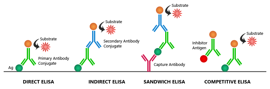
Are you familiar with the multiple methods you could use to perform an ELISA, including the wide range of commercially available elisa kits designed for different targets and sensitivity requirements? Among the standard
If you are having trouble with saturated signals in your ELISA data results, check out this table for Boster’s possible solutions to your problem:
| Possible Causes | Possible Solutions |
|---|---|
| High sample concentration | Use higher sample dilutions (Determine the optimal dilutions by titration assay). In parallel, construct an ELISA standard curve using known concentrations to more accurately map signal intensities to antigen levels. |
| Excessive substrate | Decrease concentration or amount of substrate: Follow manufacturer guidelines (The substrate provided with the ELISA kit might require further dilution) |
| Substrate color changed before use | Make substrate immediately before use |
| Non-specific antibody binding | Try different formulations in coating solutions; Ensure wells are pre-processed to prevent non-specific binding; Use affinity-purified antibody and preferably one that is pre- adsorbed; Use serum (5-10%) from same species as secondary antibody (bovine serum is also recommended). |
| Incubation time too long | Follow the manufacturer guidelines (If the problem persists, try incubating samples at 4°C overnight) |
| Excess antibody | Repeat the assay with lower antibody concentrations to find the optimal one for your experiment |
| Contaminated buffers with metals or HRP | Make and use fresh buffers |
ELISA (enzyme-linked immunosorbent assay) is a plate-based assay used to detect the concentration of a specific protein in a liquid sample and is frequently customized as part of custom assay development services tailored to specific research goals. In applications that require simultaneous measurement of multiple targets, researchers may also utilize multiplex ELISA kits to analyze several proteins within a single sample. It is also commonly performed using standardized elisa kits to ensure consistency across experiments. Three different types of data output can be obtained:

ELISA (enzyme-linked immunosorbent assay) is a convenient and simple method to quantitatively or qualitatively detect peptides, proteins, antibodies, and hormones in samples, rendering it as one of the most widely used immunoassays. Many laboratories perform these assays using standardized elisa kits, which help streamline assay setup and improve reproducibility. To derive accurate concentrations from measured signals, most ELISA protocols rely on constructing an standard curve from known standards, which serves as the reference for sample interpolation, alongside the use of quick elisa kits to support faster and more efficient assay workflows. Despite the many advantages of conducting ELISA—or more complex formats like those used in <...
ELISA (Enzyme-Linked Immunosorbent Assay) remains one of the most widely used methods for protein quantification in immunology, oncology, neuroscience, biomarker discovery, and translational research. Researchers use ELISA to measure cytokines, chemokines, growth factors, hormones, signaling proteins, and disease biomarkers across a wide range of sample types.
However, choosing an ELISA kit is often more complicated than selecting the target protein alone.
Two ELISA kits for the same analyte may differ significantly in:
These differences can directly affect data quality, quantitative reliability, and experimental reproducibility.
This guide summarizes the key factors researchers should evaluate before selecting an ELISA kit...

Accurate protein band size prediction starts with the primary sequence, but real gels reflect biology. PTMs, processing, isoforms, and experimental conditions shift apparent molecular weight. Use the estimate, then verify with the diagnostic steps below.
Western blotting separates proteins by size via gel electrophoresis (SDS-PAGE...