This website uses cookies to ensure you get the best experience on our website.


- Table of Contents
Plan ABCC1 paraffin IHC using the catalog antibody at 0.5–1 μg/ml (datasheet). Assess cytoplasmic and membranous staining while accounting for the uncertain tissue profile (HPA tissue IHC).

Expected localisation, validated protocols, controls and antibodies — the at-a-glance facts below, then the full design guide.
| Expected localisation | IHC: cytoplasmic/membranous (HPA tissue IHC); protein: basolateral membrane (UniProt) | |
| Staining pattern | Seminiferous tubules: cytoplasmic/membranous staining (HPA tissue IHC) | |
| Antigen retrieval | Citrate pH 6.0 HIER, 95–98 °C, 20 min (rule: cytoplasmic / membrane antigen) | |
| Positive control | Adipose tissue+4 more · see all | |
| Negative control | Adrenal gland+4 more · see all |
| Fixation | Keep fixation conditions consistent across sections. (standard IHC practice; not target-specific) | |
| Caveat | Staining and RNA show low consistency (HPA tissue IHC) | |
| Regulation | Expression regulation unspecified (UniProt) | |
| Isoform / epitope | 9 isoforms; check epitope coverage and membrane sidedness (UniProt) |
The catalog antibody’s IHC-P protocol is paired with two published ABCC1 IHC protocols using FFPE sections (PMC11354449; PMC3560367).
| Sample | Tissue sections; selected-image fixative not specified (standard IHC workflow) |
| Fixation | Image fixative and duration unreported (datasheet PA1634); verify before use. |
| Sectioning | 4–5 µm sections on charged slides (standard) |
| Deparaffinisation | Xylene, graded ethanol series to water (standard) |
| Antigen retrieval | Heat-induced epitope retrieval in citrate buffer, pH 6.0, 20 min at 95–98 °C (standard rule: cytoplasmic / membrane antigen) |
| Peroxidase block | 3% H2O2, 10 min, room temperature (standard) |
| Blocking | 10% normal serum of the secondary host, 30 min, room temperature (standard) |
| Primary antibody | Rabbit anti-ABCC1, 0.5-1μg/ml (datasheet PA1634) |
| Primary incubation | Overnight at 4 °C (standard) |
| Detection | HRP-polymer secondary, DAB chromogen 5–10 min (standard) |
| Counterstain | Hematoxylin, blue, dehydrate and mount (standard) |
| Expected result | ABCC1-positive staining in adipocytes of adipose tissue (HPA tissue IHC: Medium). HPA tissue profile: Cytoplasmic and membranous expression in several tissues, including seminiferous tubules in testis. No signal in the no-primary control. |
ABCC1 is a 17-pass membrane transporter at the cell and basolateral membranes (UniProt P33527 topology; UniProt P33527 subcellular location). In paraffin-section IHC, HPA reports cytoplasmic and membranous staining across several tissues, including testicular seminiferous tubules (HPA tissue IHC). Interpret the pattern cautiously: HPA rates tissue IHC reliability Uncertain because antibody staining has low consistency with RNA expression (HPA tissue IHC).
| Membranous staining in intestinal glandular cells, with some cytoplasmic staining (HPA tissue IHC). | This fits HPA's reported compartments. Duodenal, small-intestinal, rectal and appendiceal glandular cells each have Medium staining, but HPA's overall tissue IHC reliability remains Uncertain (HPA tissue IHC). A membrane-enriched pattern is also consistent with ABCC1's cell-membrane location (UniProt P33527 subcellular location). |
| Predominantly nuclear staining, without a convincing membrane-associated pattern. | Nuclear localization is outside the supplied ABCC1 locations: UniProt places it at the cell and basolateral membranes, while HPA tissue IHC describes cytoplasmic and membranous staining (UniProt P33527 subcellular location; HPA tissue IHC). Treat isolated nuclear signal as suspect and check controls before scoring it as ABCC1 (general IHC practice). |
| Strong staining in a cell population listed as Not detected by HPA, such as bronchial respiratory epithelial cells (HPA tissue IHC). | That conflicts with HPA's observation for that specific population; it does not prove the tissue lacks ABCC1 under every condition (HPA tissue IHC). Consider cross-reactivity or endogenous chromogen-generating activity, and compare a primary-antibody omission control with the same detection chemistry (general IHC practice). |
| Uniform color over cells, stroma and empty areas, obscuring cell borders. | A field-wide deposit cannot establish the cell-associated membranous or cytoplasmic distribution reported by HPA (HPA tissue IHC). Possible technical contributors include nonspecific antibody binding, inadequate washing or endogenous detection activity; assess the relevant controls and reagent conditions before interpreting tissue expression (general IHC practice). |
| No staining in HPA Medium-staining glandular cells, such as duodenal glandular cells (HPA tissue IHC). | A negative run in an expected comparison population raises a sensitivity or reagent-performance question, although HPA's Uncertain tissue IHC rating prevents treating that tissue as a definitive positive control (HPA tissue IHC). Review tissue preservation, antibody conditions, detection reagents and a concurrent positive control (general IHC practice). |
| Membrane topology and epitope position (UniProt P33527 topology) | ABCC1 has 17 transmembrane segments and both extracellular and cytoplasmic regions (UniProt P33527 topology). If an antibody's epitope is known, use its position to interpret staining; no epitope position or target-specific retrieval response is supplied here. |
| Cell population and tissue choice (HPA tissue IHC) | HPA reports Medium staining in intestinal glandular cells, kidney glomerular cells, ovarian stroma cells and adipocytes, but Low staining in lung macrophages (HPA tissue IHC). Score the named cell population; a tissue-wide average can hide the relevant pattern. |
| IHC evidence strength (HPA tissue IHC; HPA antibody validation) | HPA labels tissue IHC Uncertain and HPA002380 IHC Uncertain; HPA076011 has no supplied IHC status (HPA tissue IHC; HPA antibody validation). Treat the listed patterns as comparison observations rather than proof that every stain is specific. |
| Alternative isoforms (UniProt P33527 isoforms) | UniProt lists 9 ABCC1 isoforms (UniProt P33527 isoforms). The supplied record does not map antibody epitopes or IHC patterns to individual isoforms, so an apparent difference between specimens cannot be assigned to an isoform from staining alone. |
| Detection-system background (general IHC practice) | Chromogenic IHC can produce signal from endogenous detection activity or nonspecific binding (general IHC practice). Use reagent-appropriate controls to distinguish such color from the cell-associated pattern described for ABCC1 (HPA tissue IHC). |
| Situation | Likely cause | Next action |
|---|---|---|
| Membrane staining is weak or absent in an HPA Medium-staining cell population (HPA tissue IHC). | Antibody or detection performance, tissue preservation, or chosen retrieval conditions may limit signal (general IHC practice); no ABCC1-specific fixation or retrieval sensitivity is established by the supplied sources. | Check a concurrent positive control and reagent performance; evaluate retrieval under a validated general IHC workflow, then compare the same named cell population across runs (general IHC practice; HPA tissue IHC). |
| Only nuclear staining appears. | The compartment conflicts with the supplied membrane location and HPA's cytoplasmic/membranous IHC description (UniProt P33527 subcellular location; HPA tissue IHC). | Inspect a primary-antibody omission control and assess nonspecific signal before scoring; seek an independent specificity check if nuclear staining persists (general IHC practice). |
| The entire section has diffuse brown color. | Nonspecific binding, inadequate washing or endogenous detection activity can obscure localization (general IHC practice). | Compare a primary-antibody omission control, review blocking and washing, and check the chromogenic detection system (general IHC practice). Reassess whether cell borders can be resolved. |
| A supposedly negative tissue shows focal staining. | HPA's Not detected entries describe specified cell types, and its tissue IHC reliability is Uncertain (HPA tissue IHC). Focal signal may also reflect background or another cell population (general IHC practice). | Identify the stained cell type first, compare the matching HPA entry, and use detection controls before calling a contradiction or cross-reactivity (HPA tissue IHC; general IHC practice). |
| Different observers disagree on cytoplasmic versus membranous scoring. | HPA reports both compartments in tissue IHC, whereas UniProt places ABCC1 at the cell and basolateral membranes (HPA tissue IHC; UniProt P33527 subcellular location). | Record membrane and cytoplasmic staining separately, with the named cell population and intensity; flag ambiguous edges instead of forcing a single-compartment score (general IHC practice). |
| IF/ICC question: Should a junctional or plasma-membrane signal be expected? | HPA reports plasma membrane as the supported main ICC-IF location and cell junctions as a supported additional location (HPA subcellular ICC-IF). These observations do not validate a paraffin-section IHC result. | For IF/ICC interpretation, compare localization with HPA's supported locations and the assay's controls; use the separate IF/ICC guide for experimental conditions (HPA subcellular ICC-IF; general IF practice). |

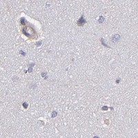
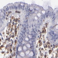


Comprehensive Human Protein Atlas IHC scoring per tissue (reliability: Uncertain — Low consistency between antibody staining and RNA expression data.). Rows are taken directly from the HPA tissue chart — click any row's HPA link to view the source.
| Tissue | Cell type | Level | Evidence | Source |
|---|---|---|---|---|
| Adipose tissue | Adipocytes | Medium | Protein (IHC) | HPA → |
| Appendix | Glandular cells | Medium | Protein (IHC) | HPA → |
| Duodenum | Glandular cells | Medium | Protein (IHC) | HPA → |
| Endometrium | Glandular cells | Medium | Protein (IHC) | HPA → |
| Kidney | Cells in glomeruli | Medium | Protein (IHC) | HPA → |
| Tissue | Cell type | Level | Evidence | Source |
|---|---|---|---|---|
| Adrenal gland | Glandular cells | Not detected | Protein (IHC) | HPA → |
| Breast | Adipocytes | Not detected | Protein (IHC) | HPA → |
| Bronchus | Respiratory epithelial cells | Not detected | Protein (IHC) | HPA → |
| Caudate | Glial cells | Not detected | Protein (IHC) | HPA → |
| Cerebellum | Cells in granular layer | Not detected | Protein (IHC) | HPA → |
Troubleshoot ABCC1 staining in paraffin sections by checking retrieval, compartment, cell type and controls before interpreting chromogenic signal.
PA1634 has a human lung cancer tissue IHC(P) image (catalog image caption) and lists human, mouse, and rat reactivity (catalog reactivity). No IF data are supplied (catalog IF images).
PA1634 will render with its human lung cancer tissue IHC(P) figure (catalog image caption). It lists IHC for human, mouse, and rat (catalog applications; catalog reactivity), with no IF image (catalog IF images).
Which to pick: Choose PA1634 for paraffin-section tissue IHC because its own figure is captioned IHC(P) in human lung cancer tissue (catalog image caption); its host is rabbit, while clonality and fixative are unreported (catalog host; catalog clone; catalog image caption). There is no IF/ICC option in this payload: PA1634 has no listed IF/ICC application, image, or dilution (catalog applications; catalog IF images; catalog dilution). For cross-species IHC, PA1634 lists human, mouse, and rat reactivity, although its displayed IHC(P) example is human only (catalog reactivity; catalog image caption).