ABCC4 / ATP-binding cassette sub-family C member 4 · IHC design guide

Design Immunohistochemistry for ABCC4

Plan chromogenic IHC on paraffin sections with the catalog antibody at 0.5–1 μg/ml (datasheet: PB9144). Assess membranous staining in prostate glandular cells and interpret unexpected staining in light of reported presumed off-target binding (HPA tissue IHC).

Evidence assembled Oct 2026 · For research use; verify linked source records and product datasheet before use
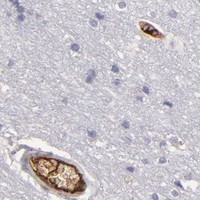
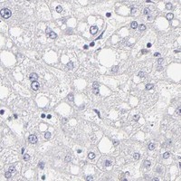
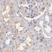
Immunohistochemistry protocol sheet for ABCC4 (IHC for ABCC4): expected localisation Membranous in prostate glands (HPA tissue IHC), antibody PB9144, validated IHC image, and IHC protocol steps
Printable ABCC4 IHC protocol sheet — expected localisation Membranous in prostate glands (HPA tissue IHC), antibody PB9144, controls and protocol steps. Open the full ABCC4 IHC guide →

ABCC4 Immunohistochemistry Experimental Design Guide

Expected localisation, validated protocols, controls and antibodies — the at-a-glance facts below, then the full design guide.

Must know before staining
Expected localisation Membranous in prostate glands (HPA tissue IHC)
Staining pattern Membranous staining in prostate glandular cells (HPA tissue IHC)
Antigen retrieval EDTA pH 8.0 HIER, heat-mediated (datasheet PB9144)
Positive control ⓘ Colon+2 more · see all
Negative control ⓘ Adipose tissue+4 more · see all
Important caveats
Reasons your staining may differ from the expected pattern.
Fixation Keep fixation consistent across paraffin sections (standard IHC practice; not target-specific)
Caveat Presumed off-target binding may complicate scoring (HPA tissue IHC)
Regulation Prostate-enriched expression (UniProt)
Isoform / epitope 4 isoforms; check whether the epitope is cytoplasmic or extracellular (UniProt)
Section 1

Recommended ABCC4 IHC & IF Protocols

Compare the catalog antibody’s IHC-P protocol (datasheet PB9144) with a published ABCC4 IHC protocol for healthy human lung tissue (PMC8527633).

Recommended immunohistochemistry (IHC-P) protocol parameters
SampleParaffin-embedded rat intestine tissue; fixative not specified (datasheet PB9144)
FixationImage fixative and duration unreported (datasheet PB9144); verify before use.
Sectioning4–5 µm sections on charged slides (standard)
DeparaffinisationXylene, graded ethanol series to water (standard)
Antigen retrievalHeat retrieval: EDTA pH 8.0 (datasheet PB9144); 20 min, 95–100 °C (standard)
Peroxidase block3% H2O2, 10 min, room temperature (standard)
Blocking10% goat serum (datasheet PB9144)
Primary antibodyRabbit anti-ABCC4, 0.5-1μg/ml (datasheet PB9144)
Primary incubationOvernight at 4 °C (datasheet PB9144)
DetectionStreptavidin-biotin complex (SABC), DAB chromogen (datasheet PB9144)
CounterstainHematoxylin, blue, dehydrate and mount (standard)
Expected resultABCC4-positive staining in enterocytes - Microvilli of colon (HPA tissue IHC: Medium). HPA tissue profile: Membranous expression in prostate glands. No signal in the no-primary control.
💡Decision noteStart with heat-mediated EDTA retrieval at pH 8.0 for the catalog antibody (datasheet PB9144); evaluate citrate at pH 6 when adapting the lung protocol (PMC8527633).
Section 2

What Is the Expected ABCC4 Staining Pattern?

ABCC4 is a 12-pass membrane transporter that can occupy apical or basolateral cell membranes, depending on tissue (UniProt O15439 topology and subcellular location). In paraffin-section IHC, expect membranous staining in prostate glandular cells and staining at colon enterocyte microvilli or fallopian-tube cilia (HPA tissue IHC). HPA rates its tissue staining “Enhanced,” while reporting medium agreement with RNA data and presumed off-target binding that was disregarded (HPA tissue IHC).

What am I looking at on my slide?
Prostate glandular cells show medium-strength staining that follows cell membranes (HPA tissue IHC).This matches HPA’s reported membranous prostate-gland pattern (HPA tissue IHC). Score the stained glandular cells and membrane distribution together; tissue identity alone does not establish that every chromogenic deposit represents ABCC4.
Colon enterocyte microvilli or fallopian-tube cilia show medium staining (HPA tissue IHC).These are HPA-observed positive structures (HPA tissue IHC). Interpret the signal against the visible epithelial architecture; a uniformly stained cytoplasm would not reproduce the reported structure-specific pattern.
The dominant signal fills nuclei or spreads uniformly through cytoplasm without a membrane pattern.That distribution conflicts with UniProt’s cell-membrane assignment and HPA’s mainly plasma-membrane ICC-IF result (UniProt O15439 subcellular location; HPA subcellular). Investigate artefact or nonspecific staining before scoring it as ABCC4.
Strong signal appears in HPA-listed adipocytes, adrenal glandular cells, or bone-marrow hematopoietic cells.Those cell populations were “Not detected” in HPA tissue IHC (HPA tissue IHC). Cross-reactivity or endogenous chromogen-generating activity is possible; confirm with appropriate negative controls and morphology rather than treating the tissue label as an absolute exclusion.
Positive tissue shows a diffuse haze that obscures cell boundaries and unstained structures.The result cannot establish the reported membranous pattern (HPA tissue IHC). General IHC causes include incomplete blocking, excessive antibody or detection signal, and insufficient washing; inspect controls before interpreting weak localized staining.
💡Expected ABCC4 appearanceCall a convincing positive when medium-strength chromogenic signal outlines prostate glandular-cell membranes or marks colon enterocyte microvilli or fallopian-tube cilia (HPA tissue IHC); dominant nuclear staining, indiscriminate cytoplasmic fill, or diffuse haze does not match the supported pattern (UniProt O15439 subcellular location; HPA tissue IHC).
How each factor affects the staining
Tissue and cell selectionProstate glandular cells, colon enterocyte microvilli, and fallopian-tube cilia are reported at medium staining; several listed cell populations are “Not detected” (HPA tissue IHC). Choose controls by the named cell population, not by an entire organ’s reputation.
Membrane polarity and topologyABCC4 has 12 transmembrane segments, with apical versus basolateral localization dependent on tissue (UniProt O15439 topology and subcellular location). Assess membrane-associated staining without requiring the same membrane face in every epithelium.
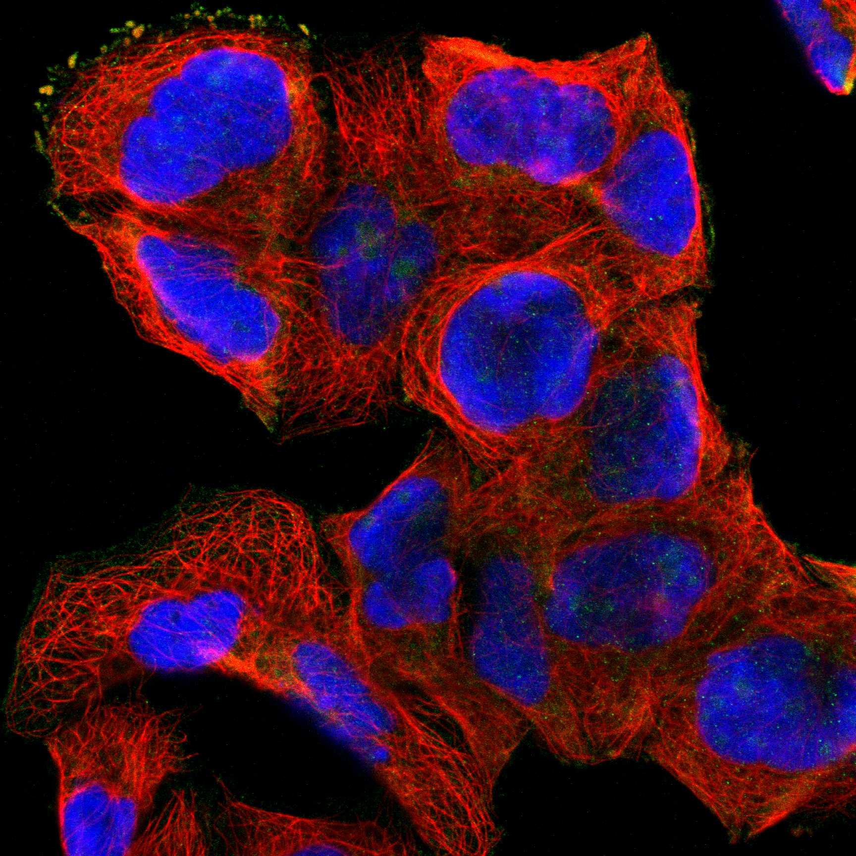
Cross-method localizationHPA ICC-IF supports plasma-membrane localization; Golgi and vesicle assignments are uncertain (HPA subcellular). Do not use those additional ICC-IF signals to redefine a convincing paraffin-section IHC positive.
Evidence and antibody interpretationHPA reports “Enhanced” tissue IHC and lists HPA002476 as IHC “Enhanced” (HPA tissue IHC; HPA antibodies). It also reports medium staining–RNA consistency and disregarded presumed off-target binding, so pattern and controls still matter (HPA tissue IHC).
Isoform coverageUniProt lists four ABCC4 isoforms (UniProt O15439 isoforms). The supplied HPA antibody record gives no epitope or isoform coverage (HPA antibodies); a negative result cannot identify which isoform is absent.
Why is my staining missing, weak or wrong?
SituationLikely causeNext action
No staining appears in prostate glandular cells.The expected medium membranous signal is missing (HPA tissue IHC); the image alone cannot distinguish absent antigen from an IHC workflow failure.Confirm glandular morphology, positive-control performance, reagent activity, retrieval conditions, and detection steps using general IHC controls; do not infer ABCC4-specific fixation sensitivity from these sources.
A faint positive is present, but its membrane distribution is hard to judge.HPA reports medium rather than universally strong staining in its listed positive cell populations (HPA tissue IHC). Heavy counterstain or weak chromogen contrast can complicate visual assessment in routine IHC.Compare a known-positive section and its negative control at matched detection settings; assess cell boundaries and the named microvillar or ciliary structures before assigning a score (HPA tissue IHC).
Nuclei dominate the stain while expected membranes are weak.Dominant nuclear localization conflicts with the membrane assignment (UniProt O15439 subcellular location; HPA subcellular); nonspecific antibody or detection signal is possible.Review the no-primary control, blocking, detection reagents, and tissue morphology using general IHC practice; score ABCC4 only where a credible membrane-associated pattern remains.
Adipocytes or other HPA-listed negative cell populations stain strongly.HPA reports “Not detected” for those specified populations and notes presumed off-target binding in its tissue assessment (HPA tissue IHC). Endogenous detection activity is another general IHC possibility.Check a no-primary control and the detection-system controls, then reassess the exact stained cell type; do not generalize a negative call from one listed population to every cell in its tissue.
Chromogen blankets the section or pools around tissue edges.Diffuse deposition obscures the expected cell-associated pattern (HPA tissue IHC). In general IHC, excess reagent, inadequate washes, drying, or endogenous detection activity can create background.Review wash and blocking steps, reagent exposure, section handling, and negative controls as general IHC checks; accept a positive only when the cell and compartment pattern can be resolved.
IF/ICC shows puncta near the Golgi as well as cell-surface signal.HPA supports plasma membrane as the main ICC-IF location but labels Golgi and vesicle locations uncertain (HPA subcellular).Use the separate IF/ICC guide for that assay; do not substitute uncertain intracellular puncta for the membrane-based paraffin IHC interpretation (HPA subcellular; HPA tissue IHC).

Sample controls for ABCC4 IHC & IF

🧪Run prostate first: glandular cells should show ABCC4 staining (HPA: Medium in prostate glandular cells). Use adipose tissue as the negative comparator, scoring its adipocytes (HPA: Not detected in adipocytes); on the prostate slide, use adjacent nonglandular stroma to assess background without assuming it is ABCC4-negative (HPA: prostate result specifies glandular cells only).
Positive control tissue: Colon (Enterocytes - Microvilli, HPA Medium)
Negative control tissue: Adipose tissue (HPA Not detected)
ICC-IF cell lines (HPA subcellular resource): HPA ICC-IF images show ABCC4 in A-431, HEL, RPTEC/TERT1, hTERT-RPE1 (serum starved), with annotated localisation: Plasma membrane (supported) (HPA subcellular).
Technical controls: Include a no-primary, secondary-only control; a rabbit control immunoglobulin matched to the primary’s isotype and concentration; and, if available, ABCC4-knockout tissue or a validated peptide-block control (caption: rabbit primary; standard IHC control practice). Check endogenous peroxidase and biotin background in prostate sections because the reported chromogenic method uses biotin, streptavidin and DAB (PB9144 tissue-IHC caption; standard IHC practice).
⚠️Feasibility: No target-specific fixation window or fixation effect is reported, and the selected PB9144 paraffin-section caption does not state a fixative (PB9144 tissue-IHC caption). That caption reports heat-mediated retrieval in EDTA at pH 8.0 for rat intestine; it does not establish that retrieval is required or optimal for prostate (PB9144 tissue-IHC caption). The supplied evidence does not establish that frozen sections or IF are easier; when scoring prostate, distinguish glandular staining from luminal chromogen deposits and background (HPA: Medium in prostate glandular cells; standard IHC practice).

HPA tissue IHC evidence for ABCC4

Comprehensive Human Protein Atlas IHC scoring per tissue (reliability: Enhanced — Medium consistency between antibody staining and RNA expression data. Presumed off target binding observed and disregarded.). Rows are taken directly from the HPA tissue chart — click any row's HPA link to view the source.

Positive expression · recommended positive controls

TissueCell typeLevelEvidenceSource
Colon Enterocytes - Microvilli Medium Protein (IHC) HPA →
Fallopian tube Ciliated cells (cilia axoneme) Medium Protein (IHC) HPA →
Prostate Glandular cells Medium Protein (IHC) HPA →

Undetected expression · recommended negative controls

TissueCell typeLevelEvidenceSource
Adipose tissue Adipocytes Not detected Protein (IHC) HPA →
Adrenal gland Glandular cells Not detected Protein (IHC) HPA →
Appendix Endocrine cells Not detected Protein (IHC) HPA →
Bone marrow Hematopoietic cells Not detected Protein (IHC) HPA →
Breast Adipocytes Not detected Protein (IHC) HPA →
Section 3

Advanced ABCC4 IHC Tips

Troubleshoot ABCC4 staining in paraffin sections by checking retrieval, membrane pattern, controls, and scoring before interpreting differences between samples.

What should I adjust when ABCC4 staining is weak after retrieval?
Start with heat-mediated retrieval in EDTA at pH 8.0 for paraffin sections stained with PB9144 (datasheet PB9144). The selected rat intestine image used this retrieval before incubation with 1 μg/ml primary antibody overnight at 4°C (caption PB9144). If staining is weak, compare heating duration on adjacent sections while holding antibody concentration, detection, and development constant; inspect whether membrane detail survives the treatment (standard IHC practice). Include a section processed by the documented method in each comparison, because altered retrieval can change both signal and tissue preservation (standard IHC practice).
Could fixation explain inconsistent ABCC4 staining between paraffin samples?
Target-specific fixation sensitivity for ABCC4 is unknown: the selected paraffin-section caption does not state a fixative (caption PB9144). Record each sample’s fixative and fixation duration, then compare matched sections using the documented EDTA retrieval at pH 8.0 (datasheet PB9144; standard IHC practice). Keep section thickness, primary incubation, and DAB development comparable so a processing difference is easier to assess (standard IHC practice). If only some samples lose membrane detail, review their morphology and handling before attributing the difference to ABCC4 expression; the available evidence does not establish a target-specific fixation effect (standard IHC practice; caption PB9144).
Should ABCC4 appear on the apical or basolateral membrane?
Interpret the stained membrane in the context of tissue architecture: ABCC4 can occupy apical or basolateral cell membranes, depending on tissue (UniProt O15439 localization). Membranous staining in prostate glands is reported, while colon enterocyte microvilli and fallopian-tube cilia axonemes show medium tissue staining (HPA tissue IHC). Examine whether DAB outlines the expected cell surface on serial sections, using a counterstain to distinguish the relevant cells from their neighbors (standard IHC practice). Diffuse staining alone is less persuasive than a reproducible membrane pattern, although additional Golgi and vesicle localization has been reported with lower certainty in subcellular imaging (HPA subcellular).
How can epitope placement affect an ABCC4 IHC result?
Check the catalog antibody’s stated immunogen or mapped epitope before interpreting absent staining, because ABCC4 has 4 isoforms and 12 transmembrane segments (UniProt O15439 isoforms and topology). Its cytoplasmic regions include residues 391–719 and 1015–1325, whereas several extracellular loops are short (UniProt O15439 topology). If the epitope is known, assess whether its sequence is present in the isoform relevant to the sample and whether retrieval exposes it in paraffin sections (standard IHC practice). Do not assign an isoform from a DAB pattern alone; the supplied evidence does not map PB9144’s epitope or establish isoform-specific staining (caption PB9144; UniProt O15439 isoforms).
How should I cross-check an IHC membrane pattern with IF?
Use IF as a separate cross-check of the IHC pattern, pairing ABCC4 with a marker for the expected cell type so adjacent cells are not mistaken for the same positive cell (standard IF practice). Choose fluorophores after checking tissue autofluorescence in unstained controls, favoring channels with clearer separation from that background (standard IF practice). If the antibody epitope is cytoplasmic, assess permeabilisation; if extracellular, first assess staining without permeabilisation, because access depends on which side of the membrane contains the epitope (UniProt O15439 topology; standard IF practice). Compare the resulting cell-surface distribution with IHC cautiously: plasma membrane localization is supported, while Golgi and vesicle assignments are uncertain (HPA subcellular).
What should I check when DAB obscures ABCC4 membrane staining?
First compare a section lacking primary antibody with a stained section to identify signal from the detection chemistry or tissue rather than ABCC4 binding (standard IHC practice). The selected procedure used 10% goat serum, a biotinylated secondary for 30 minutes at 37°C, a streptavidin–biotin complex, and DAB (caption PB9144). Check blocking, endogenous peroxidase control, and DAB development across matched sections, especially where dark deposits obscure membrane boundaries (standard IHC practice). Treat broadly diffuse staining cautiously: the tissue IHC resource reports presumed off-target binding that was disregarded in its expression assessment (HPA tissue IHC reliability).
How should I quantify ABCC4 staining across paraffin sections? ⚠ ANSWER MARKED FOR VERIFICATION
Define the compartment and eligible cells before scoring, because ABCC4’s apical or basolateral membrane position depends on tissue (UniProt O15439 localization). An H-score can combine the percentage of positive cells with ordinal membrane-staining intensity; report the percentage positive separately if intensity is difficult to standardize (standard IHC practice). Normalize counts or stained area to the number of eligible cells or evaluable tissue area, and report exclusions for folds, edges, or damaged regions (standard IHC practice). Apply the same threshold, counterstain review, and DAB development criteria across sections, since method differences can change apparent staining independently of biological expression (standard IHC practice).
When is an apparent ABCC4-positive IHC field convincing?
Look for a reproducible cell-surface pattern in the expected cells, accounting for tissue-dependent apical or basolateral placement (UniProt O15439 localization; standard IHC practice). Prostate glandular cells provide a reported membranous pattern, while colon enterocyte microvilli and fallopian-tube cilia axonemes have reported medium staining (HPA tissue IHC). Question staining confined to cut edges, necrotic areas, or the wrong cells or compartment, and use a no-primary control to investigate endogenous enzyme or detection background (standard IHC practice). Interpret an isolated positive field cautiously because the tissue IHC assessment reports medium staining–RNA consistency and presumed off-target binding, despite its Enhanced reliability designation (HPA tissue IHC reliability).
Boster reagents

Best ABCC4 / ATP-binding cassette sub-family C member 4 IHC Antibodies

The catalog includes ABCC4 antibodies for paraffin-section IHC and IF/ICC (catalog applications). PB9144 has IHC images from rat and mouse intestine and human intestinal cancer tissue (PB9144 image captions); IF images are unreported (catalog images).

Real IHC data IHC analysis of MRP4 using anti-MRP4 antibody (PB9144). MRP4 was detected in a paraffin-embedded section of rat intestine tissue. Heat mediated antigen retrieval was performed in EDTA buffer (pH 8.0, epitope retrieval solution). The tissue section was blocked with 10% goat serum. The tissue section was then incubated with 1 μg/ml rabbit anti-MRP4 Antibody (PB9144) overnight at 4°C. Biotinylated goat anti-rabbit IgG was used as secondary antibody and incubated for 30 minutes at 37°C. The tissue section was developed using Strepavidin-Biotin-Complex (SABC) (Catalog # SA1022) with DAB as the chromogen.
Anti-MRP4/ABCC4 Antibody ®
Cat # PB9144

PB9144 is listed for IHC in human, mouse, and rat, with paraffin-section images from rat and mouse intestine and human intestinal cancer tissue (PB9144 applications, reactivity, and image captions). A01596 is listed for IHC and IF/ICC in human, mouse, and rat, with no IHC or IF images supplied (A01596 applications, reactivity, and image listings).

Which to pick: For tissue IHC, choose PB9144 when a matching paraffin-section example matters; its captions document 1 μg/ml primary antibody after EDTA pH 8.0 retrieval, but do not report the fixative (PB9144 image captions). For IF/ICC, choose A01596 because those applications are listed at 2 μg/ml, without a supplied IF image (A01596 applications, dilution, and image listings). Both list human, mouse, and rat reactivity, while PB9144 alone supplies IHC images across those species; clonality is unreported for both (catalog reactivity, clone fields, and image listings).

Each figure is that product's own IHC / IF validation image from its datasheet.

References

  1. UniProt Consortium. UniProt entry O15439 (MRP4_HUMAN, ATP-binding cassette sub-family C member 4).
  2. Human Protein Atlas. ABCC4 tissue IHC expression (reliability: Enhanced).
  3. Human Protein Atlas. ABCC4 subcellular location (ICC-IF): Mainly localized to the plasma membrane. In addition localized to the Golgi apparatus and vesicles..
  4. Human Protein Atlas. ABCC4 antibody validation summary (1 antibodies).
  5. Potentiation of long-acting β(2)-agonist and glucocorticoid responses in human airway epithelial cells by modulation of intracellular cAMP. Respiratory research 2021 — PMC8527633.
  6. Suppression of the ATP-binding cassette transporter ABCC4 impairs neuroblastoma tumour growth and sensitises to irinotecan in vivo. European journal of cancer (Oxford, England : 1990) 2017 — PMC5665171.
  7. Zuo Jin Wan Reverses the Resistance of Colorectal Cancer to Oxaliplatin by Regulating the MALAT1/miR-200s/JNK Signaling Pathway. Evidence-based complementary and alternative medicine : eCAM 2022 — PMC9568309.
  8. Contribution of ABCC4-mediated gastric transport to the absorption and efficacy of dasatinib. Clinical cancer research : an official journal of the American Association for Cancer Research 2013 — PMC3752901.
  9. PubMed PMID:9661885 — UniProt-cited evidence.
  10. PubMed PMID:12105214 — UniProt-cited evidence.
  11. PubMed PMID:14702039 — UniProt-cited evidence.