This website uses cookies to ensure you get the best experience on our website.


- Table of Contents
Plan ABCG1 staining in paraffin sections using the observed cytoplasmic tissue pattern, including macrophages (HPA tissue IHC). This guide covers fixation, detection and scoring while accounting for the low agreement between antibody staining and RNA expression (HPA tissue IHC).

Expected localisation, validated protocols, controls and antibodies — the at-a-glance facts below, then the full design guide.
| Expected localisation | Cytoplasmic tissue staining (HPA tissue IHC); ER/Golgi membrane localisation (UniProt) | |
| Staining pattern | General cytoplasmic staining, including macrophages (HPA tissue IHC) | |
| Antigen retrieval | Citrate pH 6.0 HIER, 95–98 °C, 20 min (rule: cytoplasmic / membrane antigen) | |
| Positive control | Bronchus+4 more · see all | |
| Negative control | Adipose tissue+4 more · see all |
| Fixation | Keep formalin fixation consistent across sections. (standard IHC practice; not target-specific) | |
| Caveat | Low agreement between staining and RNA expression (HPA tissue IHC) | |
| Regulation | Higher in Tangier disease macrophages (UniProt) | |
| Isoform / epitope | 8 isoforms; check cytoplasmic vs extracellular epitope (UniProt) |
The catalog antibody’s IHC-P protocol is accompanied by published ABCG1 IHC methods for mouse aortic roots (PMC8510679) and tumor tissue (PMC8425206).
| Sample | FFPE human hepatocarcinoma tissue (datasheet A01227-1) |
| Fixation | Image formalin-fixed; duration unreported (datasheet A01227-1); verify before use. |
| Sectioning | 4–5 µm sections on charged slides (standard) |
| Deparaffinisation | Xylene, graded ethanol series to water (standard) |
| Antigen retrieval | Heat-induced epitope retrieval in citrate buffer, pH 6.0, 20 min at 95–98 °C (standard rule: cytoplasmic / membrane antigen) |
| Peroxidase block | 3% H2O2, 10 min, room temperature (standard) |
| Blocking | 10% normal serum of the secondary host, 30 min, room temperature (standard) |
| Primary antibody | Rabbit anti-ABCG1, 1:10-1:50 (datasheet A01227-1) |
| Primary incubation | Overnight at 4 °C (standard) |
| Detection | HRP-polymer secondary, DAB chromogen 5–10 min (standard) |
| Counterstain | Hematoxylin, blue, dehydrate and mount (standard) |
| Expected result | ABCG1-positive staining in ciliated cells (cilia axoneme) of bronchus (HPA tissue IHC: High). HPA tissue profile: General cytoplasmic expression, including macrophages. No signal in the no-primary control. |
ABCG1 is predominantly associated with intracellular ER and Golgi membranes, with cell membrane localization also reported; its six transmembrane segments support a membrane-associated pattern (UniProt P45844 topology and subcellular location). In tissue IHC, expect cytoplasmic staining that includes macrophages, with strong staining reported in selected ciliated cells and late spermatids (HPA tissue IHC). Interpret tissue intensity cautiously: HPA rates the IHC profile Approved but reports low consistency with RNA expression (HPA tissue IHC).
| Cytoplasmic staining appears in macrophages; ciliated bronchial cells show high axonemal staining (HPA tissue IHC). | This fits the reported tissue profile, while intracellular membrane staining also fits the predominant ER and Golgi localization (HPA tissue IHC; UniProt P45844 subcellular location). Assess the stained cell and compartment together: HPA's Approved profile has low consistency with RNA expression, so intensity alone is weak evidence of specificity (HPA tissue IHC). |
| The dominant signal is nuclear, although the expected ABCG1 pattern is cytoplasmic or membrane associated (HPA tissue IHC; UniProt P45844 subcellular location). | Treat dominant nuclear staining as suspect and check the counterstain, detection controls and antibody conditions (general IHC practice). Do not reject a cell edge signal solely because it differs from a Golgi-like pattern: cell membrane localization is also reported (UniProt P45844 subcellular location). |
| Strong staining is confined to adipocytes or prostate glandular cells, both listed as not detected by HPA (HPA tissue IHC). | Check for cross-reactivity or endogenous detection activity with appropriate controls (general IHC practice). These HPA observations are useful comparators, but they are not absolute biological negatives; the tissue IHC profile has low consistency with RNA expression (HPA tissue IHC). |
| Chromogen covers stroma, lumens or many cell types without a readable cellular pattern (general IHC practice). | Interpret this as background until controls establish a cellular signal (general IHC practice). The reported ABCG1 tissue profile is cytoplasmic and includes macrophages; a diffuse wash across the section cannot establish that pattern (HPA tissue IHC). |
| No staining appears in ciliated bronchial cells or late spermatids, where HPA reports high staining (HPA tissue IHC). | Check section quality, retrieval, detection reagents and the positive control before calling ABCG1 absent (general IHC practice). HPA's low staining-to-RNA consistency also means a single negative section should be interpreted cautiously (HPA tissue IHC). |
| Compartment and epitope location | ABCG1 has 6 transmembrane segments and cytoplasmic and extracellular regions (UniProt P45844 topology). The supplied evidence does not identify the antibody epitope; do not infer its accessibility in paraffin sections from topology alone. |
| Antigen retrieval | Retrieval conditions can be optimized against a positive tissue and a control section (general IHC practice). No ABCG1-specific retrieval condition or fixation sensitivity is supplied; tissue staining levels do not establish either effect (HPA tissue IHC). |
| Isoforms | UniProt lists 8 ABCG1 isoforms (UniProt P45844 isoforms). The supplied evidence does not establish which isoforms the IHC-validated antibody recognizes, so a tissue pattern cannot be assigned to one isoform. |
| Detection chemistry | Endogenous activity can produce chromogen independently of the primary antibody; use detection controls and blocking suited to the chosen chemistry (general IHC practice). Such color is not evidence of ABCG1 localization. |
| IF/ICC Q: What pattern should I expect? | A: HPA reports mainly Golgi localization, with vesicles marked uncertain, in ICC-IF (HPA subcellular). That observation can inform interpretation; IF/ICC protocol design belongs in its own guide. |
| Situation | Likely cause | Next action |
|---|---|---|
| The positive tissue is blank, including ciliated bronchial cells reported as high (HPA tissue IHC). | Section, retrieval, primary antibody or detection failure is possible (general IHC practice). | Verify section integrity and reagent activity with controls, then optimize retrieval and antibody conditions one variable at a time (general IHC practice). No ABCG1-specific retrieval setting is supplied. |
| The whole section is brown and cell boundaries are hard to score (general IHC practice). | Excess detection signal, inadequate washing or endogenous activity may contribute (general IHC practice). | Review primary-omission and detection controls; adjust dilution, washing or chemistry-specific blocking as indicated by those controls (general IHC practice). Score only a resolved cellular pattern. |
| Staining is predominantly nuclear, away from the reported cytoplasmic pattern (HPA tissue IHC). | A detection artifact or nonspecific antibody signal is possible (general IHC practice). | Compare the nuclear counterstain and control sections, then reassess antibody conditions (general IHC practice). Predominant ER and Golgi localization does not support a nuclear call (UniProt P45844 subcellular location). |
| Adipocytes stain strongly despite an HPA not detected result (HPA tissue IHC). | Cross-reactivity or endogenous detection activity is possible (general IHC practice). | Check primary-omission and detection controls, and compare a reported positive tissue in the same run (general IHC practice). Treat the HPA negative as a comparator, given its low consistency with RNA expression (HPA tissue IHC). |
| A few macrophage-like cells stain, but their identity is uncertain (HPA tissue IHC). | Cell identification from chromogen and morphology alone may be uncertain (general IHC practice). | Review morphology and the counterstain before assigning the signal to macrophages (general IHC practice). HPA describes cytoplasmic expression including macrophages but supplies no macrophage-specific intensity threshold (HPA tissue IHC). |
| A cell edge signal appears alongside intracellular staining (general IHC practice). | ABCG1 can localize to the cell membrane as well as predominantly to ER and Golgi membranes (UniProt P45844 subcellular location). | Assess whether the edge signal is cell associated and reproducible in a control tissue (general IHC practice). Do not assign a specific membrane pool from chromogenic appearance alone. |

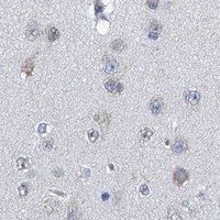
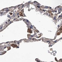
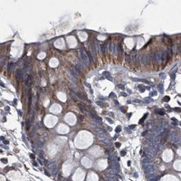
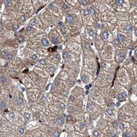


Comprehensive Human Protein Atlas IHC scoring per tissue (reliability: Approved — Low consistency between antibody staining and RNA expression data.). Rows are taken directly from the HPA tissue chart — click any row's HPA link to view the source.
| Tissue | Cell type | Level | Evidence | Source |
|---|---|---|---|---|
| Bronchus | Ciliated cells (cilia axoneme) | High | Protein (IHC) | HPA → |
| Fallopian tube | Ciliated cells (cilia axoneme) | High | Protein (IHC) | HPA → |
| Nasopharynx | Ciliated cells (cilia axoneme) | High | Protein (IHC) | HPA → |
| Testis | Elongated or late spermatids | High | Protein (IHC) | HPA → |
| Adrenal gland | Glandular cells | Medium | Protein (IHC) | HPA → |
| Tissue | Cell type | Level | Evidence | Source |
|---|---|---|---|---|
| Adipose tissue | Adipocytes | Not detected | Protein (IHC) | HPA → |
| Ovary | Ovarian stroma cells | Not detected | Protein (IHC) | HPA → |
| Prostate | Glandular cells | Not detected | Protein (IHC) | HPA → |
| Salivary gland | Glandular cells | Not detected | Protein (IHC) | HPA → |
| Soft tissue | Chondrocytes | Not detected | Protein (IHC) | HPA → |
Troubleshoot ABCG1 staining in paraffin sections by checking retrieval, compartmental pattern and cell identity before assigning biological meaning.
A01227-1 has a human IHC image; M01227 lists human and rat IHC and ICC/IF applications but supplies no IHC or IF figure (catalog: image captions, applications and reactivity).
A01227-1 renders with an image of formalin-fixed, paraffin-embedded human hepatocarcinoma stained by peroxidase and DAB (A01227-1 IHC caption). M01227 lists human and rat IHC and ICC/IF applications, but has no figure-backed card (catalog: M01227 applications, reactivity and image alts).
Which to pick: For human tissue IHC-P, choose A01227-1: its own image documents formalin-fixed, paraffin-embedded tissue, and its listed dilution is 1:10–1:50 (A01227-1 IHC caption; catalog: A01227-1 IHC dilution). For IF/ICC or rat samples, M01227 is the listed rabbit monoclonal option with human and rat reactivity; it has no IHC or IF figure, and its tissue fixative is unreported (catalog: M01227 host, clone, applications, reactivity and image alts).