This website uses cookies to ensure you get the best experience on our website.


- Table of Contents
This IHC guide identifies cytoplasmic ACKR2 staining in placenta and ciliated cells (HPA tissue IHC). It pairs that pattern with the catalog antibody’s paraffin-section workflow and 0.5–1 μg/mL working range (datasheet A05491-2).

Expected localisation, validated protocols, controls and antibodies — the at-a-glance facts below, then the full design guide.
| Expected localisation | Cytoplasmic staining in placenta and ciliated cells (HPA tissue IHC) | |
| Staining pattern | Cytoplasmic signal in syncytiotrophoblasts and ciliated cells (HPA tissue IHC) | |
| Antigen retrieval | Citrate pH 6 HIER, heat-mediated (datasheet A05491-2) | |
| Positive control | Bronchus+4 more · see all | |
| Negative control | Adrenal gland+4 more · see all |
| Fixation | Keep formalin fixation consistent across sections. (standard IHC practice; not target-specific) | |
| Caveat | Ligand stimulation can shift ACKR2 toward the cell surface (UniProt) | |
| Regulation | Ligand stimulation raises surface ACKR2 (UniProt) | |
| Isoform / epitope | No isoforms annotated; extracellular and cytoplasmic regions differ (UniProt) |
The catalog antibody’s IHC-P protocol (datasheet A05491-2) accompanies published methods for human kidney (PMC4699014) and lung cancer tissue arrays and animal sections (PMC10086750).
| Sample | Paraffin-embedded human placenta tissue; fixative not specified (datasheet A05491-2) |
| Fixation | Image fixative and duration unreported (datasheet A05491-2); verify before use. |
| Sectioning | 4–5 µm sections on charged slides (standard) |
| Deparaffinisation | Xylene, graded ethanol series to water (standard) |
| Antigen retrieval | Heat retrieval: Citrate pH 6, 20 min (datasheet A05491-2) |
| Peroxidase block | 3% H2O2, 10 min, room temperature (standard) |
| Blocking | 10% goat serum (datasheet A05491-2) |
| Primary antibody | Rabbit anti-ACKR2, 0.5-1μg/ml (datasheet A05491-2) |
| Primary incubation | Overnight at 4 °C (datasheet A05491-2) |
| Detection | Streptavidin-biotin complex (SABC), DAB chromogen (datasheet A05491-2) |
| Counterstain | Hematoxylin, blue, dehydrate and mount (standard) |
| Expected result | ACKR2-positive staining in ciliated cells (tip of cilia) of bronchus (HPA tissue IHC: High). HPA tissue profile: Cytoplasmic expression mainly in placenta and ciliated cells. No signal in the no-primary control. |
ACKR2 is a seven-pass membrane receptor found mainly in endocytic vesicles and recycling endosomes, with some cell-surface localization (UniProt O00590 topology and subcellular location). In tissue IHC, expect cell-associated cytoplasmic staining chiefly in placental syncytiotrophoblasts, plus staining in selected ciliated epithelia (HPA tissue IHC). HPA rates its tissue pattern Enhanced, while describing only medium consistency with RNA expression (HPA tissue IHC).
| Strong syncytiotrophoblast staining in placenta, or strong signal at bronchial ciliary tips and fallopian-tube cilia. | These are reported high-staining cell populations (HPA tissue IHC). Cytoplasmic or vesicular signal also fits the receptor's endosomal distribution (UniProt O00590 subcellular location). Score the named cells and structures; staining throughout neighboring cells is a different observation. |
| Predominantly nuclear staining in a paraffin section, without the expected cytoplasmic or ciliary pattern. | Treat this as discordant for tissue IHC, pending controls (HPA tissue IHC; UniProt O00590 subcellular location). HPA lists nucleoplasm only as an additional ICC-IF location, which does not establish a predominantly nuclear IHC pattern (HPA subcellular ICC-IF). |
| Strong signal in unrelated cells while expected positive cells on the same section remain unstained. | Suspect off-target binding or endogenous detection activity and check the controls (standard IHC practice). HPA's levels are cell-specific: appendix endocrine cells are not detected, although UniProt reports ACKR2 in appendix lymphatics (HPA tissue IHC; UniProt O00590 tissue specificity). |
| A uniform haze covers positive cells, negative cells, and extracellular areas. | This distribution is background rather than interpretable ACKR2 localization (standard IHC practice; UniProt O00590 subcellular location). Reassess nonspecific binding, detection background, and washing with appropriate control sections before scoring weak signal (standard IHC practice). |
| No signal appears in placental syncytiotrophoblasts on an otherwise evaluable section. | Placental syncytiotrophoblasts are a high-staining HPA reference, so this result warrants an assay check (HPA tissue IHC). A negative test section alone cannot distinguish low ACKR2 expression from failed retrieval, antibody detection, or tissue processing (standard IHC practice). |
| Compartment and trafficking (UniProt O00590 subcellular location and topology). | ACKR2 spans the membrane seven times but predominantly resides in endocytic vesicles; ligand stimulation can change membrane versus vesicle distribution (UniProt O00590). Do not require a continuous cell-surface rim to call an otherwise cell-specific cytoplasmic pattern plausible. |
| Cell-specific reference patterns (HPA tissue IHC; UniProt O00590 tissue specificity). | HPA reports high signal in placenta syncytiotrophoblasts and two ciliated-cell sites, medium signal in duodenal enterocytes, and low signal in colon and rectum endothelial cells (HPA tissue IHC). UniProt also describes lymphatic endothelium; ordinary vascular endothelium is therefore a poor blanket positive criterion (UniProt O00590 tissue specificity; HPA tissue IHC). |
| Evidence strength and antibody choice (HPA tissue IHC; HPA antibodies). | HPA calls the tissue pattern Enhanced but notes medium staining-to-RNA consistency (HPA tissue IHC). Its HPA013819 entry has Enhanced IHC validation; HPA073493 has Supported ICC validation without an IHC rating (HPA antibodies). Keep those application ratings distinct when interpreting a paraffin result. |
| Detection background (standard IHC practice). | Endogenous enzyme activity and nonspecific binding can create chromogenic signal unrelated to the target (standard IHC practice). Appropriate detection-only and tissue controls help distinguish this from cell-restricted ACKR2 staining; HPA's tissue levels do not diagnose the source of background (HPA tissue IHC; standard IHC practice). |
| IF/ICC Q: Should its distribution match paraffin IHC (HPA subcellular ICC-IF)? | A: HPA supports vesicles and cytosol in ICC-IF and additionally approves nucleoplasm (HPA subcellular ICC-IF). Those observations provide compartment context, but ICC-IF is a separate application; its additional nuclear location does not validate nuclear-dominant tissue IHC (HPA subcellular ICC-IF; HPA tissue IHC). |
| Situation | Likely cause | Next action |
|---|---|---|
| Placental syncytiotrophoblasts show no stain (HPA tissue IHC). | The expected high-signal reference failed; the cause remains undetermined from that result alone (HPA tissue IHC; standard IHC practice). | Check section quality, retrieval, antibody dilution, detection reagents, and a positive-control section; change one workflow variable at a time (standard IHC practice). |
| The whole section develops diffuse brown haze (standard IHC practice). | Nonspecific antibody binding or detection background may obscure cell-specific signal (standard IHC practice). | Review detection-only controls, blocking, washing, and development time; score only a resolved cellular pattern (standard IHC practice). |
| Nuclei stain more strongly than cytoplasm in paraffin IHC (HPA tissue IHC). | This differs from HPA's cytoplasmic tissue profile; HPA's additional nuclear observation comes from ICC-IF (HPA tissue IHC; HPA subcellular ICC-IF). | Compare positive tissue and detection controls, then reassess localization with an independently validated IHC antibody if needed (standard IHC practice; HPA antibodies). |
| Strong stain appears in an HPA-negative cell population (HPA tissue IHC). | Off-target binding or endogenous detection activity is possible; a tissue name alone may hide differences between cell populations (HPA tissue IHC; standard IHC practice). | Identify the stained cells, inspect detection controls, and compare the same cell type with HPA's entries before calling it ACKR2 (HPA tissue IHC; standard IHC practice). |
| Ciliary staining is absent, but other epithelial areas stain (HPA tissue IHC). | The result misses the high ciliary patterns reported for bronchus and fallopian tube (HPA tissue IHC). | Confirm that intact cilia and the specified cell type are present, then compare a known-positive section and background controls (HPA tissue IHC; standard IHC practice). |
| A weak endothelial signal is difficult to score (HPA tissue IHC). | HPA reports low signal in colon and rectum endothelium and no detection in cerebral-cortex endothelium; endothelial staining is context-dependent (HPA tissue IHC). | Record vessel type and tissue, compare a stronger positive reference, and avoid classifying every vessel by one endothelial result (HPA tissue IHC; standard IHC practice). |

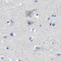
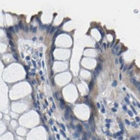
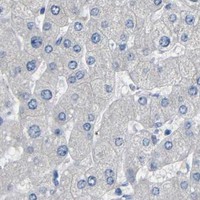
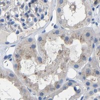


Comprehensive Human Protein Atlas IHC scoring per tissue (reliability: Enhanced — Medium consistency between antibody staining and RNA expression data.). Rows are taken directly from the HPA tissue chart — click any row's HPA link to view the source.
| Tissue | Cell type | Level | Evidence | Source |
|---|---|---|---|---|
| Bronchus | Ciliated cells (tip of cilia) | High | Protein (IHC) | HPA → |
| Fallopian tube | Ciliated cells (cilia axoneme) | High | Protein (IHC) | HPA → |
| Placenta | Syncytiotrophoblasts | High | Protein (IHC) | HPA → |
| Duodenum | Enterocytes | Medium | Protein (IHC) | HPA → |
| Nasopharynx | Ciliated cells (cell body) | Medium | Protein (IHC) | HPA → |
| Tissue | Cell type | Level | Evidence | Source |
|---|---|---|---|---|
| Adrenal gland | Glandular cells | Not detected | Protein (IHC) | HPA → |
| Appendix | Endocrine cells | Not detected | Protein (IHC) | HPA → |
| Caudate | Glial cells | Not detected | Protein (IHC) | HPA → |
| Cerebellum | Cells in granular layer | Not detected | Protein (IHC) | HPA → |
| Cerebral cortex | Endothelial cells | Not detected | Protein (IHC) | HPA → |
Troubleshoot ACKR2 staining in paraffin sections by checking retrieval, cell identity, subcellular pattern, and controls before assigning biological meaning.
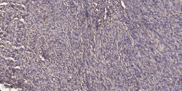
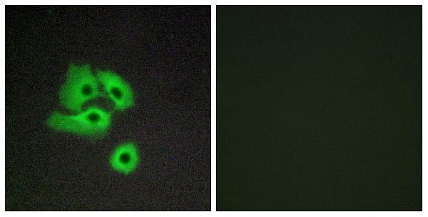
Two anti-ACKR2 antibodies have paraffin-section IHC images from human placenta and colon cancer; one also has a COS7-cell IF image (catalog image captions).
A05491-2 shows IHC on paraffin-embedded human placenta and lists Human reactivity (A05491-2 image caption; catalog applications/reactivity). A30785 shows IHC on paraffin-embedded human colon cancer and IF in COS7 cells; it lists Human, Mouse and Rat reactivity (A30785 image captions; catalog applications/reactivity).
Which to pick: For human tissue IHC, choose A05491-2 for the illustrated placenta workflow or A30785 for the illustrated colon cancer workflow (respective IHC image captions). For IF/ICC or planned Mouse or Rat work, choose polyclonal A30785 because those applications and species are listed for it (A30785 catalog dilution/application/reactivity data). The fixative is unreported in both IHC captions (A05491-2 and A30785 IHC image captions).