ADAR / Double-stranded RNA-specific adenosine deaminase · IHC design guide

Design Immunohistochemistry for ADAR

This paraffin-section ADAR IHC guide uses nuclear tissue staining as the scoring reference (HPA tissue IHC). It pairs the catalog antibody’s 2–5 μg/ml IHC range (datasheet A00869-2) with neuronal and alveolar type II cells as positive references (HPA tissue IHC).

Evidence assembled Oct 2026 · For research use; verify linked source records and product datasheet before use
Immunohistochemistry protocol sheet for ADAR (IHC for ADAR): expected localisation Nuclear staining in tissue sections (HPA tissue IHC), antibody A00869-2, validated IHC image, and IHC protocol steps
Printable ADAR IHC protocol sheet — expected localisation Nuclear staining in tissue sections (HPA tissue IHC), antibody A00869-2, controls and protocol steps. Open the full ADAR IHC guide →

ADAR Immunohistochemistry Experimental Design Guide

Expected localisation, validated protocols, controls and antibodies — the at-a-glance facts below, then the full design guide.

Must know before staining
Expected localisation Nuclear staining in tissue sections (HPA tissue IHC)
Staining pattern Widespread nuclear staining across tissues (HPA tissue IHC)
Antigen retrieval EDTA pH 8.0 HIER, heat-mediated (datasheet A00869-2)
Positive control ⓘ Caudate+4 more · see all
Negative control ⓘ Soft tissue
Important caveats
Reasons your staining may differ from the expected pattern.
Fixation Keep fixation consistent across paraffin sections (standard IHC practice; not target-specific)
Caveat Soft-tissue fibroblasts may lack signal (HPA tissue IHC)
Regulation Interferon-inducible ADAR1 (UniProt)
Isoform / epitope 5 isoforms; check antibody epitope coverage (UniProt)
Section 1

Recommended ADAR IHC & IF Protocols

The catalog antibody’s IHC-P protocol is accompanied by published ADAR staining methods for human colon and IBD tissue arrays and additional tissue sections (PMC12480066; PMC12010004).

Recommended immunohistochemistry (IHC-P) protocol parameters
SampleParaffin-embedded mouse brain tissue; fixative not specified (datasheet A00869-2)
FixationImage fixative and duration unreported (datasheet A00869-2); verify before use.
Sectioning4–5 µm sections on charged slides (standard)
DeparaffinisationXylene, graded ethanol series to water (standard)
Antigen retrievalHeat retrieval: EDTA pH 8.0 (datasheet A00869-2); 20 min, 95–100 °C (standard)
Peroxidase block3% H2O2, 10 min, room temperature (standard)
Blocking10% goat serum (datasheet A00869-2)
Primary antibodyRabbit anti-ADAR, 2-5 μg/ml (datasheet A00869-2)
Primary incubationOvernight at 4 °C (datasheet A00869-2)
DetectionStreptavidin-biotin complex (SABC), DAB chromogen (datasheet A00869-2)
CounterstainHematoxylin, blue, dehydrate and mount (standard)
Expected resultADAR-positive staining in neuronal cells of caudate (HPA tissue IHC: High). HPA tissue profile: Ubiquitous nuclear expression. No signal in the no-primary control.
💡Decision noteStart with EDTA pH 8.0 heat retrieval (datasheet: A00869-2). The published excerpts do not specify a retrieval buffer (PMC12480066; PMC12010004).
Section 2

What Is the Expected ADAR Staining Pattern?

ADAR should appear predominantly nuclear across many tissues, with high staining in selected neuronal, lung alveolar type II, respiratory epithelial, and Sertoli cells (HPA tissue IHC: ubiquitous nuclear expression; High in listed cells). Nucleoli and nucleoplasm are supported by ICC-IF, while UniProt also records cytoplasmic localization and nuclear shuttling (HPA subcellular ICC-IF; UniProt P55265 localization). The tissue IHC profile has Enhanced reliability, reflecting consistency with RNA expression (HPA tissue IHC). ADAR has no transmembrane segment (UniProt P55265 topology).

What am I looking at on my slide?
Distinct nuclear chromogen in caudate or cortical neurons, or lung alveolar type II cells, with limited surrounding background (HPA tissue IHC: High in these cells).This fits the reported IHC pattern: ubiquitous nuclear expression with high signal in these cell populations (HPA tissue IHC). Judge staining within identifiable cells rather than treating every stained area as equally informative (general IHC practice).
Predominantly membrane-like or extracellular staining, with little nuclear signal.That distribution conflicts with the nuclear tissue IHC profile and ADAR's lack of a transmembrane segment (HPA tissue IHC; UniProt P55265 topology). Suspect nonspecific staining or a detection artefact; cytoplasmic signal alone is less decisive because ADAR can shuttle (UniProt P55265 localization).
Strong staining in soft-tissue fibroblasts while nearby nuclei lack a convincing ADAR pattern (HPA tissue IHC: fibroblasts Not detected).Treat the fibroblast signal as questionable because it differs from the sampled HPA result; assess cross-reactivity or endogenous chromogenic activity with appropriate controls (HPA tissue IHC; general IHC practice). A single negative HPA observation does not prove universal absence.
Diffuse chromogen covers cells and intercellular spaces, obscuring nuclear boundaries.The expected tissue pattern is nuclear, so widespread haze cannot be scored as specific ADAR staining (HPA tissue IHC; general IHC practice). Background can arise from nonspecific antibody binding or detection chemistry; compare controls before assigning a cause (general IHC practice).
No nuclear staining in an otherwise interpretable section of lung alveolar type II cells or cortical neurons (HPA tissue IHC: High in these cells).This is discordant with the reported positive pattern, but one blank section cannot establish biological absence (HPA tissue IHC; general IHC practice). Check tissue identification, antibody and detection performance, and a known-positive control before interpreting it (general IHC practice).
💡Expected ADAR appearanceCall a convincing positive when identifiable high-expression cells show clear nuclear staining above background; dominant membrane-like staining or fibroblast-only signal is suspect (HPA tissue IHC: High in listed cells, fibroblasts Not detected; UniProt P55265 topology).
How each factor affects the staining
Tissue and cell selectionADAR is broadly nuclear in tissue IHC, with High signal reported in neuronal, alveolar type II, respiratory epithelial, and Sertoli cells; cholangiocytes, muscle cells, and adipocytes are Low (HPA tissue IHC). Select and score identifiable cell populations (general IHC practice).
Compartment and assayTissue IHC reports ubiquitous nuclear expression; ICC-IF places ADAR mainly in nucleoli and additionally in nucleoplasm (HPA tissue IHC; HPA subcellular ICC-IF). UniProt also records cytoplasmic localization and shuttling, so these assays need compartment-specific interpretation (UniProt P55265 localization).
Antibody evidenceHPA003890 and HPA051519 have Enhanced IHC and ICC validation; CAB056157 has Supported IHC validation and no supplied ICC status (HPA antibodies). These ratings support the reported applications but do not establish that every antibody or staining condition yields the same pattern.
Isoform coverageUniProt lists five ADAR isoforms, but the supplied antibody record gives no epitope positions or isoform coverage (UniProt P55265 isoforms; HPA antibodies). Differences in staining therefore cannot be assigned to a particular isoform from these data alone.
Target-specific retrieval or fixation effectsTarget-specific fixation effects are not established by the supplied assay evidence. Verify with a matched IHC source before attributing a result to fixation.
Why is my staining missing, weak or wrong?
SituationLikely causeNext action
Nuclei are blank in a known-positive cell population.The result conflicts with High staining reported for selected neurons and alveolar type II cells; antibody, retrieval, or detection performance remains uncertain (HPA tissue IHC; general IHC practice).Verify the cell population, run a positive control, and check antibody dilution, retrieval, and detection steps using the applicable IHC instructions (general IHC practice).
Chromogen forms a uniform haze that hides nuclear boundaries.Diffuse haze is inconsistent with the HPA nuclear pattern and may reflect nonspecific binding or detection background (HPA tissue IHC; general IHC practice).Compare a no-primary control, review blocking and washing, and reduce excessive antibody or chromogen development if controls indicate background (general IHC practice).
A membrane-like pattern dominates while nuclei remain faint.This conflicts with nuclear tissue IHC and the absence of a transmembrane segment; cross-reactivity or a detection artefact is plausible (HPA tissue IHC; UniProt P55265 topology; general IHC practice).Compare an independently validated antibody or suitable specificity control, and score only a reproducible cellular pattern (HPA antibodies; general IHC practice).
Soft-tissue fibroblasts appear strongly positive.HPA reports fibroblasts as Not detected in its sampled tissue; the discrepancy warrants checking, without treating that observation as a universal negative rule (HPA tissue IHC).Confirm cell identity and compare no-primary and antibody-specificity controls; investigate endogenous detection activity if control sections also develop chromogen (general IHC practice).
Nucleoli are not distinctly darker than the rest of the nucleus in paraffin IHC.The nucleolar emphasis comes from ICC-IF, whereas the supplied tissue IHC summary specifies nuclear expression without a required nucleolar contrast (HPA subcellular ICC-IF; HPA tissue IHC).Assess whether nuclei are clearly stained above controls. For an IF localization question, consult the separate IF/ICC guide; ICC-IF supports nucleoli plus nucleoplasm (HPA subcellular ICC-IF).
A low-staining population appears negative beside strongly stained cells.HPA reports Low staining in cholangiocytes, smooth muscle cells, myocytes, and adipocytes, while several other cell populations are High (HPA tissue IHC). A weak result may be difficult to distinguish from background (general IHC practice).Use the High population and a negative control to establish interpretable signal, then score the Low population separately without forcing a positive call (HPA tissue IHC; general IHC practice).

Sample controls for ADAR IHC & IF

🧪Run caudate first and score neuronal cells, which show high ADAR staining (HPA: caudate neuronal cells, High). Use soft tissue fibroblasts as the negative comparator (HPA: soft tissue fibroblasts, Not detected); no specific cell population within caudate is documented as ADAR-negative, so internal background should be judged only in cells independently verified as negative (HPA: caudate row).
Positive control tissue: Caudate (Neuronal cells, HPA High)
Negative control tissue: Soft tissue (HPA Not detected)
ICC-IF cell lines (HPA subcellular resource): HPA ICC-IF images show ADAR in A-431, U-251MG, U2OS, U2OS, siRNA 2 (10x), U2OS, siRNA 2 (40x), U2OS, scrambled (10x), U2OS, scrambled (40x), U2OS, siRNA 1 (10x), U2OS, siRNA 1 (40x), with annotated localisation: Nucleoli (enhanced) (HPA subcellular).
Technical controls: Include a no-primary (secondary-only) control, a concentration-matched rabbit IgG isotype control matched to the primary’s clonality if known, and ADAR-knockout material as a biological negative (selected-SKU IHC caption: rabbit primary; standard IHC practice). For the caudate SABC/DAB assay, quench endogenous peroxidase and block endogenous biotin (selected-SKU IHC caption: SABC/DAB; standard IHC practice).
⚠️Feasibility: No ADAR-specific fixation window or fixation effect is reported in the supplied evidence, and the selected-SKU paraffin-section mouse-brain caption does not state a fixative (selected-SKU IHC caption). The caption uses heat-mediated retrieval in EDTA at pH 8.0, but it does not establish that retrieval is required (selected-SKU IHC caption). The evidence does not show frozen sections or IF to be easier; brain autofluorescence can complicate IF assessment (standard IF practice).

HPA tissue IHC evidence for ADAR

Comprehensive Human Protein Atlas IHC scoring per tissue (reliability: Enhanced — High consistency between antibody staining and RNA expression data.). Rows are taken directly from the HPA tissue chart — click any row's HPA link to view the source.

Positive expression · recommended positive controls

TissueCell typeLevelEvidenceSource
Caudate Neuronal cells High Protein (IHC) HPA →
Cerebellum GLUC cells - nucleus High Protein (IHC) HPA →
Cerebral cortex Neuronal cells High Protein (IHC) HPA →
Hippocampus Neuronal cells High Protein (IHC) HPA →
Lung Alveolar cells type II High Protein (IHC) HPA →

Undetected expression · recommended negative controls

TissueCell typeLevelEvidenceSource
Soft tissue Fibroblasts Not detected Protein (IHC) HPA →
Section 3

Advanced ADAR IHC Tips

Troubleshoot ADAR staining in paraffin sections by checking retrieval, compartment, cell identity and controls before comparing staining intensity (UniProt P55265; HPA tissue IHC).

Which retrieval conditions should I try first for weak ADAR staining?
Start with heat-mediated retrieval in EDTA at pH 8.0 for paraffin sections (datasheet A00869-2). The selected mouse brain IHC example used this retrieval, followed by 10% goat serum blocking and 2 μg/ml primary antibody overnight at 4°C (caption A00869-2). If staining is weak, compare retrieval heating and cooling conditions on matched sections while holding antibody concentration and detection constant (standard IHC practice). Judge improvement by clearer nuclear staining in expected cells, rather than by stronger diffuse colour; HPA reports ubiquitous nuclear expression and high staining in brain neuronal cells (HPA tissue IHC).
Could fixation explain weak or uneven ADAR staining?
Target-specific fixation sensitivity is unknown because the selected paraffin-section caption does not state a fixative (caption A00869-2). Record each specimen’s fixative and fixation duration, then compare sections processed under the same conditions before changing antibody concentration (standard IHC practice). For the catalog antibody, begin with EDTA retrieval at pH 8.0 and 2 μg/ml primary antibody overnight at 4°C, as used in the selected example (caption A00869-2). If matched sections still differ, assess morphology, section thickness and staining-run controls; those observations can reveal a processing problem without assigning ADAR a fixation response that has not been demonstrated (standard IHC practice).
How should nuclear, nucleolar and cytoplasmic ADAR staining be assessed?
Score nuclear staining first: tissue IHC reports ubiquitous nuclear expression, while subcellular imaging places ADAR mainly in nucleoli with additional nucleoplasmic localisation (HPA tissue IHC; HPA subcellular). Cytoplasmic signal can be biologically plausible because ADAR shuttles between nucleus and cytoplasm (UniProt P55265 subcellular). Compare each compartment with the counterstain and cell morphology, and record nuclear and cytoplasmic scores separately when both are present (standard IHC practice; UniProt P55265 subcellular). Treat staining outside intact cells or concentrated at section edges cautiously, and check whether it persists in omission controls before assigning a compartment (standard IHC practice).
Can this antibody distinguish ADAR isoforms or reveal epitope masking?
Do not assign an IHC signal to one ADAR isoform without antibody epitope and isoform-reactivity evidence; the record lists 5 isoforms (UniProt P55265 isoforms). ADAR has Z-binding domains, 3 double-stranded RNA-binding motifs and an editase domain, so epitope position matters when assessing which variants an antibody could recognise (UniProt P55265 domains). The selected caption establishes staining conditions for A00869-2 but does not identify an epitope (caption A00869-2). If an isoform claim is essential, check the antibody’s documented epitope against isoform sequences and use an independently validated isoform-specific assay before interpreting differences between specimens (standard IHC practice; UniProt P55265 isoforms).
How can IF help check cell identity and ADAR localisation?
Use IF as a separate localisation check alongside chromogenic IHC, and multiplex ADAR with a validated marker for the cell population being assessed (standard IF practice). In brain, neuronal cells provide an evidence-based population to examine; compare ADAR with the cell marker and a nuclear counterstain (HPA tissue IHC; standard IF practice). Choose fluorophores and imaging channels after checking the section’s autofluorescence, and use single-colour controls to assess bleed-through (standard IF practice). Because ADAR is intracellular and has no transmembrane segment, optimise permeabilisation for access to the antibody’s epitope while checking that nuclear and nucleolar structure remains interpretable (UniProt P55265 topology and subcellular; standard IF practice).
What should I check when ADAR IHC has diffuse brown background?
Compare a primary-antibody omission control with the stained section to separate detection-system background from antibody-associated signal (standard IHC practice). The selected example used a biotinylated secondary, streptavidin-biotin detection and DAB, so check the detection reagents and appropriate controls when background follows that workflow (caption A00869-2; standard IHC practice). Review blocking, washes and primary concentration; the example used 10% goat serum and 2 μg/ml primary antibody overnight at 4°C (caption A00869-2). A convincing result should resolve cellular nuclear staining against the background, consistent with the tissue profile, rather than merely produce a darker section (HPA tissue IHC; standard IHC practice).
How should ADAR staining be quantified across paraffin sections? ⚠ ANSWER MARKED FOR VERIFICATION
Choose a prespecified readout such as nuclear H-score, percentage of positive cells or positive-cell density per mm², and apply the same threshold across matched staining runs (standard IHC practice). Define the cell population and region before scoring; HPA reports high staining in brain neuronal cells and lung alveolar type II cells, with no detection in soft-tissue fibroblasts (HPA tissue IHC). Normalise positive counts to the number of eligible intact cells, or density to analysed tissue area, and exclude folds and necrotic regions by consistent rules (standard IHC practice). Report nuclear and cytoplasmic measurements separately if both are scored, because ADAR can shuttle between those compartments (UniProt P55265 subcellular; standard IHC practice).
How can I distinguish credible ADAR staining from artefact?
Look for staining within intact cells, especially nuclei: HPA describes ubiquitous nuclear expression and reports nucleolar plus nucleoplasmic localisation in subcellular imaging (HPA tissue IHC; HPA subcellular). Cell identity also matters: high staining is reported in brain neuronal cells, whereas soft-tissue fibroblasts are listed as not detected (HPA tissue IHC). Be cautious with signal restricted to section edges, necrotic areas or morphologically damaged cells, and compare those regions with intact tissue and omission controls (standard IHC practice). Because the illustrated workflow uses DAB, check peroxidase blocking and detection controls before calling diffuse brown colour a positive ADAR result (caption A00869-2; standard IHC practice).
Boster reagents

Best ADAR / Double-stranded RNA-specific adenosine deaminase IHC Antibodies

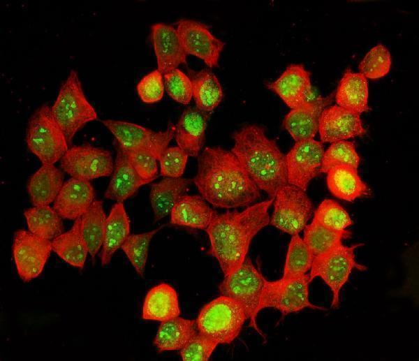
Anti-ADAR IHC images cover mouse brain and human lung cancer (A00869-2 and PB9976 IHC captions); IF/ICC images cover MCF-7 and HeLa cells (A00869-2 and M00869 IF captions).

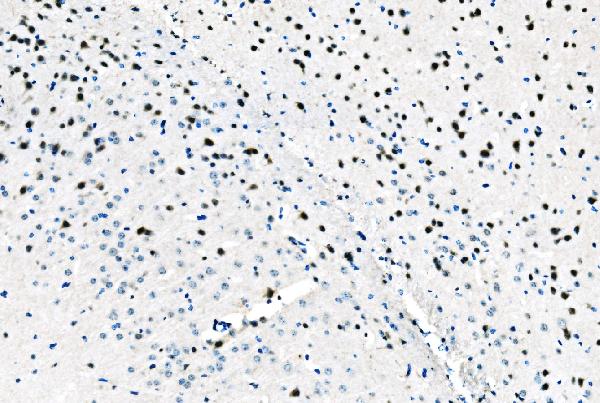
Real IHC data IHC analysis of ADAR1/ADAR using anti-ADAR1/ADAR antibody (A00869-2). ADAR1/ADAR was detected in a paraffin-embedded section of mouse brain tissue. Heat mediated antigen retrieval was performed in EDTA buffer (pH 8.0, epitope retrieval solution). The tissue section was blocked with 10% goat serum. The tissue section was then incubated with 2 μg/ml rabbit anti-ADAR1/ADAR Antibody (A00869-2) overnight at 4°C. Biotinylated goat anti-rabbit IgG was used as secondary antibody and incubated for 30 minutes at 37°C. The tissue section was developed using Strepavidin-Biotin-Complex (SABC) (Catalog # SA1022) with DAB as the chromogen.
Anti-ADAR1/ADAR Antibody ®
Cat # A00869-2
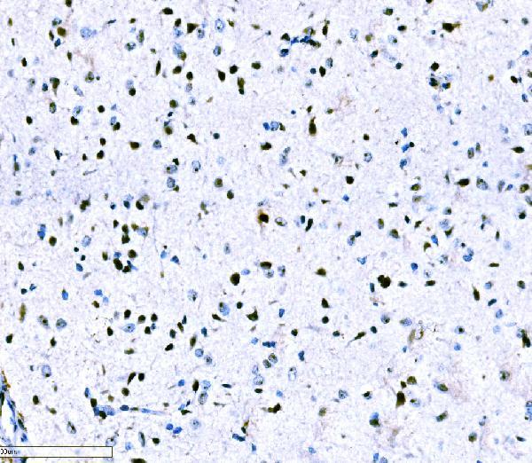
Real IHC data IHC analysis of ADAR1 using anti-ADAR1 antibody (PB9976). ADAR1 was detected in paraffin-embedded section of human lung cancer tissues. Heat mediated antigen retrieval was performed in citrate buffer (pH6, epitope retrieval solution) for 20 mins. The tissue section was blocked with 10% goat serum. The tissue section was then incubated with 1μg/ml rabbit anti-ADAR1 Antibody (PB9976) overnight at 4°C. Biotinylated goat anti-rabbit IgG was used as secondary antibody and incubated for 30 minutes at 37°C. The tissue section was developed using Strepavidin-Biotin-Complex (SABC)(Catalog # SA1022) with DAB as the chromogen.
Anti-ADAR1 Antibody ®
Cat # PB9976
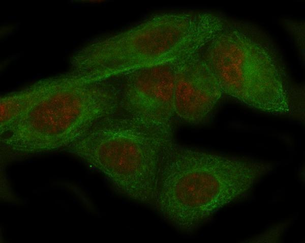
Real IF data IF analysis of ADAR using anti-ADAR antibody (M00869) and anti-Beta Tubulin antibody (M01857-3). ADAR was detected in immunocytochemical section of Hela cell. Enzyme antigen retrieval was performed using IHC enzyme antigen retrieval reagent (AR0022) for 15 mins. The cells were blocked with 10% goat serum. And then incubated at 1:100 with rabbit anti-ADAR Antibody (M00869) and mouse anti-Beta Tubulin antibody (M01857-3) overnight at 4°C. Cy3 Conjugated Goat Anti-Rabbit IgG (BA1032) and DyLight®488 Conjugated Goat Anti-Mouse IgG (BA1126) were used as secondary antibody at 1:500 dilution and incubated for 30 minutes at 37°C. Visualize using a fluorescence microscope and filter sets appropriate for the label used.
Anti-ADAR1 Monoclonal Antibody
Cat # M00869

A00869-2 has paraffin-section IHC images from mouse and rat brain and human tissues, plus an MCF-7 IF/ICC image; PB9976 has a human lung cancer paraffin-section IHC image (catalog image captions). M00869 has a HeLa IF/ICC image and lists IHC as an application, but has no IHC image caption (M00869 catalog applications and image alts).

Which to pick: For tissue IHC, choose A00869-2 for human, mouse, or rat paraffin sections with EDTA pH 8.0 retrieval, or PB9976 for human paraffin sections with citrate pH 6 retrieval (catalog reactivity; A00869-2 and PB9976 IHC captions). For IF/ICC, M00869 is a rabbit monoclonal with a HeLa cell IF image; A00869-2 also has an MCF-7 IF image (catalog host and clone; M00869 and A00869-2 IF captions). For IHC across the three listed species, A00869-2 has paraffin-section images from each, while M00869 has no IHC processing caption; the fixative is unreported in the IHC captions (A00869-2 and PB9976 IHC captions; M00869 catalog image alts).

Each figure is that product's own IHC / IF validation image from its datasheet.

References

  1. UniProt Consortium. UniProt entry P55265 (DSRAD_HUMAN, Double-stranded RNA-specific adenosine deaminase).
  2. Human Protein Atlas. ADAR tissue IHC expression (reliability: Enhanced).
  3. Human Protein Atlas. ADAR subcellular location (ICC-IF): Mainly localized to the nucleoli. In addition localized to the nucleoplasm..
  4. Human Protein Atlas. ADAR antibody validation summary (3 antibodies).
  5. A loss-of-function human ADAR variant activates innate immune response and promotes bowel inflammation. Nature communications 2025 — PMC12480066.
  6. VIRMA promotes NSCLC progression by modifying ADAR m(6)A and increasing the activity of the TGF-β signaling pathway. Scientific reports 2025 — PMC12010004.
  7. Targeting ADAR stability: a novel approach to combat glioblastoma. Frontiers in oncology 2026 — PMC13489870.
  8. ADAR1 could be a potential diagnostic target for intrauterine infection patients. Scientific reports 2024 — PMC11599928.
  9. PubMed PMID:7972084 — UniProt-cited evidence.
  10. PubMed PMID:7565688 — UniProt-cited evidence.
  11. PubMed PMID:9020165 — UniProt-cited evidence.