AIMP2 / Aminoacyl tRNA synthase complex-interacting multifunctional protein 2 · IHC design guide

Design Immunohistochemistry for AIMP2

Plan paraffin-section AIMP2 IHC with the catalog antibody at 0.5–1 μg/mL (datasheet PA1481). Use colon glandular cells as a positive comparison for cytoplasmic staining (HPA tissue IHC), and consider DNA-damage-related nuclear translocation when interpreting localisation (UniProt).

Evidence assembled Oct 2026 · For research use; verify linked source records and product datasheet before use
Immunohistochemistry protocol sheet for AIMP2 (IHC for AIMP2): expected localisation Cytoplasmic staining in tissue; nuclear localisation is possible after DNA damage (HPA tissue IHC; UniProt), antibody PA1481, validated IHC image, and IHC protocol steps
Printable AIMP2 IHC protocol sheet — expected localisation Cytoplasmic staining in tissue; nuclear localisation is possible after DNA damage (HPA tissue IHC; UniProt), antibody PA1481, controls and protocol steps. Open the full AIMP2 IHC guide →

AIMP2 Immunohistochemistry Experimental Design Guide

Expected localisation, validated protocols, controls and antibodies — the at-a-glance facts below, then the full design guide.

Must know before staining
Expected localisation Cytoplasmic staining in tissue; nuclear localisation is possible after DNA damage (HPA tissue IHC; UniProt)
Staining pattern Cytoplasmic staining in most tissues, including colon glandular cells (HPA tissue IHC)
Antigen retrieval Citrate pH 6 HIER, heat-mediated (datasheet PA1481)
Positive control ⓘ Adipose tissue+4 more · see all
Negative control ⓘ Salivary gland+2 more · see all
Important caveats
Reasons your staining may differ from the expected pattern.
Fixation Keep fixation consistent across sections. (standard IHC practice; not target-specific)
Caveat DNA damage may shift AIMP2 from cytoplasm to nucleus (UniProt)
Regulation No expression regulator is annotated (UniProt)
Isoform / epitope 2 isoforms; antibody epitope coverage is unspecified (UniProt; datasheet PA1481)
Section 1

Recommended AIMP2 IHC & IF Protocols

The catalog antibody has an IHC-P protocol (datasheet: PA1481). The published options cover AIMP2-DX2 tissue sections (PMC5941793) and frozen mouse brain sections (PMC3785563).

Recommended immunohistochemistry (IHC-P) protocol parameters
SampleParaffin-embedded human rectal cancer tissues; fixative not specified (datasheet PA1481)
FixationImage fixative and duration unreported (datasheet PA1481); verify before use.
Sectioning4–5 µm sections on charged slides (standard)
DeparaffinisationXylene, graded ethanol series to water (standard)
Antigen retrievalHeat retrieval: Citrate pH 6, 20 min (datasheet PA1481)
Peroxidase block3% H2O2, 10 min, room temperature (standard)
Blocking10% goat serum (datasheet PA1481)
Primary antibodyRabbit anti-AIMP2, 0.5-1μg/ml (datasheet PA1481)
Primary incubationOvernight at 4 °C (datasheet PA1481)
DetectionStreptavidin-biotin complex (SABC), DAB chromogen (datasheet PA1481)
CounterstainHematoxylin, blue, dehydrate and mount (standard)
Expected resultAIMP2-positive staining in adipocytes of adipose tissue (HPA tissue IHC: Medium). HPA tissue profile: Cytoplasmic expression in most tissues. No signal in the no-primary control.
💡Decision noteFor paraffin sections, start with heat-mediated citrate retrieval at pH 6 (datasheet: PA1481; PMC5941793). The mouse brain protocol uses frozen sections (PMC3785563).
Section 2

What Is the Expected AIMP2 Staining Pattern?

AIMP2 should usually appear as cytoplasmic staining in cells across many tissues (HPA tissue IHC: cytoplasmic expression in most tissues). Cytosolic localisation is consistent with its lack of a transmembrane segment (UniProt Q13155: subcellular location and topology). Colon glandular cells, adipocytes and bone marrow hematopoietic cells are examples with medium staining (HPA tissue IHC). HPA rates the tissue pattern Supported, with medium consistency between staining and RNA data (HPA tissue IHC: reliability).

What am I looking at on my slide?
Cytoplasmic staining in colon glandular cells, adipocytes or bone marrow hematopoietic cells.This fits the reported pattern: each listed cell population has medium staining (HPA tissue IHC). Judge signal in the identified cells and compartment rather than by whole-section colour alone (general IHC practice).
Predominantly nuclear staining, with little cytoplasmic signal.Review the sample context before calling it specific. AIMP2 is mainly cytosolic, but can move to the nucleus after DNA damage (HPA subcellular ICC-IF; UniProt Q13155: subcellular location). Without that context, check controls for nonspecific nuclear colour (general IHC practice).
Strong staining in salivary gland glandular cells or vaginal squamous epithelial cells.These cell populations were reported as not detected (HPA tissue IHC). Unexpected colour warrants review for cross-reactivity or endogenous detection activity; an HPA negative observation is a comparator, not proof that every specimen must be blank (general IHC practice).
Diffuse colour across cells, stroma and empty spaces, with no clear cytoplasmic boundary.A compartment-free haze does not match the reported cytoplasmic pattern (HPA tissue IHC). Compare the no-primary and tissue controls, then assess background from detection reagents, blocking or washes (general IHC practice).
No staining in colon glandular cells while the section and counterstain remain visible.Colon glandular cells are a reported medium-positive population (HPA tissue IHC). Check whether the assay controls worked before interpreting this specimen as lacking AIMP2; an absent signal alone cannot distinguish biology from assay failure (general IHC practice).
💡Expected AIMP2 appearanceCall the result positive when defined cytoplasmic colour is visible in a reported medium-positive population such as colon glandular cells (HPA tissue IHC); compartment-free haze or strong colour in an HPA not-detected population calls for control review (HPA tissue IHC; general IHC practice).
How each factor affects the staining
Cell and tissue choiceUse a reported medium-positive population, such as adipocytes or colon glandular cells, as a pattern comparator (HPA tissue IHC). Salivary gland glandular cells are reported not detected, but their status should not be treated as a universal negative guarantee (HPA tissue IHC).
Cytosol and conditional nuclear localisationCytosol is the approved main location in ICC-IF (HPA subcellular ICC-IF). UniProt reports nuclear translocation after DNA damage, so nuclear colour needs biological context and controls before interpretation (UniProt Q13155: subcellular location; general IHC practice).
Antibody evidenceHPA rates tissue staining Supported, with medium consistency against RNA data; HPA019098 and HPA020057 are each Supported for IHC (HPA tissue IHC: reliability; HPA antibody validation). This supports a pattern comparison without making any one specimen conclusive.
Isoforms and epitope coverageUniProt lists 2 isoforms (UniProt Q13155: isoforms). The supplied evidence does not map either antibody's epitope to an isoform, so do not infer which isoform produced a stain or explain a negative slide by isoform choice alone.
IF/ICC Q: What localisation is expected?A: Predominantly cytosolic signal; HPA approves cytosol as the main ICC-IF location and lists images from A-431, A-549 and U2OS (HPA subcellular ICC-IF). Nuclear localisation can occur after DNA damage (UniProt Q13155: subcellular location).
Why is my staining missing, weak or wrong?
SituationLikely causeNext action
A reported positive population has no signal.Assay failure is possible; colon glandular cells are medium positive in the HPA observations (HPA tissue IHC).Check section integrity and assay controls, then review the chosen antibody's IHC-P instructions, retrieval and detection steps (general IHC practice). Do not assign a target-specific fixation effect from this result.
Nuclei dominate the stain.AIMP2 can enter nuclei after DNA damage, while its approved main ICC-IF location is cytosol (UniProt Q13155: subcellular location; HPA subcellular ICC-IF).Record damage or treatment context and compare cytoplasmic staining and controls. If nuclear colour tracks the no-primary control, investigate detection background (general IHC practice).
Unexpected strong colour appears in salivary gland glandular cells.That cell population was not detected in HPA tissue IHC; cross-reactivity or endogenous detection activity are possible assay explanations (HPA tissue IHC; general IHC practice).Verify cell identity and compare no-primary and known-positive sections. Review endogenous activity blocking appropriate to the chromogenic detection system (general IHC practice).
Colour spreads beyond cell boundaries.Diffuse background obscures the cytoplasmic pattern reported for AIMP2 (HPA tissue IHC). Excess reagent, incomplete washes or nonspecific detection are general possibilities (general IHC practice).Inspect the no-primary control; review blocking, wash steps and detection reagent use according to the assay instructions (general IHC practice).
One population stains and a neighbouring population does not.HPA reports staining by cell population, including medium signal in colon glandular cells and no detection in vaginal squamous epithelial cells (HPA tissue IHC). Tissue-wide uniformity is not established.Identify each population on the counterstained section and compare it with the corresponding HPA observation before scoring (HPA tissue IHC; general IHC practice).
Staining is weak despite high skeletal-muscle RNA specificity.HPA calls RNA tissue enhanced in skeletal muscle, while its IHC summary says cytoplasmic expression in most tissues (HPA tissue IHC). The RNA label does not specify an IHC intensity.Judge the slide against reported IHC-positive cells and assay controls; do not set a staining threshold from RNA specificity alone (HPA tissue IHC; general IHC practice).

Sample controls for AIMP2 IHC & IF

🧪Run appendix first: glandular cells should stain for AIMP2 (HPA: Appendix, glandular cells, Medium). Use salivary gland as the negative tissue (HPA: Salivary gland, glandular cells, Not detected); any unstained neighboring cells in the appendix section should show counterstain alone, but HPA does not establish them as biological negatives.
Positive control tissue: Adipose tissue (Adipocytes, HPA Medium)
Negative control tissue: Salivary gland (HPA Not detected)
ICC-IF cell lines (HPA subcellular resource): HPA ICC-IF images show AIMP2 in A-431, A-549, U2OS, with annotated localisation: Cytosol (approved) (HPA subcellular).
Technical controls: Include a no-primary, secondary-only control, a concentration-matched rabbit IgG isotype control, and AIMP2-knockout material if available as a biological negative (PA1481 caption: rabbit primary; standard IHC practice). Quench endogenous peroxidase and check for endogenous biotin background in the appendix section when using the caption’s biotin/SABC/DAB detection method (PA1481 caption; standard IHC practice).
⚠️Feasibility: The selected PA1481 paraffin-section caption reports heat retrieval in citrate buffer at pH 6 for 20 minutes, but does not report a fixative (PA1481 tissue-IHC caption). A target-specific fixation window, fixation effect, and strict retrieval dependency are unreported; the supplied evidence also cannot establish whether frozen sections or IF are easier (PA1481 tissue-IHC caption; HPA: ICC-IF cytosolic localization). In appendix, inflammatory cells can produce endogenous peroxidase background during DAB detection (standard IHC practice).

HPA tissue IHC evidence for AIMP2

Comprehensive Human Protein Atlas IHC scoring per tissue (reliability: Supported — Medium consistency between antibody staining and RNA expression data.). Rows are taken directly from the HPA tissue chart — click any row's HPA link to view the source.

Positive expression · recommended positive controls

TissueCell typeLevelEvidenceSource
Adipose tissue Adipocytes Medium Protein (IHC) HPA →
Appendix Glandular cells Medium Protein (IHC) HPA →
Bone marrow Hematopoietic cells Medium Protein (IHC) HPA →
Bronchus Respiratory epithelial cells Medium Protein (IHC) HPA →
Caudate Neuronal cells Medium Protein (IHC) HPA →

Undetected expression · recommended negative controls

TissueCell typeLevelEvidenceSource
Salivary gland Glandular cells Not detected Protein (IHC) HPA →
Soft tissue Peripheral nerve Not detected Protein (IHC) HPA →
Vagina Squamous epithelial cells Not detected Protein (IHC) HPA →
Section 3

Advanced AIMP2 IHC Tips

These checks focus on chromogenic AIMP2 staining in paraffin sections, with one entry for planning a related IF experiment.

What retrieval should I try first for weak AIMP2 staining in paraffin sections?
Start with heat-mediated citrate retrieval at pH 6 for 20 minutes (datasheet PA1481). If staining remains weak, compare a modestly longer heating time on adjacent sections while keeping the same buffer and recording tissue damage (standard IHC practice). The documented paraffin-section workflow then used 10% goat serum and 1 µg/mL primary antibody overnight at 4°C (datasheet PA1481). Include a positive control section with cytoplasmic staining, such as colon glandular cells, and evaluate signal alongside morphology before changing several conditions at once (HPA: Medium in colon glandular cells; standard IHC practice).
Could fixation explain variable AIMP2 staining between paraffin blocks?
Target-specific fixation sensitivity is unknown because the selected paraffin-section caption does not state the fixative (datasheet PA1481). Record each block’s fixative and fixation time, then compare sections using the same pH 6 citrate retrieval and staining run (datasheet PA1481; standard IHC practice). Excessive or uneven fixation can alter antigen accessibility in IHC generally, so inspect morphology and compare internal cell populations before attributing a weak result to AIMP2 biology (standard IHC practice). Do not infer an AIMP2-specific fixation effect from its cytosolic location, lack of a transmembrane segment, or tissue staining profile (UniProt Q13155; HPA: tissue IHC profile).
Should AIMP2 staining be cytoplasmic, nuclear, or both?
Expect mainly cytoplasmic staining in routine tissue IHC: HPA reports cytoplasmic expression in most tissues, and UniProt places AIMP2 in the cytosol (HPA: tissue IHC profile; UniProt Q13155). Nuclear staining can be biologically plausible because AIMP2 translocates to the nucleus following DNA damage, but an unstated damage history cannot establish that explanation for a particular section (UniProt Q13155). Score nuclear and cytoplasmic signal separately, using the counterstain to define nuclei and checking that staining follows intact cells (standard IHC practice). Treat diffuse staining across damaged tissue or an isolated dark section edge as a technical concern requiring controls (standard IHC practice).
Can this stain distinguish AIMP2 isoforms or a modified epitope?
AIMP2 has 2 annotated isoforms, but the supplied tissue-IHC caption does not identify the antibody epitope or establish isoform-specific staining (UniProt Q13155; datasheet PA1481). Therefore, report the result as AIMP2 immunoreactivity rather than assigning either isoform from chromogenic signal alone (standard IHC interpretation). The annotated GST C-terminal domain spans residues 220–317; its presence does not locate the antibody’s binding site (UniProt Q13155). UniProt lists 0 annotated glycosylation sites and 0 modified residues in this record, which does not validate any modification-specific staining claim (UniProt Q13155). Resolve isoform or epitope questions with antibody epitope documentation or an independently validated assay (standard IHC practice).
How should I design a companion AIMP2 IF experiment?
For a companion IF experiment, pair AIMP2 with a marker that identifies the expected cell population, such as a validated glandular-cell marker when examining colon (HPA: Medium in colon glandular cells; standard IF practice). Choose spectrally separated fluorophores and test an unstained section for autofluorescence; a far-red AIMP2 channel may improve separation when tissue background is strong (standard IF practice). Because AIMP2 is cytosolic, use controlled permeabilisation for an intracellular epitope and adjust it if assessing nuclear signal after DNA damage (UniProt Q13155; standard IF practice). Include single-stain and secondary-only controls before interpreting overlap, and optimise IF fixation independently because no AIMP2-specific IF fixation condition is supplied here (standard IF practice).
How can I reduce diffuse or granular background without losing AIMP2 signal?
First compare a no-primary control with the stained section to locate signal from secondary reagents, endogenous enzyme activity, or chromogen deposition (standard chromogenic IHC practice). The documented workflow used 10% goat serum, 1 µg/mL primary antibody overnight at 4°C, a biotinylated secondary, and DAB detection (datasheet PA1481). Apply an appropriate peroxidase block and assess endogenous biotin when using a biotin-based detection system; these are general controls, not AIMP2-specific requirements (standard IHC practice). If background persists, shorten primary incubation or titrate antibody on matched sections while retaining the pH 6 citrate retrieval baseline (datasheet PA1481; standard IHC practice).
What should I measure when comparing AIMP2 IHC across sections? ⚠ ANSWER MARKED FOR VERIFICATION
Define the cell population and compartment before scoring: HPA describes predominantly cytoplasmic tissue staining, while nuclear localisation is also annotated (HPA: tissue IHC profile; UniProt Q13155). For each comparable region, record the percentage of positive cells and staining intensity, then calculate a consistent H-score if intensity categories are reproducible (standard IHC scoring practice). Normalise positive-cell counts to the number of evaluable cells in that population, or report positive-cell density per mm² of viable tissue when cell counting is impractical (standard IHC practice). Exclude folds and necrotic regions, and use identical retrieval, detection, exposure conditions, and thresholds across the comparison (standard IHC practice).
How do I distinguish credible AIMP2 staining from tissue or detection artefact?
A credible pattern should appear in intact cells with predominantly cytoplasmic distribution, consistent with HPA tissue staining and AIMP2’s annotated cytosolic location (HPA: tissue IHC profile; UniProt Q13155). Colon glandular cells offer a reported medium-staining reference, whereas salivary gland glandular cells are reported as not detected; neither reference alone proves antibody specificity (HPA: Medium in colon glandular cells; HPA: Not detected in salivary gland glandular cells; standard IHC interpretation). Examine unexpected nuclear signal with biological context because nuclear translocation is reported following DNA damage (UniProt Q13155). Discount staining confined to section edges, necrosis, or no-primary controls, and investigate endogenous peroxidase or biotin signal before assigning AIMP2 positivity (standard IHC practice).
Boster reagents

Best AIMP2 / Aminoacyl tRNA synthase complex-interacting multifunctional protein 2 IHC Antibodies

The IHC-validated anti-AIMP2 antibody PA1481 has real paraffin-section images from human rectal cancer and rat intestine (catalog: PA1481 IHC captions); its listed reactivity includes human, mouse and rat (catalog: PA1481).

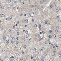
Real IHC data IHC analysis of AIMP2 using anti-AIMP2 antibody (PA1481). AIMP2 was detected in paraffin-embedded section of human rectal cancer tissues. Heat mediated antigen retrieval was performed in citrate buffer (pH6, epitope retrieval solution) for 20 mins. The tissue section was blocked with 10% goat serum. The tissue section was then incubated with 1μg/ml rabbit anti-AIMP2 Antibody (PA1481) overnight at 4°C. Biotinylated goat anti-rabbit IgG was used as secondary antibody and incubated for 30 minutes at 37°C. The tissue section was developed using Strepavidin-Biotin-Complex (SABC)(Catalog # SA1022) with DAB as the chromogen.
Anti-AIMP2/p38 Antibody ®
Cat # PA1481

PA1481 is listed for IHC and has a paraffin-section image from human rectal cancer tissue (catalog: PA1481 applications and IHC caption). PA1481 also has a paraffin-section image from rat intestine; mouse is listed as reactive but has no supplied IHC image (catalog: PA1481 reactivity and IHC captions).

Which to pick: Choose PA1481 for paraffin-section IHC, starting at 0.5–1 μg/ml; its captions document citrate retrieval and chromogenic detection, but do not report the fixative (catalog: PA1481 dilution and IHC captions). No listed SKU has IF/ICC validation, so this payload does not support an IF/ICC recommendation (catalog: PA1481 applications and IF images). PA1481 is listed as reactive with human, mouse and rat, with IHC images supplied for human and rat only (catalog: PA1481 reactivity and IHC captions).

Each figure is that product's own IHC / IF validation image from its datasheet.

References

  1. UniProt Consortium. UniProt entry Q13155 (AIMP2_HUMAN, Aminoacyl tRNA synthase complex-interacting multifunctional protein 2).
  2. Human Protein Atlas. AIMP2 tissue IHC expression (reliability: Supported).
  3. Human Protein Atlas. AIMP2 subcellular location (ICC-IF): Localized to the cytosol..
  4. Human Protein Atlas. AIMP2 antibody validation summary (2 antibodies).
  5. AIMP2-DX2 Promotes the Proliferation, Migration, and Invasion of Nasopharyngeal Carcinoma Cells. BioMed research international 2018 — PMC5941793.
  6. Pan-cancer Analysis Identifies AIMP2 as a Potential Biomarker for Breast Cancer. Current genomics 2023 — PMC10790333.
  7. Parthanatos mediates AIMP2-activated age-dependent dopaminergic neuronal loss. Nature neuroscience 2013 — PMC3785563.
  8. MTCH2 promotes the malignant progression of ovarian cancer through the upregulation of AIMP2 expression levels, mitochondrial dysfunction and by mediating energy metabolism. Oncology letters 2024 — PMC11342418.
  9. PubMed PMID:8666379 — UniProt-cited evidence.
  10. PubMed PMID:12853948 — UniProt-cited evidence.
  11. PubMed PMID:12690205 — UniProt-cited evidence.