This website uses cookies to ensure you get the best experience on our website.


- Table of Contents
Use granular cytoplasmic staining in renal tubules and hepatocytes as the tissue reference for ALDH6A1 chromogenic IHC (HPA tissue IHC). This guide covers paraffin section controls, antibody dilution and scoring by cell type and staining pattern (standard IHC practice).

Expected localisation, validated protocols, controls and antibodies — the at-a-glance facts below, then the full design guide.
| Expected localisation | Observed granular cytoplasm (HPA tissue IHC); mitochondrial protein (UniProt) | |
| Staining pattern | Renal tubules and hepatocytes: granular cytoplasmic staining (HPA tissue IHC) | |
| Antigen retrieval | EDTA pH 8.0 HIER, heat-mediated (datasheet A08868-1) | |
| Positive control | Caudate+4 more · see all | |
| Negative control | Adipose tissue+4 more · see all |
| Fixation | Keep fixation consistent across sections (standard IHC practice; not target-specific) | |
| Caveat | Staining has medium consistency with RNA expression (HPA tissue IHC) | |
| Regulation | No expression regulator annotated (UniProt) | |
| Isoform / epitope | 2 isoforms; epitope coverage unspecified (UniProt) |
Compare the catalog antibody’s IHC-P protocol (datasheet) with published ALDH6A1 staining of mouse embryo sections (PMC13509631) and renal tissue sections (PMC7038561).
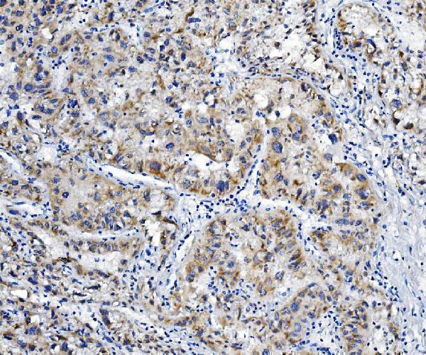
| Sample | Paraffin-embedded human liver cancer tissue; fixative not specified (datasheet A08868-1) |
| Fixation | Image fixative and duration unreported (datasheet A08868-1); verify before use. |
| Sectioning | 4–5 µm sections on charged slides (standard) |
| Deparaffinisation | Xylene, graded ethanol series to water (standard) |
| Antigen retrieval | Heat retrieval: EDTA pH 8.0 (datasheet A08868-1); 20 min, 95–100 °C (standard) |
| Peroxidase block | 3% H2O2, 10 min, room temperature (standard) |
| Blocking | 10% goat serum (datasheet A08868-1) |
| Primary antibody | Rabbit anti-ALDH6A1, 2μg/ml (datasheet A08868-1) |
| Primary incubation | Overnight at 4 °C (datasheet A08868-1) |
| Detection | Streptavidin-biotin complex (SABC), DAB chromogen (datasheet A08868-1) |
| Counterstain | Hematoxylin, blue, dehydrate and mount (standard) |
| Expected result | ALDH6A1-positive staining in glial cells of caudate (HPA tissue IHC: High). HPA tissue profile: Granular cytoplasmic expression in renal tubules and hepatocytes. No signal in the no-primary control. |
ALDH6A1 is a mitochondrial protein with no transmembrane segment (UniProt Q02252: subcellular location and topology). In paraffin section IHC, expect granular cytoplasmic staining in renal tubules and hepatocytes (HPA: tissue IHC profile). HPA rates the tissue staining profile Enhanced, while reporting medium consistency between antibody staining and RNA expression; interpret intensity alongside the cell type and compartment (HPA: tissue IHC reliability).
| Granular cytoplasmic chromogen appears in renal tubules and hepatocytes (HPA: tissue IHC profile). | This matches the observed tissue pattern and mitochondrial location (HPA: tissue IHC profile; UniProt Q02252: subcellular location). Granules are compatible with mitochondrial staining, but chromogenic IHC alone cannot establish organelle colocalisation (general IHC practice). |
| Signal is chiefly nuclear or outlines cell surfaces instead of forming cytoplasmic granules. | Treat that compartment pattern as suspect: UniProt places ALDH6A1 in mitochondria and reports no transmembrane segment (UniProt Q02252: location and topology). Check morphology and controls before interpreting it as ALDH6A1; an unexpected compartment does not identify the artefact's cause (general IHC practice). |
| Strong staining appears in adipocytes, alveolar cells or skeletal muscle myocytes (HPA: not detected in these cells). | These are poor matches to the reported IHC pattern (HPA: tissue IHC). Consider antibody cross-reactivity or endogenous detection activity, particularly if the signal persists in a no-primary control; that control helps distinguish detection background from primary-antibody staining (general IHC practice). |
| Chromogen spreads evenly across tissue, including spaces between cells, rather than marking cytoplasmic granules. | A diffuse deposit is difficult to score as ALDH6A1 because the reported pattern is granular and cytoplasmic (HPA: tissue IHC profile). Review the no-primary control and detection background, then optimise blocking, washes and chromogen development as general IHC practice. |
| No signal is visible in renal tubules or hepatocytes on an otherwise interpretable section. | The result conflicts with the reported positive pattern (HPA: tissue IHC profile), but a blank slide alone cannot distinguish absent antigen from assay failure (general IHC practice). Confirm tissue preservation and counterstain, then check the catalog antibody's validated IHC conditions and a concurrent positive control. |
| Cell type and tissue context | HPA describes granular cytoplasmic expression in renal tubules and hepatocytes, while its RNA category is group enriched in kidney and liver (HPA: tissue IHC profile and RNA specificity). Use the protein staining pattern to judge an IHC slide; the RNA category does not assign a chromogen intensity to either cell type. |
| Reported negative and high-staining cells | HPA reports no detection in adipocytes and alveolar cells, but high staining in glial cells of caudate and cerebral cortex (HPA: tissue IHC). Score the identified cells rather than treating every cell in a tissue as uniformly positive or negative (general IHC practice). |
| Evidence strength | HPA labels the tissue IHC profile Enhanced but describes medium consistency with RNA expression (HPA: tissue IHC reliability). Its listed antibodies HPA029072, HPA029073, HPA029074 and HPA029075 each have Enhanced IHC status (HPA: antibody validation); this supports the reported pattern without validating every new staining run. |
| Protein processing and isoforms | UniProt lists two isoforms and a mature chain spanning residues 34–535 (UniProt Q02252: isoforms and processing). The payload gives no antibody epitope, so these annotations cannot predict whether a given antibody distinguishes isoforms or recognises the processed protein in sections. |
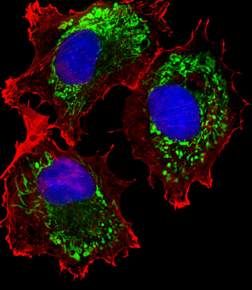
| IF/ICC Q: Where should ALDH6A1 fluorescence appear? | A: Mainly in mitochondria (HPA: subcellular ICC-IF, enhanced; UniProt Q02252: location). This is the IF/ICC expectation; use that application's own guide for its staining workflow. |
| Situation | Likely cause | Next action |
|---|---|---|
| Renal tubules and hepatocytes are both blank (HPA: expected granular cytoplasmic expression). | The assay may have failed; the blank result does not by itself identify the failed step (general IHC practice). | Run a concurrent positive section and inspect counterstain, antibody use, retrieval and detection steps against the catalog antibody's IHC-P instructions (general IHC practice). |
| Signal is mainly nuclear or confined to cell borders. | That distribution conflicts with mitochondrial ALDH6A1 and its lack of a transmembrane segment (UniProt Q02252: location and topology). | Recheck cell boundaries and the no-primary control; compare a repeat section with the reported granular cytoplasmic pattern (HPA: tissue IHC; general IHC practice). |
| Adipocytes or alveolar cells stain strongly (HPA: not detected in these cells). | Possible cross-reactivity or endogenous detection activity; the slide alone cannot separate them (general IHC practice). | Compare no-primary and positive-tissue controls, then review blocking and the detection system for background (general IHC practice). |
| Broad, diffuse colour obscures individual cells and granules. | Nonspecific background or excessive chromogen development can obscure a granular cytoplasmic pattern (general IHC practice; HPA: tissue IHC profile). | Check the no-primary control, washing and chromogen development, and rescore only where cell morphology remains clear (general IHC practice). |
| The expected cells stain, but only weakly. | HPA reports the pattern without assigning a universal intensity threshold to renal tubules or hepatocytes (HPA: tissue IHC profile). | Compare matched positive and no-primary controls before changing interpretation; follow the catalog antibody's IHC-P conditions when adjusting the assay (general IHC practice). |
| A new section differs from the HPA image pattern. | HPA's tissue profile has medium consistency with RNA expression; a mismatch needs review rather than an automatic biological explanation (HPA: reliability). | Verify the scored cell type and cytoplasmic distribution, then compare an independent IHC-validated antibody if available (HPA: antibody validation; general IHC practice). |
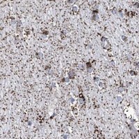

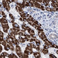


Comprehensive Human Protein Atlas IHC scoring per tissue (reliability: Enhanced — Medium consistency between antibody staining and RNA expression data.). Rows are taken directly from the HPA tissue chart — click any row's HPA link to view the source.
| Tissue | Cell type | Level | Evidence | Source |
|---|---|---|---|---|
| Caudate | Glial cells | High | Protein (IHC) | HPA → |
| Cerebellum | Bergmann glia - cytoplasm/membrane | High | Protein (IHC) | HPA → |
| Cerebral cortex | Glial cells | High | Protein (IHC) | HPA → |
| Cervix | Glandular cells | High | Protein (IHC) | HPA → |
| Duodenum | Glandular cells | High | Protein (IHC) | HPA → |
| Tissue | Cell type | Level | Evidence | Source |
|---|---|---|---|---|
| Adipose tissue | Adipocytes | Not detected | Protein (IHC) | HPA → |
| Lung | Alveolar cells | Not detected | Protein (IHC) | HPA → |
| Nasopharynx | Respiratory epithelial cells | Not detected | Protein (IHC) | HPA → |
| Oral mucosa | Squamous epithelial cells | Not detected | Protein (IHC) | HPA → |
| Placenta | Decidual cells | Not detected | Protein (IHC) | HPA → |
Troubleshoot ALDH6A1 staining in paraffin sections by checking retrieval, cytoplasmic localisation, cell identity and detection controls before comparing signal intensity (UniProt Q02252; HPA tissue IHC).
ALDH6A1 antibodies have IHC images from human paraffin sections and an IF image from MCF-7 cells (catalog image captions). Catalog reactivity covers human, mouse, and rat (catalog reactivity).
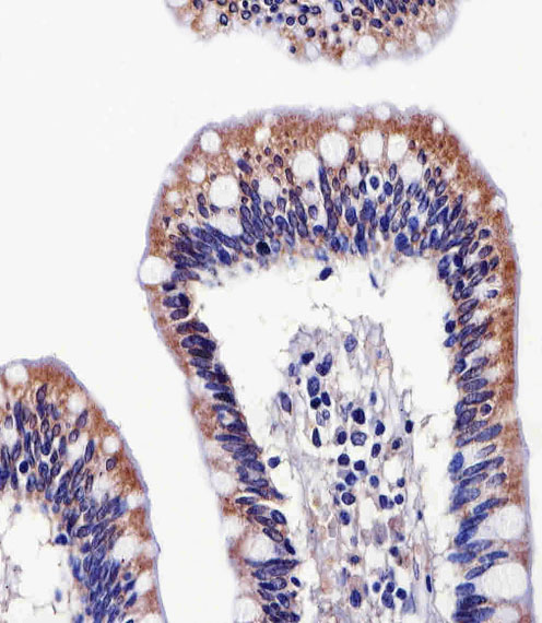
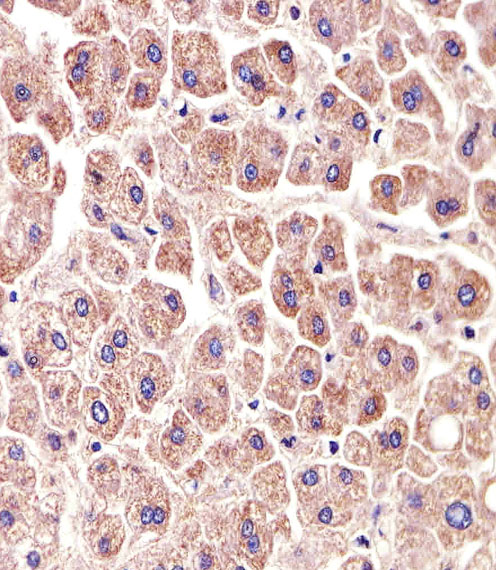
A08868-1 has an IHC image from a paraffin section of human liver cancer tissue (A08868-1 image caption). M08868 has IHC images from human colon and liver paraffin sections and an IF image from MCF-7 cells (M08868 image captions).
Which to pick: For tissue IHC, A08868-1 is a rabbit polyclonal option shown on a human liver cancer paraffin section, while M08868 is a mouse monoclonal option shown on human colon and liver paraffin sections at 1:25; the captions do not report a fixative (catalog antibody details; A08868-1 and M08868 IHC captions). For IF/ICC, choose M08868: IF is listed and its image shows MCF-7 cells stained at 1:25 (M08868 applications and IF caption). For work involving mouse or rat samples, A08868-1 lists reactivity with both, but its stated paraffin-section IHC testing and image are human only (A08868-1 catalog reactivity, dilution notes, and IHC caption).