AMPH / Amphiphysin · IHC design guide

Design Immunohistochemistry for AMPH

Plan AMPH chromogenic IHC on paraffin sections using the human-reactive catalog antibody at 1:25 (datasheet M02364). Compare staining with cerebellar GLUC cell cytoplasm/membrane and cortical neuropil patterns (HPA tissue IHC).

Evidence assembled Oct 2026 · For research use; verify linked source records and product datasheet before use
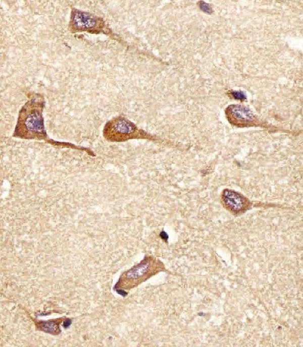
Immunohistochemistry protocol sheet for AMPH (IHC for AMPH): expected localisation Cerebellar GLUC cytoplasm/membrane; cortical neuropil (HPA tissue IHC), antibody M02364, validated IHC image, and IHC protocol steps
Printable AMPH IHC protocol sheet — expected localisation Cerebellar GLUC cytoplasm/membrane; cortical neuropil (HPA tissue IHC), antibody M02364, controls and protocol steps. Open the full AMPH IHC guide →

AMPH Immunohistochemistry Experimental Design Guide

Expected localisation, validated protocols, controls and antibodies — the at-a-glance facts below, then the full design guide.

Must know before staining
Expected localisation Cerebellar GLUC cytoplasm/membrane; cortical neuropil (HPA tissue IHC)
Staining pattern High cerebellar GLUC cytoplasm/membrane and cortical neuropil (HPA tissue IHC)
Antigen retrieval Citrate pH 6 HIER, heat-mediated (datasheet M02364)
Positive control ⓘ Cerebellum+4 more · see all
Negative control ⓘ Adipose tissue+4 more · see all
Important caveats
Reasons your staining may differ from the expected pattern.
Fixation Formaldehyde-fixed, paraffin-embedded sections (selected-SKU IHC image M02364)
Caveat Intestinal enterocyte microvilli also stain strongly (HPA tissue IHC)
Regulation CNS and peripheral nerves enriched (HPA tissue IHC)
Isoform / epitope 2 isoforms; no transmembrane segment; epitope coverage unknown (UniProt)
Section 1

Recommended AMPH IHC & IF Protocols

The catalog antibody’s IHC-P protocol (datasheet: M02364) is accompanied by two published AMPH IHC procedures (PMC6883929; PMC6010687).

Recommended immunohistochemistry (IHC-P) protocol parameters
SampleFormaldehyde-fixed, paraffin-embedded Human brain tissue (datasheet M02364)
FixationImage formalin-fixed; duration unreported (datasheet M02364); verify before use.
Sectioning4–5 µm sections on charged slides (standard)
DeparaffinisationXylene, graded ethanol series to water (standard)
Antigen retrievalHeat retrieval: Citrate pH 6 (datasheet M02364); 20 min, 95–100 °C (standard)
Peroxidase block3% H2O2, 10 min, room temperature (standard)
Blocking10% normal serum of the secondary host, 30 min, room temperature (standard)
Primary antibodyRabbit anti-AMPH, 1:25 (datasheet M02364)
Primary incubation1 hours at 37°C (datasheet M02364)
DetectionHRP-polymer secondary, DAB chromogen 5–10 min (standard)
CounterstainHematoxylin, blue, dehydrate and mount (standard)
Expected resultAMPH-positive staining in gLUC cells - cytoplasm/membrane of cerebellum (HPA tissue IHC: High). HPA tissue profile: Selective and high expression in CNS and peripheral nerves. No signal in the no-primary control.
💡Decision noteStart with heat-mediated citrate retrieval at pH 6 (datasheet: M02364); EDTA retrieval is also reported (PMC6883929).
Section 2

What Is the Expected AMPH Staining Pattern?

AMPH is associated with synaptic vesicle membranes and the cytoskeleton, and has no transmembrane segment (UniProt P49418 topology; UniProt P49418 subcellular). In paraffin-section IHC, expect cytoplasmic or membranous staining in selected neural cells and neuropil; HPA also reports strong enterocyte microvillus staining (HPA tissue IHC). Treat intensity comparisons cautiously: HPA rates tissue staining reliability Enhanced but describes only medium consistency with RNA expression (HPA tissue IHC).

What am I looking at on my slide?
Strong cytoplasmic/membranous signal in cerebellar GLUC cells, or strong cerebral cortical neuropil signal (HPA tissue IHC).These match reported high-staining compartments (HPA tissue IHC). Evaluate the stained structure against the counterstain; a positive field alone does not establish cell identity (general IHC practice).
Predominantly nuclear signal in an otherwise positive section.A nuclear pattern conflicts with UniProt vesicle/cytoskeletal localization and HPA's cytosolic/plasma-membrane ICC-IF pattern (UniProt P49418 subcellular; HPA subcellular). Check the control before interpreting it as AMPH.
Strong signal across cell populations expected to be unstained, such as adipocytes in adipose tissue (HPA tissue IHC).Consider antibody cross-reactivity or endogenous detection activity (general IHC practice). HPA reports AMPH as not detected in those adipocytes, but that finding does not guarantee every specimen is negative (HPA tissue IHC).
Uniform chromogen over cells and surrounding tissue, with little compartment definition.This is difficult to score as AMPH localization. Check nonspecific antibody binding, blocking and detection background with appropriate controls (general IHC practice); the reported pattern is selective (HPA tissue IHC).
No signal in cerebellar GLUC cells or cerebral cortical neuropil under a validated run (HPA tissue IHC).These are reported high-staining reference sites (HPA tissue IHC). First check whether the positive-control section and detection reagents worked; then review retrieval and antibody dilution (general IHC practice).
💡Expected AMPH appearanceCall a result positive when strong chromogenic signal follows cerebellar GLUC-cell cytoplasm/membrane or cerebral cortical neuropil (HPA tissue IHC); widespread nuclear staining or uniform tissue haze is suspect given the reported localization (UniProt P49418 subcellular; HPA subcellular).
How each factor affects the staining
Tissue and compartment selectionCerebellar GLUC cells, cortical neuropil and small-intestinal enterocyte microvilli are reported High; several other cell types are not detected (HPA tissue IHC). Score the specified compartment, not the whole section.
Membrane associationAMPH has no transmembrane segment, although UniProt places it at synaptic vesicle membranes (UniProt P49418 topology; UniProt P49418 subcellular). Cytoplasmic signal can therefore fit the expected pattern.
Isoforms and antibody epitopeUniProt lists two isoforms and BAR and SH3 domains (UniProt P49418 isoforms; UniProt P49418 domains). Epitope coverage is unspecified here; do not infer which isoform a stained cell contains.
Retrieval and target-specific fixation sensitivityOptimize retrieval with control sections as general IHC practice. Neither supplied source establishes an AMPH-specific fixation effect; tissue staining and topology cannot predict one (HPA tissue IHC; UniProt P49418 topology).
Q: What can ICC-IF add?A: HPA reports supported cytosol and approved additional plasma-membrane localization, with ICC approval for HPA019828 and HPA019829 (HPA subcellular; HPA antibodies). This is a localization cross-check.
Why is my staining missing, weak or wrong?
SituationLikely causeNext action
Positive-control tissue has no chromogen.The stain may have failed, or the control may lack the relevant cell compartment (general IHC practice).Confirm the expected GLUC cells or neuropil are present (HPA tissue IHC); check detection reagents, then optimize retrieval and antibody dilution using matched sections (general IHC practice).
Nuclei dominate the signal.This conflicts with reported cytosolic, vesicle-associated and cytoskeletal localization (HPA subcellular; UniProt P49418 subcellular).Compare with a positive control and a no-primary control; reassess antibody concentration and detection background before assigning the signal to AMPH (general IHC practice).
Small-intestinal microvilli stain strongly despite the CNS-enriched summary.The summary and this specific observation coexist: HPA reports High enterocyte microvillus staining (HPA tissue IHC).Record the precise compartment and compare with a control section. Do not discard a microvillus pattern solely because HPA describes overall CNS and peripheral-nerve selectivity (HPA tissue IHC).
Diffuse stain obscures cells and tissue boundaries.Nonspecific binding or detection background can reduce compartment contrast (general IHC practice).Inspect the no-primary control, blocking and washes; adjust antibody dilution if needed (general IHC practice). Score only signal with identifiable cells or structures.
Adipocytes or other reported negative populations stain strongly.HPA reports AMPH not detected in adipose-tissue adipocytes (HPA tissue IHC); cross-reactivity or endogenous detection activity is possible (general IHC practice).Check no-primary and reagent controls, then compare with a reported positive compartment in the same run (general IHC practice; HPA tissue IHC).
A weak result is being called absent or positive.HPA reports Medium staining in soft-tissue fibroblasts and caudate or hippocampal glial cells, and Low staining in specified other cells (HPA tissue IHC).Score intensity and cell type separately against run controls; reserve a negative call for signal indistinguishable from background (general IHC practice).

Sample controls for AMPH IHC & IF

🧪Run cerebellum first and look for staining in GLUC cells at the cytoplasm or membrane (HPA: High in cerebellar GLUC cells). Use adipose tissue adipocytes as the negative tissue (HPA: Not detected in adipocytes); on the cerebellum slide, assess neighboring cells outside the GLUC cell population for background staining without assuming they are AMPH-negative.
Positive control tissue: Cerebellum (GLUC cells - cytoplasm/membrane, HPA High)
Negative control tissue: Adipose tissue (HPA Not detected)
ICC-IF cell lines (HPA subcellular resource): HPA ICC-IF images show AMPH in A-431, U-251MG, U2OS, with annotated localisation: Cytosol (supported) (HPA subcellular).
Technical controls: Include no-primary (secondary-only) and primary-host- and clonality-matched isotype controls, plus AMPH knockout tissue if available as a biological negative. Check endogenous peroxidase and biotin background in the chromogenic assay, which uses a biotinylated secondary antibody (M02364 tissue-IHC caption: biotinylated secondary).
⚠️Feasibility: A target-specific fixation window or fixation effect is unreported, and the selected-SKU fixative is unreported (M02364 tissue-IHC caption: fixative unreported). The caption uses heat retrieval in citrate buffer at pH 6, but does not establish that retrieval is required (M02364 tissue-IHC caption: citrate buffer pH 6). IF/ICC can assess the reported cytosolic and additional plasma-membrane localisation (HPA: subcellular localisation); the supplied evidence does not establish that IF or frozen sections are easier than paraffin IHC.

HPA tissue IHC evidence for AMPH

Comprehensive Human Protein Atlas IHC scoring per tissue (reliability: Enhanced — Medium consistency between antibody staining and RNA expression data.). Rows are taken directly from the HPA tissue chart — click any row's HPA link to view the source.

Positive expression · recommended positive controls

TissueCell typeLevelEvidenceSource
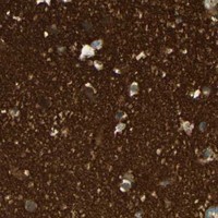
Cerebellum GLUC cells - cytoplasm/membrane High Protein (IHC) HPA →
Cerebral cortex Neuropil High Protein (IHC) HPA →
Small intestine Enterocytes - Microvilli High Protein (IHC) HPA →
Soft tissue Fibroblasts Medium Protein (IHC) HPA →
Caudate Glial cells Medium Protein (IHC) HPA →

Undetected expression · recommended negative controls

TissueCell typeLevelEvidenceSource
Adipose tissue Adipocytes Not detected Protein (IHC) HPA →
Adrenal gland Glandular cells Not detected Protein (IHC) HPA →
Appendix Endocrine cells Not detected Protein (IHC) HPA →
Bone marrow Hematopoietic cells Not detected Protein (IHC) HPA →
Breast Adipocytes Not detected Protein (IHC) HPA →
Section 3

Advanced AMPH IHC Tips

Use compartment and cell type controls when assessing AMPH staining in paraffin sections; interpret weak or diffuse signal alongside the reported tissue pattern.

What retrieval should I start with for AMPH in paraffin sections?
Start with heat-mediated citrate antigen retrieval at pH 6 for AMPH in paraffin sections (datasheet M02364). Keep section thickness, heating, cooling, and detection conditions consistent across a pilot run so that changes in staining can be attributed to retrieval. Include a brain section with an expected signal and a no-primary control; cerebral cortex neuropil has reported high staining (HPA tissue IHC). If staining remains weak, adjust heating within the laboratory's established retrieval procedure before testing an alternative buffer as a fallback, and inspect tissue morphology for damage. Record the retrieval conditions used for every section scored.
How should I troubleshoot weak AMPH staining after fixation?
Target-specific fixation sensitivity is unknown from the evidence supplied for this answer; do not assign weak AMPH staining to a particular fixative or fixation duration. Review the specimen's recorded fixation and processing history, then compare sections processed alike before changing antibody concentration or retrieval. Use a section with an expected positive pattern in the same run; high cerebral cortex neuropil staining is reported in tissue IHC (HPA tissue IHC). If the positive section also fails, examine tissue preservation, retrieval, and detection controls before interpreting a negative specimen. Keep fixation conditions documented when comparing intensity across cases.
Where should convincing AMPH chromogenic staining appear?
Assess AMPH primarily in cytoplasmic and membrane-associated structures, including synaptic vesicle-associated regions, rather than using nuclear staining as the expected pattern (UniProt P49418 subcellular location; HPA subcellular). In tissue IHC, cerebral cortex neuropil and cerebellar GLUC-cell cytoplasm or membrane show high reported staining (HPA tissue IHC). Examine cellular detail at higher magnification to distinguish cytoplasmic signal from pigment, precipitate, or staining spread beyond tissue boundaries. Compare the pattern with a no-primary control and with the expected region on the same slide when available. Treat isolated nuclear signal as a specificity concern until independently supported.
Can this stain distinguish AMPH isoforms or altered epitopes?
The AMPH record lists 2 isoforms, but the supplied antibody evidence does not map its epitope or establish isoform selectivity (UniProt P49418 isoforms; datasheet M02364). Consequently, a positive chromogenic signal cannot identify which isoform contributes to staining. AMPH contains a BAR domain at residues 24–240 and an SH3 domain at 622–695, with reported phosphorylation sites including residues 252 and 260 (UniProt P49418 domains; modified residues). Do not interpret staining differences as changes in a specific domain or phosphorylation state without an epitope map and separate validation. Compare localisation and controls before attributing differences to isoform expression.
How should I adapt the AMPH localisation check to multiplex immunofluorescence?
For IF, pair AMPH with a marker of the expected cell type and inspect whether its signal occupies the expected cytosolic or membrane-associated compartment (UniProt P49418 subcellular location; HPA subcellular). Choose spectrally separated fluorophores and assess an unstained section so tissue autofluorescence does not masquerade as colocalisation. Permeabilise when access to intracellular epitopes is needed, while recognising that the catalog antibody's precise epitope and its accessibility have not been supplied (datasheet M02364). Include single-label and no-primary controls to check bleed-through and nonspecific signal. Optimise this IF workflow independently of the antibody's paraffin-section IHC conditions.
What should I check when AMPH staining looks diffusely brown?
First inspect a no-primary control and the tissue edges to determine whether the brown deposit follows the primary antibody or the detection workflow. In chromogenic IHC, incomplete endogenous peroxidase blocking, nonspecific secondary binding, and excess chromogen development can produce background; check the relevant control at each step. Compare suspect regions with the reported high cerebral cortex neuropil pattern and with adipocytes reported as not detected (HPA tissue IHC). If background persists, optimise blocking, washing, and detection before adjusting the primary antibody. Keep exposure to chromogen consistent across comparison sections, and avoid scoring folds, damaged edges, or areas with precipitate.
How should I quantify AMPH staining across paraffin sections? ⚠ ANSWER MARKED FOR VERIFICATION
Define the tissue region and compartment before scoring: cerebral cortex neuropil has reported high staining, while cerebellar GLUC-cell cytoplasm or membrane is another reported pattern (HPA tissue IHC). For clearly bounded cells, record the percentage positive and an H-score using consistent intensity categories; for neuropil, measure positive area or signal density per mm². Normalise to evaluable tissue area or the relevant cell population, and apply the same threshold, counterstain, and imaging settings across sections. Exclude folds, necrosis, and edge artefacts by prespecified criteria. Report the scoring region and controls alongside each comparison so differences remain interpretable.
How can I distinguish a true AMPH signal from artefact?
A credible signal follows the expected cytoplasmic, vesicle-associated, or membrane-associated distribution and a plausible cell or tissue pattern (UniProt P49418 subcellular location; HPA tissue IHC). High cerebral cortex neuropil staining supports a positive control, whereas isolated strong adipocyte staining conflicts with the reported not-detected pattern (HPA tissue IHC). Examine abrupt edge staining, necrotic areas, precipitate, and nuclear-only deposits as possible artefacts, then compare them with no-primary and detection controls. An endogenous enzyme signal may persist without primary antibody in chromogenic IHC, so resolve that control before scoring the specimen. Interpret weak staining cautiously because tissue-IHC staining and RNA show medium consistency (HPA tissue IHC).
Boster reagents

Best AMPH / Amphiphysin IHC Antibodies

One human-reactive anti-AMPH antibody has a paraffin-section IHC figure (catalog: M02364 reactivity and image caption). No IF/ICC data are listed (catalog: M02364 applications and IF images).

Real IHC data M02364 staining AMPH in Human brain tissue sections by Immunohistochemistry (IHC-P -paraformaldehyde-fixed, paraffin-embedded sections). Tissue was fixed with formaldehyde and blocked with 3% BSA for 0. 5 hour at room temperature; antigen retrieval was by heat mediation with a citrate buffer (pH6). Samples were incubated with primary antibody (1/25) for 1 hours at 37°C. A undiluted biotinylated goat polyvalent antibody was used as the secondary antibody.
Anti-AMPH Antibody (Center)
Cat # M02364

M02364 is listed for human IHC-P (catalog: M02364 applications and reactivity). Its figure shows human brain tissue in paraffin sections; the caption reports formaldehyde fixation and citrate pH 6 heat retrieval (M02364 image caption).

Which to pick: Choose M02364 for human paraffin-section IHC: it is a rabbit polyclonal antibody listed for IHC-P at 1:25, with a human brain paraffin-section figure (catalog: M02364 host, dilution, applications and image caption). There is no supported IF/ICC pick because M02364 has no listed IF application or figure (catalog: M02364 applications and IF images). There is no supported cross-species pick because M02364 lists Human alone (catalog: M02364 reactivity).

Each figure is that product's own IHC / IF validation image from its datasheet.

References

  1. UniProt Consortium. UniProt entry P49418 (AMPH_HUMAN, Amphiphysin).
  2. Human Protein Atlas. AMPH tissue IHC expression (reliability: Enhanced).
  3. Human Protein Atlas. AMPH subcellular location (ICC-IF): Mainly localized to the cytosol. In addition localized to the plasma membrane..
  4. Human Protein Atlas. AMPH antibody validation summary (3 antibodies).
  5. Amphiphysin I cleavage by asparagine endopeptidase leads to tau hyperphosphorylation and synaptic dysfunction. eLife 2021 — PMC8139826.
  6. AMPH-1 As A Critical Tumor Suppressor That Inhibits Osteosarcoma Progression. Cancer management and research 2019 — PMC6883929.
  7. AMPH-1 is critical for breast cancer progression. Journal of Cancer 2018 — PMC6010687.
  8. Quantitative proteomics for identifying biomarkers for tuberculous meningitis. Clinical proteomics 2012 — PMC3572431.
  9. PubMed PMID:8076697 — UniProt-cited evidence.
  10. PubMed PMID:7757077 — UniProt-cited evidence.
  11. PubMed PMID:9513187 — UniProt-cited evidence.