ANGPTL3 / Angiopoietin-related protein 3 · IHC design guide

Design Immunohistochemistry for ANGPTL3

Plan ANGPTL3 paraffin IHC around the high cytoplasmic staining observed in liver hepatocytes (HPA tissue IHC). Interpret tissue distribution with its secreted location and processed chains in mind (UniProt).

Evidence assembled Oct 2026 · For research use; verify linked source records and product datasheet before use
Immunohistochemistry protocol sheet for ANGPTL3 (IHC for ANGPTL3): expected localisation Hepatocyte cytoplasm (HPA tissue IHC), antibody A02929-1, validated IHC image, and IHC protocol steps
Printable ANGPTL3 IHC protocol sheet — expected localisation Hepatocyte cytoplasm (HPA tissue IHC), antibody A02929-1, controls and protocol steps. Open the full ANGPTL3 IHC guide →

ANGPTL3 Immunohistochemistry Experimental Design Guide

Expected localisation, validated protocols, controls and antibodies — the at-a-glance facts below, then the full design guide.

Must know before staining
Expected localisation Hepatocyte cytoplasm (HPA tissue IHC)
Staining pattern High cytoplasmic staining in liver hepatocytes (HPA tissue IHC)
Antigen retrieval Citrate pH 6.0 HIER, 95–98 °C, 20 min (rule: cytoplasmic / membrane antigen)
Positive control ⓘ Liver+1 more · see all
Negative control ⓘ Adipose tissue+4 more · see all
Important caveats
Reasons your staining may differ from the expected pattern.
Fixation Keep fixation consistent across sections. (standard IHC practice; not target-specific)
Caveat Secreted protein may appear away from its RNA source (HPA tissue IHC)
Regulation Increased in nephrotic glomeruli (UniProt)
Isoform / epitope No isoforms; processed chains affect epitope coverage (UniProt)
Section 1

Recommended ANGPTL3 IHC & IF Protocols

Compare the catalog antibody’s IHC-P protocol (datasheet) with three published ANGPTL3 IHC protocols (PMC13551765; PMC6134083; PMC13463637).

Recommended immunohistochemistry (IHC-P) protocol parameters
SampleTissue sections; selected-image fixative not specified (standard IHC workflow)
FixationImage fixative and duration unreported (datasheet A02929-1); verify before use.
Sectioning4–5 µm sections on charged slides (standard)
DeparaffinisationXylene, graded ethanol series to water (standard)
Antigen retrievalHeat-induced epitope retrieval in citrate buffer, pH 6.0, 20 min at 95–98 °C (standard rule: cytoplasmic / membrane antigen)
Peroxidase block3% H2O2, 10 min, room temperature (standard)
Blocking10% normal serum of the secondary host, 30 min, room temperature (standard)
Primary antibodyRabbit anti-ANGPTL3, 5 μg/mL (datasheet A02929-1)
Primary incubationOvernight at 4 °C (standard)
DetectionHRP-polymer secondary, DAB chromogen 5–10 min (standard)
CounterstainHematoxylin, blue, dehydrate and mount (standard)
Expected resultANGPTL3-positive staining in hepatocytes of liver (HPA tissue IHC: High). HPA tissue profile: High cytoplasmic expression in liver. No signal in the no-primary control.
💡Decision noteStart with citrate pH 6.0 retrieval at 95–98 °C for 20 min (page retrieval); consider EDTA pH 9.0 for renal sections (PMC13551765).
Section 2

What Is the Expected ANGPTL3 Staining Pattern?

ANGPTL3 is a secreted protein with no transmembrane segment (UniProt Q9Y5C1 topology). In paraffin-section IHC, expect high cytoplasmic staining in hepatocytes (HPA: liver hepatocytes High); Sertoli cells show medium staining (HPA: testis Sertoli cells Medium). HPA rates its tissue IHC profile Approved, while reporting low agreement between staining and RNA data and disregarded presumed off-target binding (HPA: reliability description).

What am I looking at on my slide?
Strong cytoplasmic staining in hepatocytes, with tissue structure preserved.This matches the principal reported IHC pattern (HPA: High cytoplasmic expression in liver; hepatocytes High). Judge the signal against adjacent unstained areas and the assay controls (general IHC practice). Secreted ANGPTL3 can also appear outside its producing cell, so tissue location alone does not prove where it was made (UniProt Q9Y5C1: Secreted; HPA: reliability description).
Predominantly nuclear or sharply continuous membrane staining in hepatocytes.Treat this as discordant with the reported liver cytoplasmic pattern (HPA: High cytoplasmic expression in liver). ANGPTL3 has no transmembrane segment (UniProt Q9Y5C1 topology). Review morphology, antibody specificity and detection controls before assigning such staining to ANGPTL3 (general IHC practice).
Strong staining in adipocytes or another cell type reported as negative.Adipocytes in adipose tissue and breast are reported as not detected by HPA tissue IHC (HPA: negative cells). Unexpected staining may reflect cross-reactivity or endogenous detection activity; investigate with appropriate controls (general IHC practice). ANGPTL3 binding to adipocytes does not establish detectable adipocyte IHC staining (UniProt Q9Y5C1: tissue specificity).
Haze or colour spread across many cells and tissue spaces.A diffuse field that obscures cellular boundaries is difficult to score (general IHC practice). Check the no-primary control for detection background and compare the distribution with the expected hepatocyte cytoplasm (general IHC practice; HPA: liver profile). A secreted protein can have extracellular localisation, so do not dismiss every extracellular signal solely on compartment (UniProt Q9Y5C1: Secreted).
No convincing signal in hepatocytes of a liver positive-control section.This conflicts with the reported high hepatocyte staining (HPA: liver hepatocytes High). First check section quality, control performance, retrieval and detection using the established IHC workflow (general IHC practice). A failed positive control makes a negative result in the test tissue uninterpretable (general IHC practice).
💡Expected ANGPTL3 appearanceCall the result positive when hepatocytes show high cytoplasmic staining (HPA: liver hepatocytes High); widespread background or dominant nuclear staining is discordant with that pattern (HPA: liver profile; general IHC practice).
How each factor affects the staining
Secretion and tissue locationANGPTL3 is secreted and lacks a transmembrane segment (UniProt Q9Y5C1 topology). Its protein location need not match the location of RNA expression (HPA: reliability description). Interpret extracellular staining in tissue context rather than treating it as proof of production by nearby cells (general IHC practice).
Processing and epitope coverageUniProt lists a signal peptide at residues 1–16, a chain at 17–460, and N-terminal chains at 17–224 and 17–221 (UniProt Q9Y5C1: processing). Which forms an antibody detects depends on its epitope; no epitope is supplied here, so fragment-specific staining cannot be predicted (general IHC practice).
GlycosylationFour glycosylation sites are listed at residues 115, 226, 296 and 357 (UniProt Q9Y5C1: glycosylation). The supplied sources do not establish whether these sites alter this antibody’s IHC staining or retrieval requirements; use the observed control-tissue pattern to assess assay performance (general IHC practice; HPA: liver hepatocytes High).
Tissue evidence and antibody validationHPA reports an Approved tissue IHC profile for HPA038097, with low staining-to-RNA consistency and presumed off-target binding disregarded (HPA: antibody validation; reliability description). Approved is the reported rating; it does not make unexpected staining automatically specific (general IHC practice).
ICC/IF question: Is a cellular pattern established?HPA lists ANGPTL3 as Secreted but gives no main ICC/IF location or cell-line images (HPA: subcellular summary). UniProt reports colocalisation with HSPG2 and activated ITGB3 on podocytes (UniProt Q9Y5C1: subcellular location). Neither source establishes a general ICC/IF staining pattern or an IF protocol for this section.
Why is my staining missing, weak or wrong?
SituationLikely causeNext action
Liver positive control is blank or faint.The assay may have failed; high hepatocyte staining is expected (HPA: liver hepatocytes High).Check the established positive-control run, retrieval, primary incubation and detection reagents before scoring test sections (general IHC practice). No ANGPTL3-specific retrieval condition is supplied.
Signal appears chiefly nuclear or outlines every hepatocyte membrane.That distribution disagrees with HPA’s high cytoplasmic liver profile (HPA: liver profile); ANGPTL3 lacks a transmembrane segment (UniProt Q9Y5C1 topology).Check the no-primary control and tissue morphology, then reassess antibody specificity before recording a positive result (general IHC practice).
Adipocytes show strong chromogenic staining.HPA reports adipocytes as not detected in adipose tissue and breast (HPA: negative cells). Cross-reactivity or endogenous detection activity is possible (general IHC practice).Run appropriate no-primary and detection controls, and compare with the liver positive control (general IHC practice; HPA: liver hepatocytes High).
Diffuse colour obscures hepatocyte boundaries.Background from the detection workflow can prevent cellular scoring (general IHC practice); ANGPTL3 is also secreted (UniProt Q9Y5C1: subcellular location).Inspect no-primary controls, blocking and washes, then score only interpretable cellular staining (general IHC practice). Do not assume all extracellular signal is artefactual (UniProt Q9Y5C1: Secreted).
Testis staining is weaker than liver staining.That relative intensity matches HPA’s reported medium Sertoli-cell and high hepatocyte levels (HPA: tissue IHC).Compare each section with its own controls and score the named cell type; do not require testis to equal liver intensity (general IHC practice; HPA: tissue IHC).
Staining differs across sections processed in separate runs.Run conditions can affect IHC signal (general IHC practice). The supplied sources report no ANGPTL3-specific fixation sensitivity or retrieval requirement.Compare sections processed with the same established workflow and controls before attributing the difference to biology (general IHC practice).

Sample controls for ANGPTL3 IHC & IF

🧪Run liver first and expect hepatocyte staining (HPA: High in hepatocytes); run adipose tissue as the negative tissue and expect no adipocyte staining (HPA: Not detected in adipocytes). On the liver slide, compare hepatocytes with non-hepatocyte areas for background, but do not treat those areas as confirmed ANGPTL3-negative cells because the supplied HPA row specifies only hepatocytes (HPA: liver row).
Positive control tissue: Liver (Hepatocytes, HPA High)
Negative control tissue: Adipose tissue (HPA Not detected)
ICC-IF cell lines (HPA subcellular resource): HPA carries no ICC-IF cell line for ANGPTL3; derive a cell-line control from the positive tissue's cell type (Hepatocytes) and confirm it by RNA or western blot first.
Technical controls: Include a no-primary, secondary-only control and an irrelevant antibody matched to the primary antibody’s host species and isotype; use confirmed ANGPTL3-knockout material or an epitope peptide block if available as a target-specific control (standard IHC practice). Quench endogenous peroxidase in liver sections for chromogenic detection, and block endogenous biotin if using avidin–biotin detection (standard IHC practice).
⚠️Feasibility: A target-specific fixation window or fixation effect is unreported, and the rat-heart IHC caption for SKU A02929-1 does not state a fixative (supplied target/application evidence; selected-SKU caption). Antigen-retrieval dependence is unreported, so assess retrieval empirically on paraffin sections; the supplied evidence does not establish that frozen sections or IF would be easier (supplied target/application evidence). Because ANGPTL3 is secreted, distinguish hepatocyte-associated signal from extracellular staining when scoring liver, while checking pigment and endogenous peroxidase background (UniProt Q9Y5C1 subcellular location; standard IHC practice).

HPA tissue IHC evidence for ANGPTL3

Comprehensive Human Protein Atlas IHC scoring per tissue (reliability: Approved — Low consistency between antibody staining and RNA expression data. Secreted protein, tissue location of RNA and protein is expected to differ. Presumed off target binding observed and disregarded.). Rows are taken directly from the HPA tissue chart — click any row's HPA link to view the source.

Positive expression · recommended positive controls

TissueCell typeLevelEvidenceSource
Liver Hepatocytes High Protein (IHC) HPA →
Testis Sertoli cells Medium Protein (IHC) HPA →

Undetected expression · recommended negative controls

TissueCell typeLevelEvidenceSource
Adipose tissue Adipocytes Not detected Protein (IHC) HPA →
Adrenal gland Glandular cells Not detected Protein (IHC) HPA →
Appendix Glandular cells Not detected Protein (IHC) HPA →
Bone marrow Hematopoietic cells Not detected Protein (IHC) HPA →
Breast Adipocytes Not detected Protein (IHC) HPA →
Section 3

Advanced ANGPTL3 IHC Tips

Troubleshoot chromogenic ANGPTL3 IHC in paraffin sections using tissue context, controls, and the antibody’s documented evidence.

How should I retrieve ANGPTL3 in paraffin sections when staining is weak?
Start with citrate buffer at pH 6.0, heated to 95–98 °C for 20 min (page retrieval setting). Allow sections to cool in buffer, then compare a liver positive control with a no-primary control processed in the same run (HPA: high hepatocyte staining; standard IHC practice). If staining remains weak, check section adhesion, deparaffinisation, primary-antibody concentration, and detection reagents before changing retrieval (standard IHC practice). Record retrieval temperature and timing across runs so differences in staining can be traced to processing (standard IHC practice). Do not treat stronger staining alone as proof of specificity, because presumed off-target binding has been reported (HPA: reliability description).
Can fixation account for inconsistent ANGPTL3 staining between paraffin blocks?
Target-specific fixation sensitivity is unknown from the supplied evidence, and the selected A02929-1 rat-heart caption does not state a fixative (selected tissue-IHC caption). Compare blocks only after documenting their fixative, time in fixative, processing schedule, and storage history (standard IHC practice). Run a known positive liver section alongside each batch and apply identical retrieval and detection conditions (HPA: high hepatocyte staining; standard IHC practice). If older and newer blocks differ, repeat staining together and inspect tissue preservation before attributing the difference to ANGPTL3 biology (standard IHC practice). Keep fixation-related conclusions provisional until matched specimens support them (standard IHC practice).
Where should convincing ANGPTL3 staining appear in tissue sections?
Expect strong cytoplasmic staining in liver hepatocytes as the principal tissue reference (HPA: high cytoplasmic expression in liver; UniProt Q9Y5C1: principally expressed in liver). ANGPTL3 is secreted and has no transmembrane segment, so extracellular or cell-associated staining requires careful anatomical interpretation (UniProt Q9Y5C1: subcellular location and topology). Podocyte lamellipodial localisation and colocalisation with HSPG2 and activated ITGB3 have been reported in the specified kidney context (UniProt Q9Y5C1: subcellular location). Assess staining against cell morphology and adjacent compartments rather than assigning a secreted signal automatically to its producing cell (standard IHC practice). Diffuse nuclear staining would warrant a specificity check against controls and expected localisation (UniProt Q9Y5C1: subcellular location; standard IHC practice).
Could ANGPTL3 processing change which cells this antibody detects?
First obtain the antibody’s immunogen or mapped epitope; none is supplied for the selected A02929-1 caption (selected tissue-IHC caption). The precursor has a 1–16 signal peptide, reported 17–224 and 17–221 chains, and a fibrinogen C-terminal domain at 237–455 (UniProt Q9Y5C1: processing and domains). An N-terminal and a C-terminal epitope could therefore sample different processed species, but staining cannot establish which species is present without epitope information (UniProt Q9Y5C1: processing; standard IHC interpretation). Four glycosylation sites are annotated at 115, 226, 296, and 357 (UniProt Q9Y5C1: glycosylation). Compare independently mapped antibodies on serial sections if domain-specific interpretation is essential (standard IHC practice).
How can IF help resolve ambiguous ANGPTL3 localisation seen by IHC?
Use IF as a follow-up localisation assay and multiplex ANGPTL3 with a validated hepatocyte identity marker when assessing liver signal (HPA: high hepatocyte staining; standard IF practice). Choose spectrally separated red or far-red fluorophores after checking tissue autofluorescence and single-colour controls (standard IF practice). For an intracellular secretory pool, test mild permeabilisation; for extracellular or cell-bound antigen, compare a nonpermeabilised preparation (UniProt Q9Y5C1: secreted, no transmembrane segment; standard IF practice). Keep fixation and permeabilisation conditions documented for each comparison because the supplied rat-heart IHC caption gives no IF/ICC fixation evidence (selected tissue-IHC caption). Confirm any apparent overlap with optical sections and appropriate secondary-only controls (standard IF practice).
How do I distinguish weak ANGPTL3 signal from chromogenic background?
Compare test sections with a no-primary control and an expected positive liver section in the same staining run (HPA: high hepatocyte staining; standard IHC practice). Block endogenous peroxidase before peroxidase-based detection, and inspect the no-primary section for staining generated by the detection system (standard IHC practice). Optimise antibody concentration and blocking conditions if widespread staining persists, while keeping retrieval and development time constant during each comparison (standard IHC practice). Treat pigment, tissue folds, damaged edges, and necrotic areas as potential sources of misleading chromogen deposits (standard IHC practice). Because HPA reports presumed off-target binding, require the expected cellular pattern and reproducible controls before calling faint signal positive (HPA: reliability description).
What scoring method best compares ANGPTL3 across liver sections? ⚠ ANSWER MARKED FOR VERIFICATION
Define hepatocyte regions before scoring and exclude damaged tissue and obvious staining artefacts using the same criteria for every slide (HPA: high hepatocyte staining; standard IHC practice). Record the percentage of positive hepatocytes and staining intensity, or calculate an H-score using fixed intensity categories (standard IHC practice). Normalise cell-based scores to the number of evaluable hepatocytes; for area-based measurements, report positive area or cell density per mm² of viable tissue (standard IHC practice). Keep section thickness, imaging settings, thresholds, and batch controls consistent across comparisons (standard IHC practice). Report extracellular deposits separately from hepatocyte cytoplasmic staining because ANGPTL3 is secreted (UniProt Q9Y5C1: subcellular location; HPA: liver profile).
What findings would challenge a positive ANGPTL3 IHC result?
A strong hepatocyte cytoplasmic pattern is consistent with the liver reference, but it does not by itself establish antibody specificity (HPA: liver profile and reliability description). Be cautious with dominant nuclear staining, staining confined to section edges or necrosis, and signal reproduced in no-primary controls (UniProt Q9Y5C1: subcellular location; standard IHC practice). Examine unexpected cell types against morphology and matched controls, since a secreted protein can appear away from its site of synthesis (UniProt Q9Y5C1: secreted; HPA: RNA–protein consistency note). Adipocytes were not detected in the HPA tissue-IHC survey, although ANGPTL3 can bind adipocytes (HPA: adipose tissue; UniProt Q9Y5C1: tissue specificity). Interpret discrepancies in light of HPA’s reported presumed off-target binding (HPA: reliability description).
Boster reagents

Best ANGPTL3 / Angiopoietin-related protein 3 IHC Antibodies

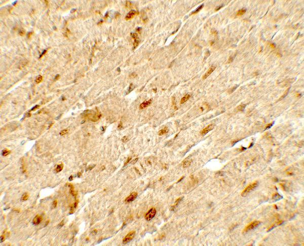
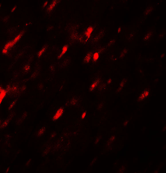
A02929-1 has rat heart IHC and IF images (catalog: A02929-1 image captions); its listed reactivity covers human, mouse, and rat (catalog: A02929-1 reactivity).

Real IHC data Immunohistochemistry of ANGPTL3 in rat heart tissue with ANGPTL3 antibody at 5 μg/mL.
Anti-Angiopoietin-related protein 3 ANGPTL3 Antibody
Cat # A02929-1

A02929-1 is the sole rendered SKU and lists IHC-P and IF applications (cards payload; catalog: A02929-1 applications). Its images show rat heart IHC at 5 μg/mL and IF at 20 μg/mL (catalog: A02929-1 image captions).

Which to pick: Choose A02929-1 for paraffin-section tissue IHC: IHC-P is listed, and its own caption shows rat heart staining at 5 μg/mL; the fixative is unreported (catalog: A02929-1 applications and IHC caption). For tissue IF, A02929-1 has a rat heart image; for ICC/IF, M02929-1 is a rabbit monoclonal, clone 24A86, with ICC and IF listed but no image supplied (catalog: A02929-1 IF caption; catalog: M02929-1 clone, applications, and image alts). Both list human, mouse, and rat reactivity, while the supplied tissue images document rat samples only (catalog: A02929-1 and M02929-1 reactivity; catalog: A02929-1 image captions).

Each figure is that product's own IHC / IF validation image from its datasheet.

References

  1. UniProt Consortium. UniProt entry Q9Y5C1 (ANGL3_HUMAN, Angiopoietin-related protein 3).
  2. Human Protein Atlas. ANGPTL3 tissue IHC expression (reliability: Approved).
  3. Human Protein Atlas. ANGPTL3 subcellular location (ICC-IF): Secreted.
  4. Human Protein Atlas. ANGPTL3 antibody validation summary (1 antibodies).
  5. Urinary ANGPTL3: a novel noninvasive biomarker for podocyte injury in pediatric glomerular diseases. Journal of translational medicine 2026 — PMC13551765.
  6. Angiopoietin-like protein 3 blocks nuclear import of FAK and contributes to sorafenib response. British journal of cancer 2018 — PMC6134083.
  7. ANGPTL3 is involved in kidney injury in high-fat diet-fed mice by suppressing ACTN4 expression. Lipids in health and disease 2022 — PMC9487085.
  8. Anti-ANGPTL3 antibody and IL-22 fusion protein mitigate nephrotic syndrome via mitochondrial protection, anti-apoptosis, and autophagy suppression. Antibody therapeutics 2026 — PMC13463637.
  9. PubMed PMID:10644446 — UniProt-cited evidence.
  10. PubMed PMID:12975309 — UniProt-cited evidence.
  11. PubMed PMID:14702039 — UniProt-cited evidence.