ANP32E / Acidic leucine-rich nuclear phosphoprotein 32 family member E · IHC design guide

Design Immunohistochemistry for ANP32E

Plan chromogenic IHC-P for ANP32E using the catalog antibody at a starting concentration of 2 μg/mL (datasheet). Assess nuclear staining against the widespread tissue pattern while accounting for the reported specificity caveat (HPA tissue IHC).

Evidence assembled Oct 2026 · For research use; verify linked source records and product datasheet before use
Immunohistochemistry protocol sheet for ANP32E (IHC for ANP32E): expected localisation Nuclear staining in tissue (HPA tissue IHC); cytoplasm listed (UniProt), antibody A07456, validated IHC image, and IHC protocol steps
Printable ANP32E IHC protocol sheet — expected localisation Nuclear staining in tissue (HPA tissue IHC); cytoplasm listed (UniProt), antibody A07456, controls and protocol steps. Open the full ANP32E IHC guide →

ANP32E Immunohistochemistry Experimental Design Guide

Expected localisation, validated protocols, controls and antibodies — the at-a-glance facts below, then the full design guide.

Must know before staining
Expected localisation Nuclear staining in tissue (HPA tissue IHC); cytoplasm listed (UniProt)
Staining pattern Widespread nuclear staining across tissue cells (HPA tissue IHC)
Antigen retrieval Tris-EDTA pH 9.0 HIER, 95–98 °C, 20 min (rule: nuclear antigen)
Positive control ⓘ Adrenal gland+4 more · see all
Negative control ⓘ None in HPA (detected in all 45 tissues); use no-primary + isotype controls
Important caveats
Reasons your staining may differ from the expected pattern.
Fixation Matched tissue-IHC evidence does not establish the fixation claim. Validate the specimen-specific method before use. (selected-SKU IHC image A07456)
Caveat Staining may reflect proteins from more than one gene (HPA tissue IHC)
Regulation Low tissue specificity; no regulator established (HPA tissue IHC)
Isoform / epitope 3 isoforms; epitope effects are unknown (UniProt)
Section 1

Recommended ANP32E IHC & IF Protocols

Compare the catalog antibody’s IHC-P protocol with published ANP32E protocols for kidney sections (PMC9576520) and tissue sections (PMC13194895).

Recommended immunohistochemistry (IHC-P) protocol parameters
SampleTissue sections; selected-image fixative not specified (standard IHC workflow)
FixationImage fixative and duration unreported (datasheet A07456); verify before use.
Sectioning4–5 µm sections on charged slides (standard)
DeparaffinisationXylene, graded ethanol series to water (standard)
Antigen retrievalHeat-induced epitope retrieval in Tris-EDTA buffer, pH 9.0, 20 min at 95–98 °C (standard rule: nuclear antigen)
Peroxidase block3% H2O2, 10 min, room temperature (standard)
Blocking10% normal serum of the secondary host, 30 min, room temperature (standard)
Primary antibodyRabbit anti-ANP32E, 2 μg/mL (datasheet A07456)
Primary incubationOvernight at 4 °C (standard)
DetectionHRP-polymer secondary, DAB chromogen 5–10 min (standard)
CounterstainHematoxylin, blue, dehydrate and mount (standard)
Expected resultANP32E-positive staining in glandular cells of adrenal gland (HPA tissue IHC: High). HPA tissue profile: Ubiquitous nuclear expression. No signal in the no-primary control.
💡Decision noteStart with Tris-EDTA pH 9.0 HIER at 95–98 °C for 20 min (page retrieval); PMC13194895 also uses pH 9.0.
Section 2

What Is the Expected ANP32E Staining Pattern?

ANP32E should stain mainly nuclei across many cell types in paraffin sections; HPA describes ubiquitous nuclear expression and rates its tissue IHC evidence Supported, with medium agreement between staining and RNA data (HPA tissue IHC). High staining is reported in colon glandular cells, bronchial respiratory epithelial cells and bone marrow hematopoietic cells (HPA tissue IHC). UniProt also places ANP32E in the cytoplasm and reports no transmembrane segment (UniProt Q9BTT0).

What am I looking at on my slide?
Predominantly nuclear chromogen in colon glandular cells, with clear cell boundaries.This fits the reported high glandular-cell staining and ubiquitous nuclear profile (HPA tissue IHC). Judge intensity against the chosen tissue and its negative control; a high HPA category is a reference observation, not an absolute staining threshold (HPA tissue IHC; general IHC practice).
Strong staining confined to cell membranes, with little or no nuclear signal.Membrane-only staining conflicts with the reported nuclear tissue profile and the absence of a transmembrane segment (HPA tissue IHC; UniProt Q9BTT0). Treat it as an unexpected pattern and investigate nonspecific staining or detection artefact before interpreting it as ANP32E (general IHC practice).
Signal appears chiefly in an unexpected cell population while expected nuclei are blank.Compare the cell identity with the annotated population for that tissue; HPA reports high staining in bronchial respiratory epithelial cells and bone marrow hematopoietic cells (HPA tissue IHC). An isolated unexpected pattern may reflect cross-reactivity or endogenous detection activity (general IHC practice). HPA also cautions that the antibodies target proteins from more than one gene (HPA tissue IHC).
Diffuse chromogen obscures nuclei and extends over stroma or empty spaces.This prevents a confident compartment call (general IHC practice). Check the negative control and inspect blocking, washing and detection background before assigning positive cells (general IHC practice). HPA's nuclear profile cannot validate a diffuse field-wide deposit (HPA tissue IHC).
No staining is visible in the expected cells of a high-staining reference tissue.A blank colon glandular-cell or bronchial epithelial-cell reference conflicts with the reported high staining in those populations (HPA tissue IHC). First check that the reference section contains the annotated cells, then assess the IHC run and controls; absence alone does not establish absent ANP32E (general IHC practice).
💡Expected ANP32E appearanceCall positive when identifiable cells show predominantly nuclear chromogen, including strong staining in an HPA high category such as colon glandular cells; membrane-only or diffuse field-wide colour is an unexpected result requiring control review (HPA tissue IHC; UniProt Q9BTT0; general IHC practice).
How each factor affects the staining
Compartment and processingTarget-specific fixation effects are not established by the supplied assay evidence. Verify with a matched IHC source before attributing a result to fixation.
Isoforms and epitope coverageThree isoforms are listed (UniProt Q9BTT0). Without an antibody epitope or isoform coverage in the supplied evidence, a stain cannot identify which isoform contributes to the signal; interpret the result as antibody-dependent ANP32E-family staining rather than an isoform-specific measurement (UniProt Q9BTT0; HPA tissue IHC caveat).
Evidence strength and specificityHPA calls the tissue IHC pattern Supported, describes medium consistency with RNA, and warns that the antibodies target proteins from more than one gene; external verification is pending (HPA tissue IHC). Its three listed antibodies have Supported IHC status, which supports pattern comparison but does not resolve every unexpected positive cell (HPA antibodies; HPA tissue IHC).
IF/ICC question: should cytosolic or centrosomal signal be expected?HPA's ICC-IF summary calls nucleoplasm the main approved location and cytosol and centrosome additional approved locations, with a cross-gene antibody caution (HPA subcellular). That cellular localisation context does not replace the predominantly nuclear tissue IHC expectation (HPA tissue IHC).
Why is my staining missing, weak or wrong?
SituationLikely causeNext action
High-staining reference tissue gives no nuclear signal.The section may lack the annotated cells, or the IHC run may have failed; HPA reports high staining in colon glandular cells and bronchial respiratory epithelial cells (HPA tissue IHC; general IHC practice).Confirm cell identity and section quality, then review the positive control, antibody incubation, chromogen development and run records (general IHC practice). Do not infer a target-specific fixation defect from this result; none is documented in the supplied sources (HPA tissue IHC; UniProt Q9BTT0).
Membrane-only or predominantly extracellular staining appears.This distribution does not match ubiquitous nuclear tissue staining or the reported nonmembrane topology (HPA tissue IHC; UniProt Q9BTT0). Nonspecific staining or detection artefact is possible (general IHC practice).Compare the negative control and repeat interpretation in identifiable cells; assess blocking and detection background before scoring (general IHC practice). Retain any persistent discordant pattern as unresolved, given HPA's cross-gene caution (HPA tissue IHC).
Unexpected cell types stain more strongly than the annotated population.The signal could reflect tissue-dependent expression, cross-reactivity or endogenous detection activity; HPA's tissue annotations and cross-gene caution limit a specificity claim (HPA tissue IHC; general IHC practice).Identify the stained cells morphologically, compare the corresponding HPA tissue entry, and examine a negative control for endogenous or nonspecific colour (HPA tissue IHC; general IHC practice). Avoid assigning the unexpected cells as ANP32E-positive from intensity alone (general IHC practice).
Diffuse brown background makes nuclei hard to score.Nonspecific binding, insufficient washing or detection background can obscure chromogenic IHC interpretation (general IHC practice). HPA reports a nuclear tissue profile, not a diffuse background pattern (HPA tissue IHC).Review the negative control, blocking and wash steps, then assess whether individual nuclei remain distinguishable (general IHC practice). If they do not, mark the field uninterpretable and repeat the run under controlled conditions (general IHC practice).
Cytoplasmic colour accompanies clear nuclear staining.UniProt lists both nucleus and cytoplasm; HPA ICC-IF additionally approves cytosol, although tissue IHC is described as ubiquitously nuclear (UniProt Q9BTT0; HPA subcellular; HPA tissue IHC).Score the nuclear component separately and check that cytoplasmic colour exceeds the negative-control background (general IHC practice). Report mixed localisation descriptively; do not use the ICC-IF finding as proof that a cytoplasm-only tissue result is specific (HPA subcellular; HPA tissue IHC).
A tissue listed as low shows faint staining.HPA lists low staining in caudate neuronal cells, oral mucosa squamous epithelial cells and stomach glandular cells; low is not a negative-control designation (HPA tissue IHC).Compare the relevant cell population and nuclear compartment with the HPA entry, then judge faint colour against the run's negative control (HPA tissue IHC; general IHC practice). Report weak interpretable staining separately from background or a completely blank result (general IHC practice).

Sample controls for ANP32E IHC & IF

🧪Run bone marrow first; hematopoietic cells should stain (HPA: High in bone marrow hematopoietic cells). HPA detects ANP32E in all 45 scored tissues, so use no-primary and isotype controls for the negative; any cells without specific signal on the positive slide should show counterstain only, but no cell type is established as an internal negative (HPA: no negative tissue rows).
Positive control tissue: Adrenal gland (Glandular cells, HPA High)
Negative control tissue: None in HPA: ANP32E is detected in all 45 scored tissues. Use a no-primary (secondary-only) and an isotype control instead.
ICC-IF cell lines (HPA subcellular resource): HPA ICC-IF images show ANP32E in MCF-7, Rh30, U2OS, HEK293, SK-MEL-30, with annotated localisation: Nucleoplasm (approved) (HPA subcellular).
Technical controls: Include a no-primary, secondary-only control, an isotype control matched to the primary antibody’s host species and immunoglobulin class (or matched nonimmune host immunoglobulin for a polyclonal antibody), and ANP32E knockout material as a biological negative (standard IHC practice). For chromogenic bone marrow IHC, block endogenous peroxidase and check the no-primary slide for residual signal in hematopoietic cells (standard IHC practice).
⚠️Feasibility: A target-specific fixation window and the effect of antigen retrieval are unreported in the supplied evidence; the selected A07456 small-intestine IHC caption also leaves the fixative unreported (caption: fixative not stated). ICC-IF images exist for ANP32E, but the supplied evidence does not establish whether frozen sections or IF are easier than paraffin IHC (HPA: ICC-IF images in MCF-7, Rh30, U2OS, HEK293 and SK-MEL-30). Endogenous peroxidase in bone marrow hematopoietic cells can complicate chromogenic interpretation (standard IHC practice).

HPA tissue IHC evidence for ANP32E

Comprehensive Human Protein Atlas IHC scoring per tissue (reliability: Supported — Medium consistency between antibody staining and RNA expression data. Caution, targets protein from more than one gene. Pending external verification.). Rows are taken directly from the HPA tissue chart — click any row's HPA link to view the source.

Positive expression · recommended positive controls

TissueCell typeLevelEvidenceSource
Adrenal gland Glandular cells High Protein (IHC) HPA →
Bone marrow Hematopoietic cells High Protein (IHC) HPA →
Breast Glandular cells High Protein (IHC) HPA →
Bronchus Respiratory epithelial cells High Protein (IHC) HPA →
Cerebellum Cells in molecular layer High Protein (IHC) HPA →

Undetected expression · recommended negative controls

TissueCell typeLevelEvidenceSource
None in HPA: ANP32E is detected in all 45 scored tissues. Use a no-primary (secondary-only) and an isotype control instead.
Section 3

Advanced ANP32E IHC Tips

Troubleshoot ANP32E staining in paraffin sections by checking retrieval, compartment, controls and cell-level scoring before interpreting chromogenic signal.

How should I retrieve ANP32E in paraffin sections with weak nuclear staining?
Start with Tris-EDTA at pH 9.0 for heat-induced retrieval at 95–98 °C for 20 minutes (page retrieval rule: nuclear antigen). Allow sections to cool consistently, then compare staining with a known positive section processed in the same run (standard IHC practice; HPA: ubiquitous nuclear expression). If signal remains weak, compare a milder retrieval condition on serial sections while holding antibody concentration and detection constant; no alternative buffer is established for this antibody here (standard IHC practice; supplied evidence: no antibody-specific retrieval result). Judge improvement by sharper nuclear signal without increased diffuse or edge staining, and keep the chosen condition fixed across specimens (HPA: ubiquitous nuclear expression; standard IHC practice).
Could fixation explain variable ANP32E staining between paraffin blocks?
ANP32E-specific sensitivity to fixation is unknown from the supplied evidence; the small-intestine image caption reports 2 µg/mL antibody but does not state a fixative (catalog antibody A07456 caption). Record fixation duration and processing history for each block before comparing staining, because variable processing can change epitope accessibility in paraffin IHC (standard IHC practice). Compare serial sections from blocks with documented processing in one staining run, using identical retrieval, antibody incubation and chromogen development (standard IHC practice). Treat any association between processing and signal as a finding to validate with controls, rather than inferring a target-specific fixation effect from ANP32E localisation or tissue expression (UniProt Q9BTT0: nucleus and cytoplasm; HPA: tissue IHC profile).
How should I interpret nuclear versus cytoplasmic ANP32E staining?
Prioritise crisp nuclear staining when evaluating paraffin sections, since the tissue profile describes ubiquitous nuclear expression and the subcellular profile places ANP32E mainly in the nucleoplasm (HPA: tissue IHC profile; HPA: subcellular profile). Cytoplasmic signal is biologically plausible because UniProt lists both nucleus and cytoplasm, while HPA also reports cytosol (UniProt Q9BTT0: subcellular location; HPA: subcellular profile). Score nuclear and cytoplasmic compartments separately across matched cell types, and compare each with background in the same section (standard IHC practice). Interpret isolated centrosomal-looking puncta cautiously: that HPA observation uses antibodies targeting proteins from multiple genes and cannot establish this antibody's specificity (HPA: subcellular caution).
Can this antibody distinguish ANP32E isoforms or detect every cellular pool?
The record lists 3 ANP32E isoforms, but supplies no epitope map or isoform-reactivity data for the catalog antibody (UniProt Q9BTT0: isoforms; supplied antibody evidence). Therefore, neither pan-isoform detection nor isoform-specific staining follows from a positive paraffin section (supplied antibody evidence: epitope unspecified; standard IHC interpretation). ANP32E has no transmembrane segment or signal peptide and contains an LRRCT region at residues 123–161, yet those annotations do not locate this antibody's epitope (UniProt Q9BTT0: topology, processing and domain). If isoform discrimination matters, use independently validated isoform-specific reagents or orthogonal expression measurements and report the antibody's unknown coverage (standard IHC practice; supplied antibody evidence).
How can IF help check the cell type and compartment behind ANP32E IHC staining?
For a separate IF experiment, multiplex ANP32E with a validated marker for the cell type being examined and a nuclear counterstain, then check whether the signal occupies the expected cells and nucleoplasm (HPA: mainly nucleoplasmic; standard IF practice). Choose fluorophores in channels with low measured tissue autofluorescence, and include single-stain and no-primary controls before interpreting overlap (standard IF practice). Permeabilise for intracellular access, because ANP32E has no transmembrane segment and is reported in nucleus and cytoplasm; optimise detergent exposure so nuclear structure remains readable (UniProt Q9BTT0: topology and location; standard IF practice). Treat cytosolic or centrosomal IF patterns as provisional because the HPA subcellular evidence carries a multi-gene antibody caution (HPA: subcellular profile).
What should I change when ANP32E chromogenic staining is diffuse or patchy?
Check a no-primary control and examine whether colour follows tissue edges, folds or endogenous pigment rather than cell nuclei (standard IHC practice; HPA: ubiquitous nuclear expression). For peroxidase-based detection, apply the routine endogenous-peroxidase block and confirm that its control removes enzyme-derived colour; this is a general workflow measure, not ANP32E-specific evidence (standard chromogenic IHC practice). Titrate the catalog antibody around its reported 2 µg/mL small-intestine image concentration while keeping retrieval and DAB development fixed; that caption does not specify fixation (catalog antibody A07456 caption; standard IHC practice). If diffuse signal persists, compare blocking and wash conditions on serial sections and retain only conditions preserving clear compartment contrast (standard IHC practice; UniProt Q9BTT0: nucleus and cytoplasm).
How should I quantify ANP32E staining across paraffin sections? ⚠ ANSWER MARKED FOR VERIFICATION
Choose a prespecified cell population and score nuclear ANP32E as percentage positive or an H-score using fixed intensity thresholds across the staining run (HPA: ubiquitous nuclear expression; standard IHC quantification practice). Report cytoplasmic staining separately if measured, because ANP32E is also annotated in cytoplasm and combining compartments can obscure a change in distribution (UniProt Q9BTT0: subcellular location; standard IHC quantification practice). Normalise cell counts to the number of eligible cells, or positive-cell density to analysed viable area in mm², after excluding folds and necrosis (standard IHC quantification practice). Include matched controls and document threshold settings, since HPA reports medium RNA–protein consistency and a multi-gene antibody caution (HPA: tissue IHC reliability).
What makes an apparent ANP32E-positive cell convincing rather than artefactual?
A convincing result shows reproducible cell-associated staining with a clear nuclear component, consistent with the HPA tissue profile and ANP32E's reported nuclear location (HPA: ubiquitous nuclear expression; UniProt Q9BTT0: subcellular location). Compare the cell identity and compartment with adjacent morphology and controls; an unexpected isolated cell type needs independent confirmation because this antibody's specificity is not established by the supplied caption (standard IHC interpretation; catalog antibody A07456 caption). Discount staining confined to section edges, folds or necrotic areas, and check no-primary sections for endogenous enzyme colour (standard chromogenic IHC practice). Avoid equating intensity with H2A.Z removal: that is an ANP32E function, whereas staining measures antibody-detected protein distribution (UniProt Q9BTT0: function; standard IHC interpretation).
Boster reagents

Best ANP32E / Acidic leucine-rich nuclear phosphoprotein 32 family member E IHC Antibodies

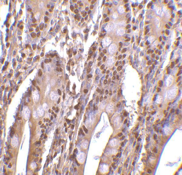
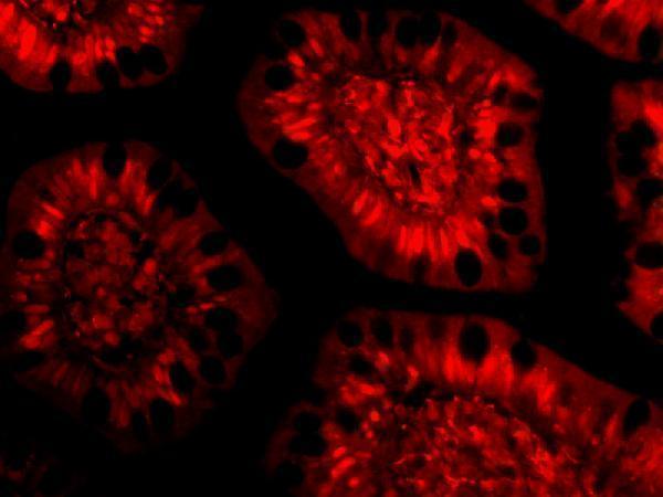
A07456 provides human small-intestine IHC and IF images (catalog image captions), with IHC-P and IF applications and Human, Mouse, and Rat reactivity listed (catalog applications and reactivity).

Real IHC data Immunohistochemistry of PHAP III in human small intestine tissue with PHAP III antibody at 2 μg/mL.
Anti-PHAP III ANP32E Antibody
Cat # A07456

A07456 has an IHC image of human small-intestine tissue at 2 μg/mL (catalog IHC image caption). Its IF image shows human small-intestine cells at 10 μg/mL (catalog IF image caption); the application list includes IHC-P and IF (catalog applications).

Which to pick: Choose A07456 for paraffin-section tissue IHC: IHC-P is listed, and its own IHC image shows human small-intestine tissue at 2 μg/mL (catalog applications; catalog IHC image caption). For IF/ICC planning, A07456 has an IF application and a human small-intestine cell image at 10 μg/mL; ICC processing is unreported (catalog applications; catalog IF image caption). A07456 lists Human, Mouse, and Rat reactivity, while its clone and the IHC specimen’s fixative are unreported (catalog reactivity; catalog clone field; catalog IHC image caption).

Each figure is that product's own IHC / IF validation image from its datasheet.

References

  1. UniProt Consortium. UniProt entry Q9BTT0 (AN32E_HUMAN, Acidic leucine-rich nuclear phosphoprotein 32 family member E).
  2. Human Protein Atlas. ANP32E tissue IHC expression (reliability: Supported).
  3. Human Protein Atlas. ANP32E subcellular location (ICC-IF): Mainly localized to the nucleoplasm. In addition localized to the cytosol and centrosome. Caution: Based on antibodies targeting proteins from multiple genes..
  4. Human Protein Atlas. ANP32E antibody validation summary (3 antibodies).
  5. Inhibiting ANP32E protects against acute kidney injury by regulating autophagy via the AMPK pathway. iScience 2025 — PMC12275872.
  6. Anp32e promotes renal interstitial fibrosis by upregulating the expression of fibrosis-related proteins. International journal of biological sciences 2022 — PMC9576520.
  7. ANP32E drives lung adenocarcinoma progression via GSK3β-mediated glycolytic reprogramming. Cell death & disease 2026 — PMC13194895.
  8. ANP32E induces tumorigenesis of triple-negative breast cancer cells by upregulating E2F1. Molecular oncology 2018 — PMC5983205.
  9. PubMed PMID:12438741 — UniProt-cited evidence.
  10. PubMed PMID:14702039 — UniProt-cited evidence.
  11. PubMed PMID:17974005 — UniProt-cited evidence.