This website uses cookies to ensure you get the best experience on our website.


- Table of Contents
Plan ARHGEF3 paraffin-section IHC around the reported nuclear tissue staining (HPA tissue IHC) and its cytoplasmic annotation (UniProt). Compare positive cell populations carefully because staining and RNA expression show low consistency (HPA tissue IHC).

Expected localisation, validated protocols, controls and antibodies — the at-a-glance facts below, then the full design guide.
| Expected localisation | Nuclear staining in tissue (HPA tissue IHC); cytoplasmic annotation (UniProt) | |
| Staining pattern | Nuclear staining in glandular, hematopoietic and neuronal cells (HPA tissue IHC) | |
| Antigen retrieval | Tris-EDTA pH 9.0 HIER, heat-mediated (datasheet A09012-1) | |
| Positive control | Appendix+4 more · see all | |
| Negative control | None in HPA (detected in all 45 tissues); use no-primary + isotype controls |
| Fixation | Keep paraffin-section fixation consistent. (standard IHC practice; not target-specific) | |
| Caveat | Tissue staining has low consistency with RNA expression (HPA tissue IHC) | |
| Regulation | Widely expressed; regulation unspecified (UniProt) | |
| Isoform / epitope | 4 isoforms; variant epitope coverage is unknown (UniProt) |
Compare the catalog antibody’s IHC-P protocol (datasheet A09012-1) with a published ARHGEF3 protocol for nasopharyngeal carcinoma tissue microarrays (PMC5041948).
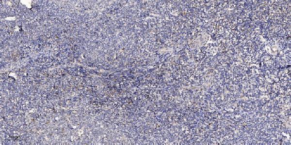
| Sample | Paraffin-embedded human tonsil tissue; fixative not specified (datasheet A09012-1) |
| Fixation | Image fixative and duration unreported (datasheet A09012-1); verify before use. |
| Sectioning | 4–5 µm sections on charged slides (standard) |
| Deparaffinisation | Xylene, graded ethanol series to water (standard) |
| Antigen retrieval | Heat retrieval: Tris-EDTA pH 9.0 (datasheet A09012-1); 20 min, 95–100 °C (standard) |
| Peroxidase block | 3% H2O2, 10 min, room temperature (standard) |
| Blocking | 10% normal serum of the secondary host, 30 min, room temperature (standard) |
| Primary antibody | Rabbit anti-ARHGEF3, 1:50 recommended; image 1:200 (datasheet A09012-1) |
| Primary incubation | Overnight at 4 °C (standard) |
| Detection | HRP-polymer secondary, DAB chromogen 5–10 min (standard) |
| Counterstain | Hematoxylin, blue, dehydrate and mount (standard) |
| Expected result | ARHGEF3-positive staining in glandular cells of appendix (HPA tissue IHC: High). HPA tissue profile: Nuclear expression in all tissues. No signal in the no-primary control. |
ARHGEF3 is a cytoplasmic protein with no transmembrane segment (UniProt Q9NR81). Cytosolic signal is also supported by approved ICC-IF localisation (HPA: cytosol, approved). In paraffin IHC, examine the named cells in HPA high-staining tissues, including appendix glandular cells and cerebral cortex neuronal cells (HPA: High). HPA reports nuclear staining across tissues, but rates its tissue IHC pattern Uncertain because antibody staining and RNA data have low consistency (HPA: IHC Uncertain).
| Cytoplasmic staining in appendix glandular cells or cerebral cortex neuronal cells. | This fits the reported protein compartment and named high-staining cells (UniProt Q9NR81: cytoplasm; HPA: cytosol approved; HPA: High in these cells). Interpret the slide alongside controls: HPA's tissue IHC reliability remains Uncertain, so a matching appearance alone does not establish antibody specificity (HPA: IHC Uncertain). |
| Predominantly nuclear staining, especially when it appears across many cell types. | Treat this as a discordant compartment and investigate a possible staining artefact (UniProt Q9NR81: cytoplasm; HPA: cytosol approved). HPA does report nuclear expression in all tissues, but labels that tissue IHC pattern Uncertain; nuclear signal therefore needs independent confirmation before it is assigned to ARHGEF3 (HPA: tissue IHC). |
| Strong signal in adipocytes while the expected glandular or neuronal cells show little staining. | Adipocytes are listed as low-staining, whereas appendix glandular and cerebral cortex neuronal cells are High (HPA: tissue IHC). Check cell identification and controls; an inverted pattern can reflect cross-reactivity or endogenous detection activity (general IHC practice). HPA's Uncertain IHC rating limits how firmly any one tissue comparison can establish specificity (HPA: IHC Uncertain). |
| Diffuse colour across cells, stroma, and the empty areas of a section. | A pattern without clear cellular boundaries cannot be scored as ARHGEF3 localisation (general IHC practice). Check whether the same colour appears in a no-primary control, and review blocking, washes, and chromogen development before comparing tissue staining levels (general IHC practice). |
| No discernible signal in the named high-staining cells of an otherwise interpretable section. | First check the positive control and detection workflow (general IHC practice). HPA lists several high-staining cell populations, but its tissue IHC reliability is Uncertain; an HPA High call is a useful comparison, not a guarantee that every section or antibody run will stain (HPA: tissue IHC). |
| Compartment evidence | UniProt places ARHGEF3 in the cytoplasm, and HPA approves cytosolic ICC-IF localisation (UniProt Q9NR81; HPA: ICC-IF approved). HPA's nuclear tissue IHC profile has Uncertain reliability, so the two observations should be reported separately rather than merged into a single expected compartment (HPA: tissue IHC). |
| Tissue and cell selection | HPA calls appendix glandular, bone marrow hematopoietic, and cerebral cortex neuronal cells High, while adipocytes and soft-tissue fibroblasts are Low (HPA: tissue IHC). These provide relative staining comparisons; HPA reports low tissue RNA specificity and an Uncertain IHC profile, so neither group is an absolute positive or negative control (HPA: tissue IHC). |
| Antibody evidence | The listed tissue IHC antibody HPA034715 is rated Uncertain, whereas HPA064945 is approved for ICC-IF and has no supplied IHC rating (HPA: antibody validation). An ICC-IF approval cannot establish performance of a paraffin IHC assay; assess the IHC-validated antibody and its controls in the intended detection workflow (general IHC practice). |
| Protein form and epitope | UniProt lists 4 isoforms, a 1–526 chain, no signal peptide or propeptide, and no transmembrane segment (UniProt Q9NR81). No epitope location or isoform-specific staining evidence is supplied; these sequence facts cannot predict which isoforms an antibody detects or justify a processing-related tissue pattern. |
| Detection chemistry | In chromogenic IHC, endogenous enzyme activity can produce colour independent of primary-antibody binding (general IHC practice). A no-primary control helps identify that contribution; it does not resolve whether primary-dependent staining comes from ARHGEF3 or another bound antigen (general IHC practice). |
| Situation | Likely cause | Next action |
|---|---|---|
| Nuclear colour dominates while cytoplasm is faint. | The slide conflicts with UniProt cytoplasmic and HPA approved cytosolic localisation, although HPA reports an Uncertain nuclear tissue IHC pattern (UniProt Q9NR81; HPA: ICC-IF approved; HPA: IHC Uncertain). | Check the no-primary control and nuclear counterstain, then verify any primary-dependent nuclear pattern with independent specificity evidence before calling it ARHGEF3 (general IHC practice). |
| Adipocytes stain more strongly than named High cell populations. | This reverses the supplied HPA contrast and may reflect mistaken cell assignment, cross-reactivity, or endogenous detection activity (HPA: adipocytes Low; HPA: selected glandular and neuronal cells High; general IHC practice). | Recheck morphology and compare matched controls and tissue compartments on the same run; report the discordance without treating HPA's Uncertain profile as a definitive exclusion rule (general IHC practice; HPA: IHC Uncertain). |
| Diffuse colour obscures cell boundaries. | Nonspecific reagent binding, insufficient washing, or excessive chromogen development can produce background in chromogenic IHC (general IHC practice). | Compare a no-primary section, review blocking and wash steps, and reduce detection development as appropriate; score only signal with identifiable cellular localisation (general IHC practice). |
| No signal appears in appendix glandular or cerebral cortex neuronal cells. | These cells are reported High by HPA, but a failed control, detection step, or antibody run can also yield a blank slide (HPA: tissue IHC; general IHC practice). | Confirm that an appropriate positive control stained, then review the catalog antibody's IHC-P instructions for retrieval and dilution and inspect the detection reagents (general IHC practice). No ARHGEF3-specific retrieval condition is supplied. |
| Colour appears on a no-primary section. | The colour does not require the primary antibody; endogenous detection activity or reagent background is possible (general IHC practice). | Address the detection system's endogenous-activity block and reagent background, then repeat the no-primary control before interpreting ARHGEF3 staining (general IHC practice). |
| A positive control stains, but the test section remains negative. | The control confirms that detection worked in that run, while the test section's cell composition or antigen signal may differ (general IHC practice). HPA's High categories do not guarantee staining in every specimen (HPA: IHC Uncertain). | Verify the expected cell population on the counterstained section and document the negative result with the control outcome; seek independent evidence before concluding that ARHGEF3 is absent (general IHC practice). |


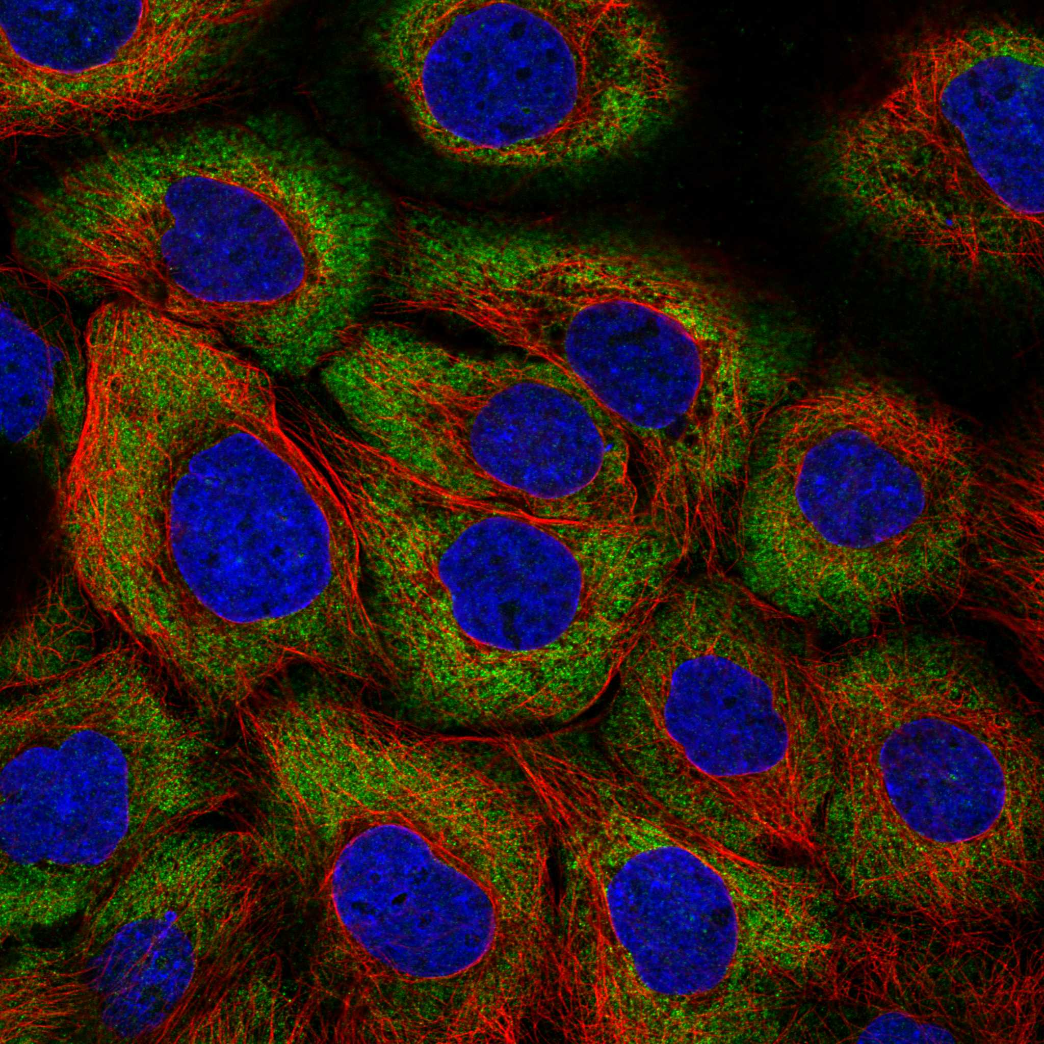
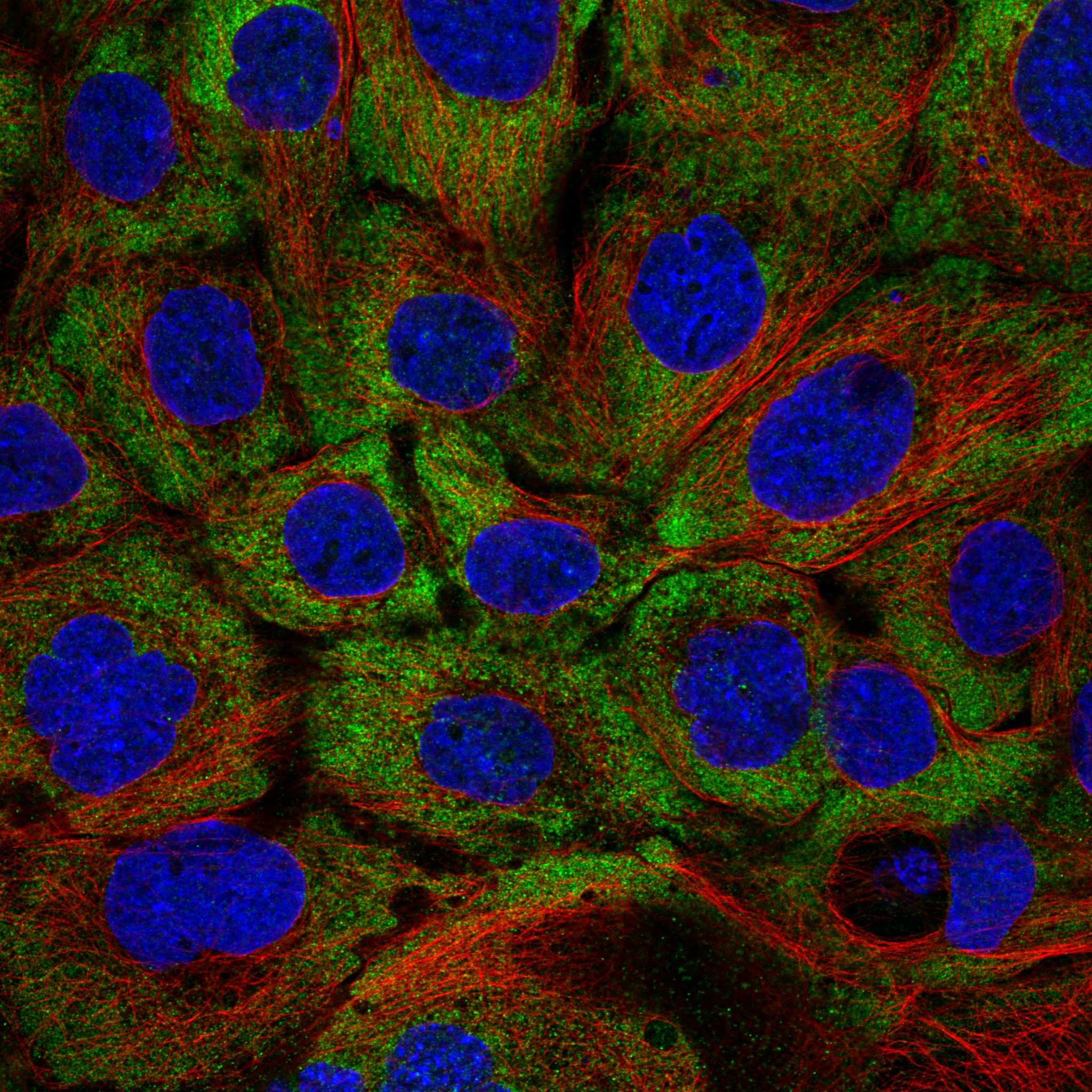
Comprehensive Human Protein Atlas IHC scoring per tissue (reliability: Uncertain — Low consistency between antibody staining and RNA expression data.). Rows are taken directly from the HPA tissue chart — click any row's HPA link to view the source.
| Tissue | Cell type | Level | Evidence | Source |
|---|---|---|---|---|
| Appendix | Glandular cells | High | Protein (IHC) | HPA → |
| Bone marrow | Hematopoietic cells | High | Protein (IHC) | HPA → |
| Breast | Glandular cells | High | Protein (IHC) | HPA → |
| Cerebral cortex | Neuronal cells | High | Protein (IHC) | HPA → |
| Cervix | Squamous epithelial cells | High | Protein (IHC) | HPA → |
| Tissue | Cell type | Level | Evidence | Source |
|---|---|---|---|---|
| None in HPA: ARHGEF3 is detected in all 45 scored tissues. Use a no-primary (secondary-only) and an isotype control instead. | ||||
Troubleshoot ARHGEF3 chromogenic IHC in paraffin sections by checking retrieval, compartment, controls and scoring; use IF as a secondary localisation check.
A09012-1 has a paraffin-section IHC image from human tonsil (image caption). IF is listed for a human-, mouse- and rat-reactive antibody, but no IF image is supplied (catalog applications/reactivity; IF image alts).
A09012-1 is shown staining paraffin-embedded human tonsil by IHC, with antibody diluted 1:200 and Tris-EDTA retrieval at pH 9.0 (image caption). A09012-1 also lists IF and human, mouse and rat reactivity; no IF specimen or image is provided (catalog applications/reactivity; IF image alts).
Which to pick: For tissue IHC, choose A09012-1 for the demonstrated paraffin-section use; the fixative is unreported (image caption). For IF/ICC, A09012-1 lists IF, while ICC use and an IF image are unreported (catalog applications; IF image alts). For work across species, A09012-1 lists human, mouse and rat reactivity, although its IHC image shows only human tissue and clonality is unreported (catalog reactivity/clone; image caption).