This website uses cookies to ensure you get the best experience on our website.


- Table of Contents
Plan chromogenic IHC for ASF1B in paraffin sections with the catalog antibody at 1:100–1:300 (datasheet A05211). Assess nuclear staining in testis spermatogonia and use adipose tissue as a negative reference (HPA tissue IHC).

Expected localisation, validated protocols, controls and antibodies — the at-a-glance facts below, then the full design guide.
| Expected localisation | Nuclear staining in tissue IHC (HPA tissue IHC) | |
| Staining pattern | Nuclear staining in testis spermatogonia (HPA tissue IHC) | |
| Antigen retrieval | Tris-EDTA pH 9.0 HIER, heat-mediated (datasheet A05211) | |
| Positive control | Cerebral cortex+4 more · see all | |
| Negative control | Adipose tissue+4 more · see all |
| Fixation | Matched tissue-IHC evidence does not establish the fixation claim. Validate the specimen-specific method before use. (selected-SKU IHC image A05211) | |
| Caveat | Antibody staining and RNA show medium consistency (HPA tissue IHC) | |
| Regulation | Testis-high; lower in colon and thymus (UniProt) | |
| Isoform / epitope | No annotated isoforms or processing; one 1–202 chain (UniProt) |
The catalog antibody protocol (datasheet A05211) is accompanied by a published ASF1B IHC protocol for mouse tumor sections (PMC9782623).
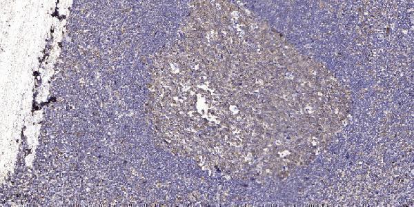
| Sample | Paraffin-embedded human tonsil tissue; fixative not specified (datasheet A05211) |
| Fixation | Image fixative and duration unreported (datasheet A05211); verify before use. |
| Sectioning | 4–5 µm sections on charged slides (standard) |
| Deparaffinisation | Xylene, graded ethanol series to water (standard) |
| Antigen retrieval | Heat retrieval: Tris-EDTA pH 9.0 (datasheet A05211); 20 min, 95–100 °C (standard) |
| Peroxidase block | 3% H2O2, 10 min, room temperature (standard) |
| Blocking | 10% normal serum of the secondary host, 30 min, room temperature (standard) |
| Primary antibody | Rabbit anti-ASF1B, 1:100 - 1:300 (datasheet A05211) |
| Primary incubation | Overnight at 4 °C (standard) |
| Detection | HRP-polymer secondary, DAB chromogen 5–10 min (standard) |
| Counterstain | Hematoxylin, blue, dehydrate and mount (standard) |
| Expected result | ASF1B-positive staining in neuronal cells of cerebral cortex (HPA tissue IHC: Medium). HPA tissue profile: Nuclear expression in several tissues, including testis. No signal in the no-primary control. |
ASF1B staining should be predominantly nuclear in IHC, especially in testis spermatogonia and glandular cells of the duodenum and small intestine (HPA tissue IHC: medium staining; reliability Enhanced). HPA reports medium consistency between staining and RNA data (HPA tissue IHC). ASF1B is also annotated in the cytosol and has no transmembrane segment, so limited cytoplasmic signal is biologically plausible (UniProt Q9NVP2: subcellular location and topology).
| Nuclear staining in testis spermatogonia, with nuclei distinguishable from the counterstain (HPA tissue IHC). | This matches the reported ASF1B pattern: HPA scores spermatogonial staining as Medium and describes nuclear expression in several tissues, including testis (HPA tissue IHC). Judge the signal by its location and cell type; the HPA score does not require every cell on the section to stain. |
| Predominantly cytoplasmic staining with little nuclear contrast in a known-positive section. | Recheck the result because HPA describes nuclear tissue staining and nucleoplasmic ICC-IF localization (HPA tissue IHC; HPA subcellular). Cytosolic ASF1B is annotated, so cytoplasmic signal alone does not prove an artefact (UniProt Q9NVP2). Compare with a no-primary control and the nuclear pattern in the same tissue (general IHC practice). |
| Strong staining in adipocytes while the expected positive cells are faint. | Consider nonspecific antibody binding or endogenous chromogenic activity: HPA reports ASF1B as Not detected in adipocytes, but Medium in testis spermatogonia (HPA tissue IHC). A negative tissue result is a comparison point, not proof that every stained adipocyte is cross-reactivity; inspect controls and staining location (general IHC practice). |
| A diffuse deposit covers nuclei and surrounding tissue, obscuring cell boundaries. | The slide cannot reliably show the reported nuclear pattern (HPA tissue IHC). Check whether a no-primary section also develops color, then review blocking, washes and chromogen development for background (general IHC practice). Do not score diffuse deposit as ASF1B-positive cells. |
| No nuclear signal is visible in testis spermatogonia. | This conflicts with the Medium staining reported for that cell type (HPA tissue IHC). First check that the spermatogonia are identifiable and the staining run worked; then review antibody application, retrieval and detection using routine IHC controls (general IHC practice). A single blank section cannot establish biological absence. |
| Tissue and cell selection | Testis spermatogonia and duodenal or small-intestinal glandular cells are reported at Medium staining; colon glandular cells are Low, and adipocytes are Not detected (HPA tissue IHC). Compare like cell populations when judging a weak or absent result. |
| Antibody evidence | HPA lists IHC validation as Enhanced for HPA069385, while HPA054036 has no listed IHC status and has Supported ICC status (HPA antibodies). The tissue profile has Enhanced reliability with medium staining-to-RNA consistency (HPA tissue IHC); neither label validates an untested antibody. |
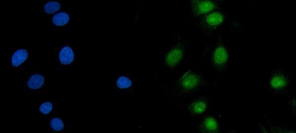
| IF/ICC Q: Where should fluorescence appear? | A: HPA reports enhanced nucleoplasmic localization in ICC-IF (HPA subcellular). This supports a nuclear localization check, while the IHC tissue profile supplies the cell-type comparisons for this paraffin-section guide (HPA tissue IHC). |
| Situation | Likely cause | Next action |
|---|---|---|
| Known-positive testis has no visible nuclear staining. | The run may have failed, or the intended cells may be missed; HPA reports Medium staining in spermatogonia (HPA tissue IHC). | Verify cell identity and run controls, then review the applied antibody, retrieval and detection steps (general IHC practice). |
| Only weak staining appears in colon glandular cells. | Low staining is reported for that population, so a faint result can fit the reference profile (HPA tissue IHC). | Compare with testis spermatogonia or small-intestinal glandular cells reported at Medium, using the same run (HPA tissue IHC; general IHC practice). |
| Cytoplasmic color dominates with little nuclear detail. | HPA reports nuclear tissue staining, although UniProt also annotates cytosolic ASF1B (HPA tissue IHC; UniProt Q9NVP2). | Compare nuclear contrast and a no-primary control before calling the cytoplasmic signal specific (general IHC practice). |
| Background persists across multiple tissue regions. | Nonspecific binding or detection background can obscure a nuclear readout (general IHC practice). | Inspect a no-primary section; review blocking, washing and chromogen development against the run controls (general IHC practice). |
| Adipocytes stain as strongly as the expected positive cells. | That conflicts with HPA's Not detected adipocyte result and may reflect cross-reactivity or endogenous activity (HPA tissue IHC; general IHC practice). | Check the no-primary control, staining compartment and antibody specificity before assigning ASF1B positivity (general IHC practice). |
| Two antibodies give different IHC patterns. | Their listed validation differs: HPA069385 is IHC Enhanced; HPA054036 has no listed IHC status (HPA antibodies). | Compare each result with the HPA nuclear tissue pattern and appropriate run controls; do not transfer ICC validation to IHC (HPA tissue IHC; HPA antibodies; general IHC practice). |


Comprehensive Human Protein Atlas IHC scoring per tissue (reliability: Enhanced — Medium consistency between antibody staining and RNA expression data.). Rows are taken directly from the HPA tissue chart — click any row's HPA link to view the source.
| Tissue | Cell type | Level | Evidence | Source |
|---|---|---|---|---|
| Cerebral cortex | Neuronal cells | Medium | Protein (IHC) | HPA → |
| Duodenum | Glandular cells | Medium | Protein (IHC) | HPA → |
| Epididymis | Glandular cells | Medium | Protein (IHC) | HPA → |
| Oral mucosa | Squamous epithelial cells | Medium | Protein (IHC) | HPA → |
| Small intestine | Glandular cells | Medium | Protein (IHC) | HPA → |
| Tissue | Cell type | Level | Evidence | Source |
|---|---|---|---|---|
| Adipose tissue | Adipocytes | Not detected | Protein (IHC) | HPA → |
| Bone marrow | Hematopoietic cells | Not detected | Protein (IHC) | HPA → |
| Bronchus | Respiratory epithelial cells | Not detected | Protein (IHC) | HPA → |
| Caudate | Glial cells | Not detected | Protein (IHC) | HPA → |
| Cerebellum | Cells in granular layer | Not detected | Protein (IHC) | HPA → |
Troubleshoot ASF1B staining by assessing nuclear signal, tissue context and controls alongside the catalog antibody’s paraffin-section IHC conditions.
Anti-ASF1B options include paraffin-section IHC in human tonsil (A05211 image caption) and IF/ICC in MCF-7 cells (A05211-2 image caption); catalog reactivity covers human, mouse and rat (A05211 catalog).
A05211 will render with human tonsil paraffin-section IHC data (A05211 image caption); its listed reactivity is human, mouse and rat (A05211 catalog). A05211-2 will render with IF/ICC data from MCF-7 cells (A05211-2 image caption) and is listed as human-reactive (A05211-2 catalog).
Which to pick: Choose A05211 for tissue IHC: its own image shows paraffin-embedded human tonsil with Tris-EDTA retrieval at pH 9.0 and primary antibody at 1:200 overnight (A05211 image caption); the fixative is unreported (A05211 image caption). Choose A05211-2 for IF/ICC in human cells because its own image documents MCF-7 staining at 5 μg/mL (A05211-2 image caption) and IHC is absent from its application list (A05211-2 catalog). For mouse or rat samples, A05211 is the option with listed reactivity (A05211 catalog); both antibodies are polyclonal (catalog dilution data).