ASIC2 / Acid-sensing ion channel 2 · IHC design guide

Design Immunohistochemistry for ASIC2

Plan ASIC2 chromogenic IHC in paraffin sections using the observed CNS cytoplasmic pattern as a staining reference (HPA tissue IHC). The guide covers neuronal membrane topology (UniProt), control tissue selection (HPA tissue IHC), and interpretation of possible off-target staining (HPA tissue IHC).

Evidence assembled Oct 2026 · For research use; verify linked source records and product datasheet before use
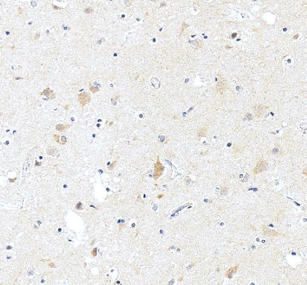
Immunohistochemistry protocol sheet for ASIC2 (IHC for ASIC2): expected localisation CNS cytoplasm in tissue IHC; neuronal plasma membrane is the molecular location (HPA tissue IHC; UniProt), antibody A04055-1, validated IHC image, and IHC protocol steps
Printable ASIC2 IHC protocol sheet — expected localisation CNS cytoplasm in tissue IHC; neuronal plasma membrane is the molecular location (HPA tissue IHC; UniProt), antibody A04055-1, controls and protocol steps. Open the full ASIC2 IHC guide →

ASIC2 Immunohistochemistry Experimental Design Guide

Expected localisation, validated protocols, controls and antibodies — the at-a-glance facts below, then the full design guide.

Must know before staining
Expected localisation CNS cytoplasm in tissue IHC; neuronal plasma membrane is the molecular location (HPA tissue IHC; UniProt)
Staining pattern Cytoplasmic CNS staining in neurons and neuropil (HPA tissue IHC)
Antigen retrieval EDTA pH 8.0 HIER, heat-mediated (datasheet A04055-1)
Positive control ⓘ Cerebral cortex+3 more · see all
Negative control ⓘ Adipose tissue+4 more · see all
Important caveats
Reasons your staining may differ from the expected pattern.
Fixation Keep paraffin tissue fixation consistent across specimens. (selected-SKU IHC image A04055-1)
Caveat Presumed off-target binding can complicate interpretation (HPA tissue IHC)
Regulation Expression regulation is unreported (UniProt)
Isoform / epitope 2 isoforms; check whether the epitope is extracellular or cytoplasmic (UniProt)
Section 1

Recommended ASIC2 IHC & IF Protocols

Compare the catalog antibody’s IHC-P protocol (datasheet A04055-1) with a published colorectal cancer tissue protocol (PMC5606037).

Recommended immunohistochemistry (IHC-P) protocol parameters
SampleParaffin-embedded human brain tissue; fixative not specified (datasheet A04055-1)
FixationImage fixative and duration unreported (datasheet A04055-1); verify before use.
Sectioning4–5 µm sections on charged slides (standard)
DeparaffinisationXylene, graded ethanol series to water (standard)
Antigen retrievalHeat retrieval: EDTA pH 8.0 (datasheet A04055-1); 20 min, 95–100 °C (standard)
Peroxidase block3% H2O2, 10 min, room temperature (standard)
Blocking10% goat serum (datasheet A04055-1)
Primary antibodyRabbit anti-ASIC2, 2-5μg/ml (datasheet A04055-1)
Primary incubationOvernight at 4 °C (datasheet A04055-1)
DetectionHRP-conjugated secondary, DAB chromogen (datasheet A04055-1)
CounterstainHematoxylin, blue, dehydrate and mount (standard)
Expected resultASIC2-positive staining in neuropil of cerebral cortex (HPA tissue IHC: High). HPA tissue profile: Cytoplasmic expression in CNS. No signal in the no-primary control.
💡Decision noteStart with EDTA pH 8.0 heat retrieval for the catalog antibody (datasheet A04055-1); consider citrate pH 6.0 as a published alternative (PMC5606037).
Section 2

What Is the Expected ASIC2 Staining Pattern?

ASIC2 is a two-pass membrane channel localized to neuronal plasma membranes, including the soma and punctate peripheral processes (UniProt Q16515 topology and subcellular location). In paraffin-section IHC, expect staining in CNS neuropil and neuronal populations: HPA reports high staining in cerebral cortex neuropil and hippocampal neurons (HPA tissue IHC). HPA rates its tissue pattern Enhanced, while noting that presumed off-target binding was disregarded and external verification is pending (HPA tissue IHC).

What am I looking at on my slide?
Cerebral cortex shows strong neuropil staining; hippocampal neuronal cells stain strongly.This matches the reported high-staining populations (HPA tissue IHC). Assess distribution within the section: ASIC2 is assigned to neuronal plasma membranes, soma and punctate peripheral processes (UniProt Q16515 subcellular location). A chromogenic section may show a broader neuropil pattern than a sharply outlined cell border (general IHC interpretation).
Caudate neurons or cerebellar granular-layer cells stain less strongly than the main positive controls.Medium staining in those populations is consistent with HPA observations; high staining is reported for hippocampal neurons and cerebral cortex neuropil (HPA tissue IHC). Compare like tissue regions and use the same scoring criteria across sections (general IHC practice).
Nuclei dominate the signal, with little staining in neuronal processes or soma.A nuclear-dominant result conflicts with ASIC2's membrane assignment and reported neuronal distribution (UniProt Q16515 subcellular location; HPA tissue IHC). Treat it as suspect, check the counterstain and detection controls, and reassess antibody specificity (general IHC practice).
Strong staining appears in an HPA-negative cell population, such as cervical glandular cells.HPA reports cervical glandular cells as not detected by tissue IHC, despite group-enriched cervical RNA (HPA tissue IHC and RNA specificity). Unexpected chromogen may reflect cross-reactivity or endogenous detection activity; the discrepancy needs controls before it is called ASIC2 (general IHC interpretation).
Signal is diffuse across the section, or absent from cerebral cortex neuropil and hippocampal neurons.Diffuse colour lacks the reported cell and region pattern; absent signal misses two high-staining positive populations (HPA tissue IHC). Interpret either result alongside tissue preservation and assay controls, rather than inferring ASIC2 expression from colour alone (general IHC practice).
💡Expected ASIC2 appearanceCall a result positive when high staining tracks cerebral cortex neuropil or hippocampal neuronal cells, with a neuronal soma/process distribution consistent with membrane-localized ASIC2; prominent nuclear or HPA-negative glandular staining is suspect (HPA tissue IHC; UniProt Q16515 subcellular location).
How each factor affects the staining
Positive and comparison tissueCerebral cortex neuropil and hippocampal neurons are high-staining reference populations; caudate neurons and cerebellar granular-layer cells are medium (HPA tissue IHC). Adipocytes and cervical glandular cells are reported not detected, so they can help reveal nonspecific signal without serving as proof of specificity (HPA tissue IHC; general IHC interpretation).
Antibody validationThe reported tissue pattern has Enhanced reliability, and HPA052112 is rated Enhanced for IHC (HPA tissue IHC; HPA antibodies). HPA also says presumed off-target binding was disregarded and external verification is pending; interpret unusual staining cautiously (HPA tissue IHC).
Channel topology and epitope locationASIC2 has cytoplasmic residues 1–42 and 440–512, an extracellular region at 65–424, and two membrane spans (UniProt Q16515 topology). The supplied evidence does not locate the antibody epitope; topology alone cannot select an antigen-retrieval condition or predict epitope access in paraffin sections.
Isoforms and modificationsUniProt lists 2 isoforms and glycosylation sites at residues 365 and 392 (UniProt Q16515). Without an antibody epitope or isoform-reactivity record, do not assign a staining difference to an isoform or glycosylation state.
IF/ICC Q&A: what localization should be expected?HPA summarizes subcellular localization as membrane but supplies no ICC-IF image cell lines or main-location detail; its listed antibody has no ICC validation entry (HPA subcellular; HPA antibodies). Neuronal soma and punctate peripheral-process membrane staining is the UniProt expectation, not an IF/ICC-validated pattern (UniProt Q16515 subcellular location).
Why is my staining missing, weak or wrong?
SituationLikely causeNext action
No stain in the high-staining CNS reference sections.The result conflicts with high cerebral cortex neuropil and hippocampal neuronal staining (HPA tissue IHC); an assay failure remains possible (general IHC practice).Check tissue integrity, primary-antibody application and chromogen controls; review the catalog antibody's IHC-P instructions before adjusting dilution or retrieval (general IHC practice). The supplied evidence gives no ASIC2-specific retrieval condition.
Strong staining appears mainly in nuclei.Nuclear dominance conflicts with the neuronal membrane location (UniProt Q16515 subcellular location); counterstain or detection artefact is possible (general IHC interpretation).Inspect a no-primary control and the chromogen/counterstain balance; repeat localization scoring in neuronal soma and processes (general IHC practice). Do not score nuclear colour as confirmed ASIC2.
Diffuse colour covers positive and negative tissue alike.A uniform pattern lacks the HPA CNS distribution and may indicate nonspecific detection or inadequate blocking or washing (HPA tissue IHC; general IHC practice).Compare a no-primary control, review blocking and washes, and score cell-specific staining after background is controlled (general IHC practice). HPA-negative cell populations provide a useful comparison (HPA tissue IHC).
Cervical glandular cells or adipocytes stain strongly.HPA reports both cell populations as not detected by IHC; cervical RNA group enrichment does not itself establish glandular protein staining (HPA tissue IHC and RNA specificity).Check no-primary and detection controls for endogenous activity, then reassess antibody specificity against the CNS positive pattern (general IHC practice; HPA tissue IHC).
Caudate or cerebellum stains more weakly than hippocampus.This ordering may be expected: HPA reports medium staining in caudate neurons and cerebellar granular-layer cells versus high staining in hippocampal neurons (HPA tissue IHC).Score the named cell populations separately and compare sections processed together before treating lower intensity as assay failure (general IHC practice).
IF/ICC membrane signal is used to validate a paraffin-section IHC result.HPA gives an IF subcellular summary but no ICC-IF image cell lines or ICC antibody validation entry (HPA subcellular; HPA antibodies).Judge the IHC slide against its tissue distribution and IHC controls (HPA tissue IHC; general IHC practice). Treat the IF membrane summary as localization context, not independent IHC validation.

Sample controls for ASIC2 IHC & IF

🧪Start with cerebral cortex and require neuropil staining (HPA: High in cerebral-cortex neuropil); use adipose tissue as the negative tissue (HPA: Not detected in adipocytes). Adjacent unstained cells on the cortex slide can show background, but HPA does not establish those cells as ASIC2-negative (HPA: High in cerebral-cortex neuropil).
Positive control tissue: Cerebral cortex (Neuropil, HPA High)
Negative control tissue: Adipose tissue (HPA Not detected)
ICC-IF cell lines (HPA subcellular resource): HPA carries no ICC-IF cell line for ASIC2; derive a cell-line control from the positive tissue's cell type (Neuropil) and confirm it by RNA or western blot first.
Technical controls: Include a no-primary, secondary-only control and a matched rabbit immunoglobulin isotype control (selected-SKU caption: rabbit primary antibody). Use ASIC2 knockout tissue as a target-specific control if available; quench endogenous peroxidase for chromogenic detection and check brain autofluorescence if adapting the assay to IF (selected-SKU caption: brain tissue, HRP/DAB).
⚠️Feasibility: The selected-SKU paraffin-section caption uses heat retrieval in EDTA at pH 8.0, but does not report the fixative (selected-SKU caption: EDTA pH 8.0; fixative unreported). A target-specific fixation window and fixation effect are unreported, and the supplied evidence does not establish whether frozen sections or IF are easier; HPA lists no ICC-IF image-bearing cell lines (HPA subcellular: no ICC-IF cell lines). For brain IF, assess tissue autofluorescence before interpreting the neuronal signal (HPA: High in cerebral-cortex neuropil and hippocampal neuronal cells).

HPA tissue IHC evidence for ASIC2

Comprehensive Human Protein Atlas IHC scoring per tissue (reliability: Enhanced — Presumed off target binding observed and disregarded. Pending external verification.). Rows are taken directly from the HPA tissue chart — click any row's HPA link to view the source.

Positive expression · recommended positive controls

TissueCell typeLevelEvidenceSource
Cerebral cortex Neuropil High Protein (IHC) HPA →
Hippocampus Neuronal cells High Protein (IHC) HPA →
Caudate Neuronal cells Medium Protein (IHC) HPA →
Cerebellum Cells in granular layer Medium Protein (IHC) HPA →

Undetected expression · recommended negative controls

TissueCell typeLevelEvidenceSource
Adipose tissue Adipocytes Not detected Protein (IHC) HPA →
Adrenal gland Glandular cells Not detected Protein (IHC) HPA →
Appendix Glandular cells Not detected Protein (IHC) HPA →
Bone marrow Hematopoietic cells Not detected Protein (IHC) HPA →
Breast Adipocytes Not detected Protein (IHC) HPA →
Section 3

Advanced ASIC2 IHC Tips

Troubleshoot ASIC2 staining in paraffin sections using the catalog antibody’s tissue result, ASIC2 topology, and tissue expression as reference points.

Which antigen retrieval conditions should I try first for ASIC2 IHC?
Start with heat-mediated retrieval in EDTA pH 8.0 for paraffin sections (datasheet A04055-1). This is the retrieval used for the catalog antibody’s human brain section stained at 2 μg/ml; the caption does not report the fixative (datasheet A04055-1). If staining is weak, compare a carefully controlled retrieval time series on adjacent sections while holding antibody concentration, detection, and development constant (standard IHC practice). Excessive retrieval can damage section morphology or increase nonspecific signal, so judge each condition against tissue preservation, no-primary controls, and the expected neuronal pattern (standard IHC practice; UniProt Q16515 localisation).
How should I troubleshoot fixation-related loss of ASIC2 staining?
Target-specific fixation sensitivity is unknown: the catalog image identifies a paraffin-embedded human brain section but does not state its fixative (datasheet A04055-1). Record fixative, fixation duration, specimen thickness, and processing history for each section, then compare matched samples before attributing weaker staining to fixation (standard IHC practice). Use EDTA pH 8.0 retrieval and the reported 2 μg/ml starting concentration when reproducing that image’s staining conditions (datasheet A04055-1). If samples differ in fixation history, assess morphology and no-primary background alongside ASIC2 staining, and avoid treating a change in intensity alone as a biological difference (standard IHC practice).
What staining distribution fits ASIC2, and when is cytoplasmic signal concerning?
ASIC2 is annotated at the neuronal plasma membrane, including the soma and punctate peripheral processes (UniProt Q16515 localisation). The tissue atlas also reports cytoplasmic expression in the CNS, with high staining in cerebral cortex neuropil and hippocampal neuronal cells (HPA tissue IHC). Examine whether apparent cytoplasmic DAB follows neuronal somata or dense processes, using matched counterstain and serial sections to resolve crowded tissue (standard IHC practice; HPA tissue IHC). A diffuse signal across unrelated cell types or section edges warrants review of blocking, detection controls, and morphology before it is scored as ASIC2 (standard IHC practice; UniProt Q16515 localisation).
How does epitope location affect interpretation of ASIC2 staining?
ASIC2 has 2 annotated isoforms, while the supplied antibody caption does not identify its recognized isoform or epitope (UniProt Q16515 isoforms; datasheet A04055-1). Its topology places residues 1–42 and 440–512 on the cytoplasmic side and residues 65–424 extracellularly, between 2 membrane segments (UniProt Q16515 topology). Accordingly, document whether any independently established epitope is shared across isoforms before calling absent staining isoform-specific (standard IHC practice; UniProt Q16515 isoforms). Glycosylation sites at 365 and 392 and phosphorylation sites at 8 and 11 are annotated, but their effects on this antibody’s IHC signal are unreported (UniProt Q16515 modifications; datasheet A04055-1).
How should I adapt ASIC2 localisation checks for multiplex immunofluorescence?
For a separate IF experiment, pair ASIC2 with a validated neuronal marker and assess signal in neuronal somata and processes, its annotated location (UniProt Q16515 localisation; standard IF practice). Select spectrally separated fluorophores after checking tissue autofluorescence in unstained and secondary-only sections, particularly where weak puncta could be mistaken for signal (standard IF practice). Set permeabilisation according to the verified epitope: intracellular termini lie at residues 1–42 and 440–512, whereas residues 65–424 are extracellular (UniProt Q16515 topology; standard IF practice). The supplied 2 μg/ml concentration and EDTA pH 8.0 retrieval describe a paraffin-section chromogenic IHC image, so establish IF conditions with IF controls (datasheet A04055-1; standard IF practice).
What controls help diagnose diffuse or speckled ASIC2 DAB background?
The catalog tissue image used 10% goat serum blocking, an overnight 4°C primary incubation at 2 μg/ml, and DAB detection (datasheet A04055-1). Compare a no-primary section with the stained section to identify detection-system or endogenous peroxidase signal, and include a peroxidase-block step in the chromogenic workflow (standard IHC practice). If background persists, titrate primary concentration and DAB development while keeping retrieval at the reported EDTA pH 8.0 starting condition (standard IHC practice; datasheet A04055-1). Inspect edge staining, folds, and damaged regions separately from neuronal soma and neuropil staining before adjusting the protocol (standard IHC practice; HPA tissue IHC).
How should I quantify ASIC2 IHC across brain sections? ⚠ ANSWER MARKED FOR VERIFICATION
Define anatomical regions and cell compartments before scoring, since high staining is reported in cerebral cortex neuropil and hippocampal neuronal cells (HPA tissue IHC). For neuronal somata, report percent positive cells or an H-score from prespecified intensity categories; for neuropil, report DAB-positive area or optical density per tissue area in mm² (standard IHC practice). Normalise cell counts to counterstain-defined eligible cells and area measurements to intact region area, excluding folds, edges, and damaged tissue by the same rules for every section (standard IHC practice). Keep retrieval, imaging, DAB development, and threshold settings consistent, and analyse biological samples separately from technical fields (standard IHC practice).
How can I distinguish true ASIC2 staining from tissue artefact?
Give greatest weight to staining aligned with neuronal somata or peripheral processes, consistent with ASIC2’s neuronal membrane annotation and reported CNS tissue pattern (UniProt Q16515 localisation; HPA tissue IHC). Interpret broad staining in unrelated cells cautiously: the tissue atlas notes presumed off-target binding that was disregarded and says external verification is pending (HPA tissue IHC). Compare stained and no-primary sections for endogenous enzyme signal, and reject edge effects, folds, and necrotic areas when scoring DAB (standard IHC practice). Concordance with a neuronal pattern supports interpretation, but morphology and an independent specificity control remain necessary before assigning unexpected compartments or cell types to ASIC2 (standard IHC practice; UniProt Q16515 localisation).
Boster reagents

Best ASIC2 / Acid-sensing ion channel 2 IHC Antibodies

ASIC2 antibody A04055-1 has human brain paraffin-section IHC and A431-cell IF images; human, mouse and rat reactivity is listed (catalog image captions; catalog reactivity).

Real IHC data IHC analysis of ASIC2 using anti-ASIC2 antibody (A04055-1). ASIC2 was detected in a paraffin-embedded section of human brain tissue. Heat mediated antigen retrieval was performed in EDTA buffer (pH 8.0, epitope retrieval solution). The tissue section was blocked with 10% goat serum. The tissue section was then incubated with 2 μg/ml rabbit anti-ASIC2 Antibody (A04055-1) overnight at 4°C. Peroxidase Conjugated Goat Anti-rabbit IgG was used as secondary antibody and incubated for 30 minutes at 37°C. The tissue section was developed using HRP Conjugated Rabbit IgG Super Vision Assay Kit (Catalog # SV0002) with DAB as the chromogen.
Anti-ACCN1/ASIC2 Antibody ®
Cat # A04055-1

A04055-1 will render with a human brain paraffin-section IHC image using 2 μg/mL primary antibody (A04055-1 IHC image caption). The same SKU lists IHC, IF and ICC and has an A431-cell IF image; human, mouse and rat reactivity is listed (catalog applications/reactivity; A04055-1 IF image caption).

Which to pick: For tissue IHC, choose A04055-1: its own image documents a human brain paraffin section with EDTA retrieval at pH 8.0; the fixative is unreported (A04055-1 IHC image caption). For IF/ICC, the same SKU lists both applications and shows A431-cell IF (catalog applications; A04055-1 IF image caption). For cross-species work, A04055-1 lists human, mouse and rat reactivity, while its supplied IHC image documents human tissue (catalog reactivity; A04055-1 IHC image caption).

Each figure is that product's own IHC / IF validation image from its datasheet.

References

  1. UniProt Consortium. UniProt entry Q16515 (ASIC2_HUMAN, Acid-sensing ion channel 2).
  2. Human Protein Atlas. ASIC2 tissue IHC expression (reliability: Enhanced).
  3. Human Protein Atlas. ASIC2 subcellular location (ICC-IF): Membrane.
  4. Human Protein Atlas. ASIC2 antibody validation summary (1 antibodies).
  5. The acid-sensing ion channel, ASIC2, promotes invasion and metastasis of colorectal cancer under acidosis by activating the calcineurin/NFAT1 axis. Journal of experimental & clinical cancer research : CR 2017 — PMC5606037.
  6. Potential Implications of Acid-Sensing Ion Channels ASIC2 and ASIC4 in Gonadal Differentiation of Dicentrarchus labrax Subjected to Water Temperature Increase during Gonadal Development. Animals : an open access journal from MDPI 2024 — PMC11010900.
  7. Expression Profiles of ASIC1/2 and TRPV1/4 in Common Skin Tumors. International journal of molecular sciences 2021 — PMC8199644.
  8. Expression of Acid-Sensing Ion Channels in Renal Tubular Epithelial Cells and Their Role in Patients with Henoch-Schönlein Purpura Nephritis. Medical science monitor : international medical journal of experimental and clinical research 2017 — PMC5408900.
  9. PubMed PMID:8626462 — UniProt-cited evidence.
  10. PubMed PMID:8631835 — UniProt-cited evidence.
  11. PubMed PMID:9037075 — UniProt-cited evidence.